Shoulder Pain
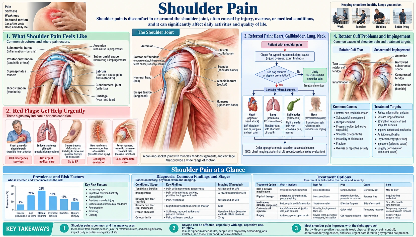
Table of Contents
- What Shoulder Pain Feels Like
- Red Flags: Get Help Urgently
- Referred Pain: Heart, Gallbladder, Lung, Neck
- Rotator Cuff Problems and Impingement
- Frozen Shoulder (Adhesive Capsulitis)
- Arthritis and Inflammatory Causes
- Other Common Causes
- How Shoulder Pain Is Evaluated
- What Actually Helps
- What to Expect Over Time
- Connections
- Key Research Papers
- Featured Videos
What Shoulder Pain Feels Like
Shoulder pain rarely announces itself with one clear injury. It creeps in: you cannot reach the top shelf without wincing, and then — the detail almost everyone mentions — you cannot sleep on that side anymore. Point prevalence in adults runs between 7% and 26%, and primary care sees about 15 new shoulder complaints per 1,000 patients a year. Where the pain sits, and what provokes it, tells you most of what you need:
- Deep ache on the outer upper arm, worst lifting sideways between about 60 and 120 degrees, worse at night — the rotator cuff pattern, and by far the commonest.
- Severe pain first, stiffness later, the shoulder refusing to rotate outward so you cannot reach behind your back — frozen shoulder.
- A pinpoint sore spot on the bony bump on top, sore reaching across your chest — the acromioclavicular (AC) joint.
- Deep pain with grinding, and motion limited in every direction rather than only overhead — arthritis of the ball-and-socket joint.
- Pain from the neck shooting past the elbow, with numbness in specific fingers — a pinched nerve, not a shoulder problem.
- Pain unrelated to how you move your arm, arriving with exertion, nausea or sweating — referred pain, from the heart, gallbladder, lung or diaphragm.
Nearly all shoulder pain is mechanical, and nearly all of it improves. But the shoulder is one of the body's favourite places to display a warning from an internal organ, and telling the two apart is learnable.
Red Flags: Get Help Urgently
Most shoulder pain can safely wait for a routine appointment. The following cannot.
Call emergency services now
- Shoulder or arm pain with chest pressure, breathlessness, cold sweat, nausea or lightheadedness. Possible heart attack — and it does not require crushing chest pain. In a registry analysis of over 430,000 confirmed heart attacks, about one in three had no chest pain at all, and they died more often because recognition was slower — women, people with diabetes and adults over 75 most of all.
- Pain at the very tip of the shoulder with abdominal pain, faintness, or after abdominal trauma. Blood or infection under the diaphragm refers pain to the shoulder tip via the phrenic nerve (Kehr's sign) — a ruptured spleen, a perforated ulcer, a ruptured ectopic pregnancy. Left shoulder-tip pain with abdominal pain and a missed period needs a pregnancy test today.
- A hot, swollen, exquisitely painful shoulder with fever that you cannot bear to have moved — possible septic arthritis, which destroys cartilage within days and needs urgent aspiration and intravenous antibiotics. Also urgent: sudden tearing pain between the shoulder blades with unequal pulses in the two arms (possible aortic dissection), deformity after a fall with movement impossible, or a cold, pale, pulseless arm.
See a doctor within a day or two
- Right upper abdominal pain radiating to the right shoulder blade after a fatty meal, lasting 30 minutes to several hours with nausea — biliary colic from gallstones; fever, chills or yellow eyes make it urgent.
- New shoulder pain with cough, coughing blood or weight loss, especially in a smoker. A tumour at the top of the lung (Pancoast tumour) can present as shoulder pain months before any chest symptom — look for a drooping eyelid and small pupil on that side, and wasting of the small hand muscles.
- Bilateral shoulder and hip girdle aching with over 45 minutes of morning stiffness, over age 50 — the signature of polymyalgia rheumatica. Roughly one in six also has giant cell arteritis, so a new headache, scalp tenderness, jaw pain while chewing or any visual change is an emergency: untreated, it blinds.
- You cannot lift the arm at all after an injury, or it drops when you lower it slowly — a large acute cuff tear under age 65 is one of the few shoulder problems where earlier surgery gives better results. And night pain that no longer varies with position, with fever, weight loss or any cancer history, needs checking: bone metastases hurt at rest.
Referred Pain: Heart, Gallbladder, Lung, Neck
Nerves from internal organs and nerves from skin and muscle converge on the same neurons in the spinal cord, so your brain gets a signal from the C3–C5 or T1–T4 levels and cannot tell whether it began in the heart, the diaphragm or your shoulder — and it guesses the shoulder. This viscerosomatic convergence is why a heart attack can hurt in your arm.
Four questions separate the two. Does moving the arm change it? — the most useful by far: mechanical pain has a movement that provokes it and a position that relieves it, while referred pain does not care where your arm is. Is there a tender spot? — cuff, bursal and AC joint pain almost always have one you can find with a fingertip. What brings it on? — reaching and lying on it for mechanical pain, whole-body exertion for cardiac pain, eating for biliary pain. What comes with it? — sweating, nausea and shortness of breath point to the heart; none of that belongs to a torn tendon.
The heart
Cardiac pain enters the cord at roughly T1 to T4 and is referred to the chest, the left shoulder and inner arm, the neck, the jaw and between the shoulder blades. It is usually pressure or a heavy weight rather than a sharp stab, and diffuse rather than pinpoint. Pain predictable with exertion and relieved by rest within 1–5 minutes suggests angina; pain lasting over 15–20 minutes with sweating or nausea should be treated as a possible heart attack. Pain reproducible by arm movement and unaffected by walking is far more likely musculoskeletal — but a cuff problem does not protect you from coronary disease. And the left shoulder is classic, not exclusive: right shoulder, both shoulders and isolated jaw pain are all documented heart attack presentations. An ECG takes five minutes and a troponin test an hour — both cheap compared with missing a heart attack.
The gallbladder and diaphragm
The gallbladder sits under the liver, directly beneath the diaphragm, which is supplied by the phrenic nerve from C3, C4 and C5 — the same spinal levels that supply the skin over the shoulder. Hence the pattern of gallbladder disease: pain in the right upper abdomen radiating to the right shoulder blade or shoulder tip, beginning 30 to 60 minutes after a fatty meal and often at night, holding steady for 30 minutes to a few hours, with nausea and tenderness under the right ribs — and crucially, moving the arm does nothing to it.
Any diaphragmatic irritation does the same: internal bleeding, air from laparoscopic surgery (a harmless cause of shoulder ache for a day or two after gallbladder removal), pneumonia at a lung base, a pneumothorax or a pulmonary embolism. There the giveaway is that the pain changes with breathing, not arm position.
The neck
A pinched nerve root in the neck, most often C5 or C6, gives pain over the outer shoulder that is easy to mistake for a cuff problem. It often extends below the elbow into specific fingers, comes with numbness or tingling, worsens when you tip your head toward the painful side and press down on the crown (Spurling's test), and eases when you rest your hand on your head. Passive shoulder motion is usually full — the opposite of frozen shoulder.
Rotator Cuff Problems and Impingement
The shoulder trades stability for range of motion: the head of the arm bone sits against a dish covering only a quarter to a third of the ball, so it is held in place almost entirely by the rotator cuff, four muscles whose tendons blend into one sleeve over the ball. Their job is not raising the arm (the deltoid does that) but holding the ball centred while the deltoid pulls. When the cuff weakens the ball rides upward and the space beneath the bony roof above it, the acromion, narrows, squeezing the supraspinatus tendon and the subacromial bursa — the painful arc between 60 and 120 degrees.
This family accounts for most shoulder pain seen in primary care, now grouped as subacromial pain syndrome — a term that deliberately avoids claiming to know which structure hurts. The spectrum runs from subacromial bursitis (inflammation of the lubricating sac, often the most acutely painful stage), through cuff tendinopathy — degeneration of the tendon substance, where biopsies show disorganised collagen and failed healing rather than inflammation, which is why anti-inflammatories ease the pain but do not fix the tendon — to partial- and full-thickness rotator cuff tears.
Typical symptoms: ache over the outer upper arm just below the bony tip of the shoulder; pain reaching into a back pocket, fastening a bra or reaching a seatbelt; night pain lying on that side, often relieved by sleeping semi-upright; and weakness that may be genuine (a tear) or pain-inhibited. In a full supraspinatus tear there is a drop-arm sign — you can hold the arm up if someone lifts it there, but you cannot lower it smoothly. Age is the dominant risk factor, followed by repeated overhead work (painters, electricians, hairdressers), overhead sport, smoking, diabetes and high cholesterol; a single fall onto an outstretched arm can tear an already degenerated tendon.
The finding that changed how doctors think about this
A pooled analysis of imaging in people with no shoulder symptoms at all found rotator cuff abnormalities in fewer than 10% of those in their twenties, rising to roughly 62% of people in their eighties. A torn cuff on an MRI is a normal finding in an older adult, much as grey hair is — so an MRI showing a tear does not prove the tear is causing your pain. The corollary: in a multicentre study of 452 patients with atraumatic full-thickness tears, a structured 6-to-12 week physical therapy program allowed roughly three-quarters to avoid surgery, and that held at two years. Therapy does not close the hole in the tendon — it retrains the remaining muscles to keep the ball centred, and the pain resolves anyway.
Frozen Shoulder (Adhesive Capsulitis)
Frozen shoulder is a distinct condition, and mistaking it for a cuff problem leads to the wrong treatment. The capsule around the joint inflames and then fibroses — it thickens, tightens and shrinks. The shoulder is not weak; it is physically shorter on the inside, and forcing it makes the inflammation worse. It affects roughly 2% to 5% of the population, rising to something like 10% to 20% in people with diabetes, in whom it is more stubborn and more often bilateral. Other associations: thyroid disease, Parkinson's disease, Dupuytren's contracture, immobilisation after surgery, and a previous frozen shoulder on the other side. It peaks between 40 and 60.
The single most useful sign is loss of passive external rotation. Tuck your elbow against your side, bend it to 90 degrees, then rotate the forearm outward away from your belly. In frozen shoulder this is lost early and badly — often to 20 degrees or less — even when someone else moves the arm while you relax. If a doctor can move your arm through a range you cannot, that argues for a cuff problem; if the arm simply stops, that argues for a frozen capsule.
Phases, outlook and treatment
It moves through three phases: freezing (roughly 2 to 9 months, pain dominating and worst at night while motion is progressively lost), frozen (4 to 12 months, pain settling but stiffness at its worst), and thawing (5 to 24 months, motion returning mostly on its own). Total duration is typically one to three years, and the teaching that it always resolves completely is optimistic: at a mean of about four and a half years in a long-term follow-up study, roughly 59% had normal or near-normal shoulders, 35% mild ongoing symptoms and 6% severe residual limitation.
The UK FROST trial randomised 503 patients to early physiotherapy plus a steroid injection, manipulation under anaesthesia plus injection, or arthroscopic capsular release, and at 12 months all three ended up in broadly similar territory — so start with the least invasive option. Ask for a fasting glucose or HbA1c too: frozen shoulder is sometimes the first visible sign of undiagnosed diabetes.
Arthritis and Inflammatory Causes
Osteoarthritis of the shoulder and AC joint
Cartilage wear in the ball-and-socket joint is less common than in the knee or hip — the shoulder is not weight-bearing — but it happens, particularly after dislocations, fractures or long-standing large cuff tears. The pattern is a deep, dull ache inside the joint, grinding you can hear and feel, and crucially loss of motion in every direction rather than only overhead; X-rays show narrowed joint space and bone spurs (see osteoarthritis). The small AC joint on top of the shoulder wears out earlier, especially after years of bench pressing or overhead work: a sharply localised tender point on the bump.
Rheumatoid arthritis and polymyalgia rheumatica
Rheumatoid arthritis involves the shoulder in most people with long-standing disease, though it usually starts in the hands and feet. What marks inflammatory rather than mechanical joint pain: morning stiffness over 30 to 60 minutes (mechanical stiffness eases in minutes); pain that improves with movement through the day; multiple joints, often symmetrically; swelling and warmth; fatigue, low-grade fever and weight loss; and raised ESR and CRP.
In anyone over 50 with new bilateral shoulder girdle pain, polymyalgia rheumatica belongs high on the list: aching and profound morning stiffness across both shoulders and often both hips, severe enough that people cannot roll over in bed, with markedly raised inflammatory markers. It responds to low-dose prednisone (around 15 mg daily) so dramatically, often within 48 to 72 hours, that the response is itself part of the diagnosis.
Calcific tendinitis, crystal and infectious arthritis
Calcium hydroxyapatite crystals deposit in the cuff tendon, usually supraspinatus. Many deposits are silent, but when one begins to resorb it can produce some of the most severe shoulder pain in medicine — sudden, relentless agony over hours to days in someone with no injury at all, typically aged 30 to 50. It shows on a plain X-ray and is self-limiting. Gout and calcium pyrophosphate disease occasionally attack the shoulder too, producing a red, hot, swollen joint — overlapping exactly with septic arthritis, which is why an acutely inflamed shoulder with fever gets aspirated rather than guessed at.
Other Common Causes
- Biceps tendinopathy, labral tears and instability. The long head of the biceps runs in a groove at the front of the humerus, so its pain is anterior and tender in the groove; rupture may bring a pop and the muscle bunching lower in the arm, and is well tolerated. Labral tears give deep pain and catching in throwers, swimmers and after a fall on an outstretched hand. After a first dislocation, especially before age 25, the symptom becomes apprehension — a feeling the shoulder will slip when you raise and rotate the arm.
- Scapular dyskinesis, myofascial pain and fractures. Desk work with rounded shoulders changes how the shoulder blade sits and narrows the subacromial space functionally though nothing is torn — often the real driver behind stubborn "impingement", and what scapular strengthening fixes. Tight bands in the trapezius and infraspinatus refer pain in patterns that pressing the band reproduces exactly. And a proximal humerus fracture after a fall in an older adult with osteoporosis is easily mistaken for bursitis, so any real fall with new shoulder pain over 65 deserves an X-ray.
How Shoulder Pain Is Evaluated
A competent assessment is mostly history and hands; imaging comes later and answers narrower questions than most patients expect. Worth preparing: whether there was an injury; where the pain is when you point with one finger; whether it goes past the elbow and whether there is numbness; what provokes and relieves it, and above all whether moving the arm changes it at all; night pain; how long morning stiffness lasts (minutes means mechanical, an hour means inflammatory); and your background, including diabetes, thyroid disease, smoking, cancer and cardiac risk factors.
The examination. Your clinician looks for muscle wasting, then measures active range of motion (watching for a painful arc and for shrugging to compensate) and passive range while you relax. That comparison is the crux of the exam. Passive much greater than active suggests cuff weakness or tear; passive equally restricted, especially external rotation, suggests frozen shoulder or arthritis. Strength testing follows, with palpation of the AC joint and biceps groove and a neck screen. A large systematic review found most famous named tests have disappointing accuracy in isolation, so clusters read alongside the history beat any single manoeuvre.
Blood tests are not part of a routine mechanical workup; they answer specific questions. ESR, CRP and a CBC when inflammatory arthritis, polymyalgia rheumatica, giant cell arteritis or infection is considered — the markers run strikingly high in polymyalgia rheumatica and in joint infection. Fasting glucose or HbA1c in frozen shoulder and stubborn tendinopathy, plus TSH, since thyroid disease is linked to frozen shoulder (see hypothyroidism). Rheumatoid factor and anti-CCP when multiple joints are involved, and troponin with an ECG when the story raises a cardiac question — not a shoulder test, but the omission that causes real harm. Joint aspiration is mandatory, not optional, for an acutely hot swollen joint.
Imaging. A plain X-ray in three views (AP, axillary, scapular Y) is the reasonable first image, showing arthritis, calcific deposits, fractures, dislocation and a high-riding humeral head that hints at a large chronic tear. Ultrasound is excellent for cuff and bursa, comparable to MRI for full-thickness tears in experienced hands, and useful for guiding injections. MRI is best for the labrum, partial tears and muscle quality — reserve it for when the answer will change what you do, because an early scan often creates a finding that leads to a procedure that was never going to help. Add a chest X-ray if a Pancoast tumour is suspected, and abdominal ultrasound when the story is biliary.
What Actually Helps
The evidence here is clear and humbling: for most shoulder pain, graded exercise beats almost everything, and several popular procedures do not outperform placebo. Complete rest and a sling are almost always the wrong instinct outside of fractures: immobility produces stiffness, and stiffness is harder to fix than pain. What works is relative rest: stop the provocative activity, usually repeated overhead reaching, while keeping the shoulder moving through whatever range is comfortable, every day.
Exercise therapy and self-care
Exercise is first-line for subacromial pain, cuff tendinopathy and most non-traumatic tears, and a progressive program runs 6 to 12 weeks before you judge it: pendulum swings (lean forward, let the arm hang, let gravity swing it in small circles) for motion without loading the tendon; scapular retraction, squeezing the shoulder blades down and back; resisted external and internal rotation with a band, elbow tucked at the side, the single most valuable cuff exercise; then rows and progressive loading, because tendons remodel in response to load and not to rest. Pain clearly worse the next morning means the load was too high.
At home, the change most people value most is the sleep setup: do not sleep on the painful side, put a pillow under the affected elbow so the arm cannot drop backward, and use a recliner or wedge of pillows in the acute phase. Ice for 15 to 20 minutes after activity, heat before exercise, and move the load, not the shoulder. Stopping smoking matters — it impairs tendon healing and raises the rate of failed cuff repair — as does glucose control.
Medication, and when it is warranted
- Acetaminophen (paracetamol) for mild pain, safer than NSAIDs in older adults and in kidney disease. Oral NSAIDs (ibuprofen, naproxen) are useful for a short course of one to two weeks in a flare, chiefly because they let you sleep and start exercising — they do not repair tendon, and stomach, kidney and cardiovascular effects need watching. Topical NSAIDs suit the AC joint.
- Corticosteroid injection relieves pain mostly in the first 6 to 8 weeks, with little left by 6 months. Use it as a window to do the exercises properly; repeated injections around a tendon are associated with tendon deterioration, so most clinicians limit them to two or three a year. Opioids are not appropriate here.
Surgery, and what the placebo-controlled trials showed
Two randomised trials tested arthroscopic subacromial decompression — shaving bone from the acromion to make more room — against a placebo operation. CSAW (313 UK patients) found the difference between real decompression and sham arthroscopy small and not clinically important; FIMPACT (210 Finnish patients) found no clinically relevant difference at 24 months, persisting at five years. So this operation for this diagnosis mostly delivers what time and exercise deliver, and it is fair to ask about these trials if it is proposed to you. Surgery genuinely is the answer for an acute traumatic full-thickness cuff tear in a younger, active person (promptly, since results decline as the muscle becomes fatty); for a cuff tear with real weakness after a proper 3-to-6 month rehabilitation program; for recurrent dislocation; for advanced arthritis, where shoulder replacement is durable; and for displaced fractures.
Among add-ons, manual therapy gives modest short-term benefit alongside exercise and shockwave therapy has evidence for calcific tendinitis only; platelet-rich plasma, magnesium and collagen are unproven in cuff disease.
What to Expect Over Time
Most shoulder pain improves, but slowly — shoulders are not ankles. A classic primary care cohort following new episodes found only about 21% had fully recovered at 6 months, rising to roughly 49% at 18 months. Knowing that prevents two errors: concluding after three weeks that treatment has failed, and concluding after three months that something must be seriously wrong. But slow is not permanent — improvement continues over a year and beyond, and the people who keep up their exercises after the pain fades are the ones who stay better.
- Subacromial pain / cuff tendinopathy — noticeable improvement in 6 to 12 weeks of consistent exercise; substantial resolution over 3 to 6 months.
- Non-traumatic full-thickness tear treated conservatively — roughly three-quarters do well enough never to need surgery, judged over two years. After a cuff repair, expect a sling for several weeks, active motion around 6 to 12 weeks, strengthening from 3 months, and full recovery commonly at 6 to 12 months.
- Frozen shoulder — 1 to 3 years overall, about 59% ending up normal or near-normal; polymyalgia rheumatica eases within days of starting steroids but is treated for one to two years.
Recovery is slower with delay before starting treatment, higher baseline pain and greater loss of motion, poorly controlled diabetes, smoking, heavy overhead demands at work, involvement of the neck, and depression, anxiety or low expectation of recovery — among the strongest predictors of persistent musculoskeletal pain, which is not a claim the pain is imagined but a reason to treat the whole person.
Go back to your doctor if there is no improvement after 6 to 12 weeks of a genuine exercise program; if weakness worsens or new numbness, tingling or hand weakness appears; if you develop fever, weight loss or night sweats; if pain at rest no longer varies with position; or if any red flag above appears — above all any episode where pain arrives with exertion, sweating, breathlessness or nausea rather than with movement of the arm.
Connections
Key Research Papers
Key Research Papers
- Luime JJ, et al. Prevalence and incidence of shoulder pain in the general population: a systematic review. Scand J Rheumatol. 2004;33(2):73-81. — Search PubMed
- Croft P, Pope D, Silman A. The clinical course of shoulder pain: prospective cohort study in primary care. BMJ. 1996;313(7057):601-602. — Search PubMed
- Teunis T, et al. Prevalence of rotator cuff disease with increasing age: a systematic review and pooled analysis. J Shoulder Elbow Surg. 2014;23(12):1913-1921. — Search PubMed
- Kuhn JE, et al. Effectiveness of physical therapy in treating atraumatic full-thickness rotator cuff tears. J Shoulder Elbow Surg. 2013;22(10):1371-1379. — Search PubMed
- Beard DJ, et al. Arthroscopic subacromial decompression for subacromial shoulder pain (CSAW): a placebo-controlled randomised surgical trial. Lancet. 2018;391(10118):329-338. — PubMed PMID: 29169668
- Paavola M, et al. Subacromial decompression versus diagnostic arthroscopy for shoulder impingement: placebo surgery controlled trial. BMJ. 2018;362:k2860. — Search PubMed
- Rangan A, et al. Management of adults with primary frozen shoulder in secondary care (UK FROST): a three-arm randomised trial. Lancet. 2020;396(10256):977-989. — PubMed PMID: 33010843
- Hand C, Clipsham K, Rees JL, Carr AJ. Long-term outcome of frozen shoulder. J Shoulder Elbow Surg. 2008;17(2):231-236. — PubMed PMID: 17993282
- Hegedus EJ, et al. Which physical examination tests provide clinicians with the most value when examining the shoulder? Br J Sports Med. 2012;46(14):964-978. — Search PubMed
- Canto JG, et al. Prevalence, clinical characteristics, and mortality among patients with myocardial infarction presenting without chest pain. JAMA. 2000;283(24):3223-3229. — Search PubMed