Aortic Dissection
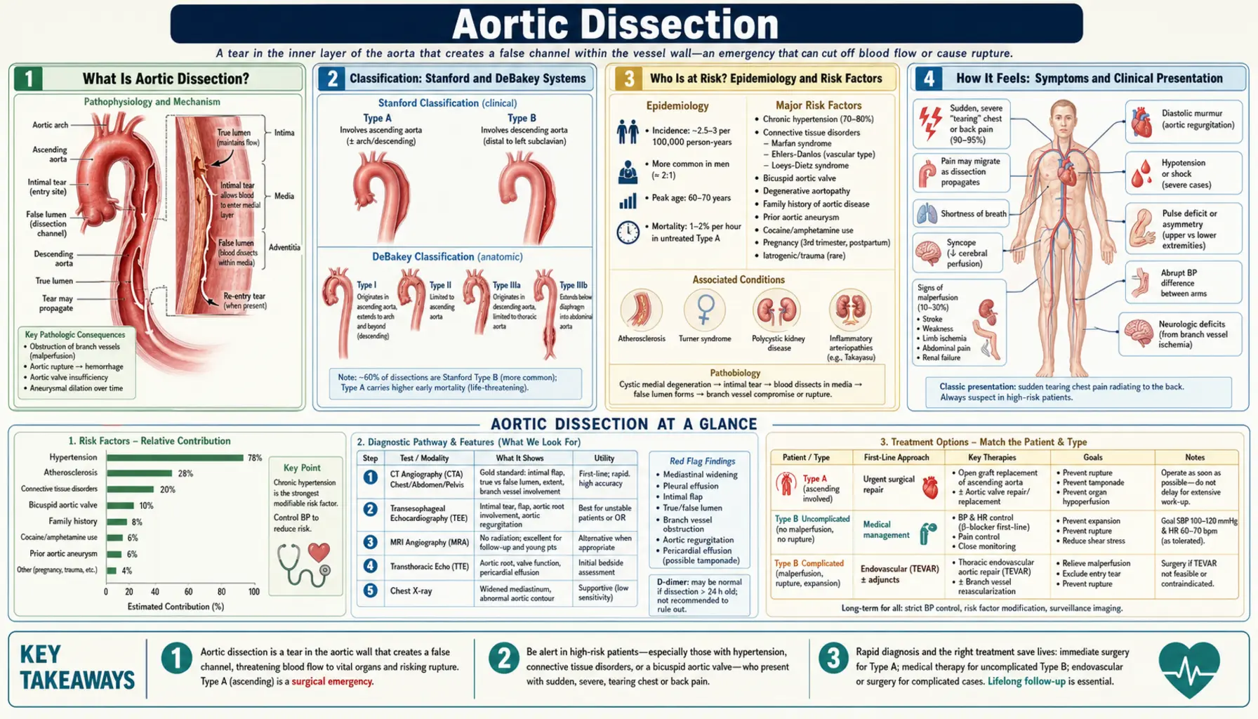
Aortic dissection is a tear in the inner lining of the aorta that lets blood enter the aortic wall and split its layers apart. It is the most common of the acute aortic syndromes, and the 2022 American Heart Association/American College of Cardiology (ACC/AHA) aortic guideline describes untreated dissection of the ascending aorta as highly lethal, with an early mortality of 1% to 2% per hour after symptom onset. This page summarizes what the guideline, the International Registry of Acute Aortic Dissection (IRAD) and the main trials report about how dissection develops, how it presents, and how it is treated.
Table of Contents
- What Is Aortic Dissection?
- Classification: Stanford and DeBakey Systems
- Who Is at Risk? Epidemiology and Risk Factors
- How It Feels: Symptoms and Clinical Presentation
- Diagnosis: Imaging and Laboratory Workup
- Treatment: Type A Emergency Surgery and Type B Medical Management
- Complications and Prognosis
- Living After Aortic Dissection: Long-Term Management
- Key Research Papers
- Connections
- Featured Videos
What Is Aortic Dissection?
The aorta is the body's largest artery, carrying blood from the heart to the rest of the body. The 2022 ACC/AHA guideline describes its wall as three layers: a thin inner intima, a thicker central media (smooth muscle cells, elastic fibers and collagen arranged in more than 50 elastic lamellae, which give the wall its strength), and a thin outer adventitia.
In a dissection, an intimal tear lets blood pass into the media, splitting the wall lengthwise and creating a dissection flap that divides the original channel (the true lumen) from a newly formed false lumen. The guideline notes that the flap can propagate forward or backward along the aorta. Blood in the false lumen may tear back into the true lumen (a re-entry tear); if it instead tears through the outer media and adventitia, the aorta ruptures.
Aortic dissection belongs to the acute aortic syndromes, which also include intramural hematoma (bleeding within the media, with or without an intimal tear) and penetrating atherosclerotic ulcer. The guideline notes that all three can lead to rupture.
The guideline defines dissection as "acute" during the first 2 weeks after symptom onset and "chronic" after that, and describes a newer four-stage scheme (hyperacute, acute, subacute, chronic) proposed by IRAD investigators and by two surgical societies.
Back to Table of ContentsClassification: Stanford and DeBakey Systems
The 2022 ACC/AHA guideline describes two commonly used anatomic systems. The distinction matters because it separates the dissections treated with emergency surgery from those first treated with medication.
Stanford Classification
Stanford Type A covers all dissections involving the ascending aorta, wherever the intimal tear began. The guideline lists the sequelae that make it life-threatening:
- Rupture causing cardiac tamponade
- Acute severe aortic regurgitation causing heart failure or shock
- Compromise of the coronary artery openings causing myocardial ischemia
- Malperfusion (blocked flow to branch arteries) causing ischemia or infarction of organs, including stroke
Stanford Type B covers all dissections that do not involve the ascending aorta, including those that involve the aortic arch but spare the ascending aorta. The guideline also mentions a 2019 European consensus category, "non-A non-B," for dissections whose flap begins in the arch, and a more detailed zone-based scheme proposed in 2020 by two surgical societies.
DeBakey Classification
- DeBakey Type I: The tear originates in the ascending aorta and extends to include the arch and typically the descending aorta.
- DeBakey Type II: The tear is confined to the ascending aorta.
- DeBakey Type III: The tear originates in the descending thoracic aorta and usually extends downstream (IIIa: confined to the descending thoracic aorta; IIIb: extends below the diaphragm).
Who Is at Risk? Epidemiology and Risk Factors
The 2022 ACC/AHA guideline estimates the incidence of aortic dissection at 5 to 30 cases per million people per year, with men more commonly affected and most dissections occurring between ages 50 and 70; people with Marfan syndrome, bicuspid aortic valve, Loeys-Dietz syndrome or vascular Ehlers-Danlos syndrome present younger. In a prospective population study in Oxfordshire, United Kingdom (2002–2012), the incidence was 6 per 100,000 per year, the mean age was 72, and 48.6% of people with a type A dissection died before reaching hospital assessment (Howard 2013, PubMed PMID: 23599348). The authors note that hospital-based registries underestimate both incidence and case fatality. In the first IRAD report of 464 patients, the mean age was 63 and 65.3% were men (Hagan 2000).
High Blood Pressure
In the Oxfordshire study, 67.3% of patients were taking blood-pressure medication before the event, 46.0% had at least one systolic reading of 180 mmHg or higher in their primary-care records over the preceding 5 years, and on average 56.0% of their recorded blood pressures were in the hypertensive range (above 140/90 mmHg). The authors concluded that uncontrolled hypertension remains the most significant treatable risk factor (Howard 2013).
Bicuspid Aortic Valve
The guideline describes bicuspid aortic valve (two valve leaflets instead of three) as a common congenital condition affecting about 1% of the population, with a 2 to 3:1 male-to-female ratio, and lists it among the conditions in which dissection presents at younger ages.
Heritable Connective Tissue and Aortic Disorders
- Marfan syndrome (FBN1 gene): affects about 1 in 5,000 people, according to the guideline. See the Marfan syndrome page.
- Loeys-Dietz syndrome (TGFBR1, TGFBR2, SMAD3, TGFB2, TGFB3): the guideline states that dissection with TGFBR1, TGFBR2 or SMAD3 variants may occur at smaller aortic diameters than in Marfan syndrome, and that TGFBR1 and TGFBR2 variants carry a risk of type A dissection at younger ages.
- Vascular Ehlers-Danlos syndrome (COL3A1): affects 1 in 50,000 to 100,000 people and leads to spontaneous aortic and arterial dissections, aneurysms and rupture at young ages, per the guideline.
- Familial thoracic aortic disease without a syndrome: the guideline reports that 13% to 20% of patients with thoracic aortic disease who lack Marfan or Loeys-Dietz features have a similarly affected first-degree relative.
Other Risk Factors Reported
- Cocaine: in 3,584 IRAD patients (1996–2012), cocaine use was implicated in 1.8% of acute dissections; these patients were younger, more often male, and more often smoked (Dean 2014, PubMed PMID: 24835037).
- Pregnancy: the guideline describes pregnancy as a risk factor in women with an underlying aortopathy, with most pregnancy-related dissections in the third trimester or up to 12 weeks after delivery.
- Turner syndrome: affects 1 in 2,500 liveborn girls; about half have cardiovascular defects, including bicuspid valve (15%–30%) and aortic coarctation (7%–18%), and the guideline lists aortic dilation, bicuspid valve, coarctation and hypertension as dissection risk factors in this group.
- Conditions in the guideline's Aortic Dissection Detection Risk Score: Marfan syndrome or another connective tissue disease, a family history of aortic disease, known aortic valve disease, recent aortic manipulation, and a known thoracic aortic aneurysm.
How It Feels: Symptoms and Clinical Presentation
The guideline notes that the diagnosis can be difficult because the symptoms overlap with more common emergency complaints, and IRAD's first report concluded that dissection "presents with a wide range of manifestations, and classic findings are often absent" (Hagan 2000).
Pain
According to the guideline, the textbook description is "tearing" or "ripping" pain, but patients more commonly report the abrupt onset of severe "sharp" or "stabbing" pain in the chest or back (sometimes the abdomen), maximal at the start, that sometimes radiates. Across 17 years of IRAD data (4,428 patients), severe or worst-ever pain was reported by 93% of type A and 94% of type B patients, and chest pain by 83% and 71% respectively (Pape 2015, PubMed PMID: 26205591).
Signs That Point to Dissection
The guideline lists these signs and symptoms of acute aortic syndromes, with their causes:
- Blood pressure difference of more than 20 mmHg between limbs — compromised branch-artery flow
- New murmur of aortic regurgitation — the dissection disturbing the aortic valve
- Shortness of breath — heart failure from aortic regurgitation, pericardial effusion, or blood in the chest
- Myocardial ischemia or infarction — dissection involving a coronary artery
- Stroke symptoms — carotid or vertebral artery involvement
- Paraplegia — loss of blood supply to the spinal cord through the intercostal arteries
- Bowel ischemia or gastrointestinal bleeding — celiac or superior mesenteric artery malperfusion
- Oliguria or visible blood in the urine — renal artery malperfusion
- Lower-limb ischemia — iliac artery malperfusion
- Hoarseness, Horner's syndrome, difficulty swallowing — compression of the recurrent laryngeal nerve, the sympathetic chain, or the esophagus
- Shock — cardiac tamponade, blood in the chest, aortic rupture, acute severe aortic regurgitation, or severe myocardial ischemia
In the first IRAD report, the classic findings of aortic regurgitation and pulse deficit were noted in only 31.6% and 15.1% of patients (Hagan 2000). The guideline describes measuring blood pressure in both arms and both legs.
Syncope
In 728 IRAD patients, 13% reported fainting (syncope). Those who fainted were more likely to have cardiac tamponade (28% vs 8%) and stroke (18% vs 4%) and more likely to die in hospital (34% vs 23%); once tamponade, stroke and dissection type were accounted for, syncope itself was not independently linked to death (Nallamothu 2002, PubMed PMID: 12427495).
Delays in Recognition
In an IRAD analysis of type A dissection, the median time from emergency-department arrival to diagnosis was 4.3 hours. Diagnosis was delayed in women, in people whose symptoms were atypical (pain that was not abrupt, or not in the chest or back, or no pain at all), in those without a pulse deficit or low blood pressure, and in those first seen at a non-tertiary hospital (Harris 2011).
Back to Table of ContentsDiagnosis: Imaging and Laboratory Workup
CT Angiography
The 2022 ACC/AHA guideline recommends CT for initial imaging of a suspected acute aortic syndrome, given its wide availability, accuracy, speed and anatomic detail. It shows the full extent of the dissection and sometimes the entry tear, involvement and patency of branch vessels, signs of malperfusion, pericardial effusion and blood around the heart, and mediastinal hematoma. ECG-synchronized CT is described for accurately depicting the proximal aorta and coronary openings. In IRAD, CT use for type A diagnosis rose from 46% to 73% over 17 years (Pape 2015).
Transesophageal Echocardiography and MRI
The guideline rates transesophageal echocardiography (TEE) and MRI as reasonable alternatives. TEE can be done at the bedside in the emergency department or in the operating room and is preferred over transthoracic echocardiography because of its higher sensitivity and resolution. MRI is described as the third choice in the acute setting (less available, longer acquisition times, and harder to care for unstable patients inside the scanner) and is used mostly for follow-up, although it may be the study of choice for a stable patient who cannot receive iodinated contrast.
Chest X-Ray and ECG
The guideline states that a plain chest X-ray is neither sensitive nor specific enough to diagnose an acute aortic syndrome; suggestive findings include mediastinal widening, a disrupted aortic-knob contour, and the "calcium sign" (intimal calcium separated from the aortic wall by more than 5 mm). In the first IRAD report, the initial chest X-ray showed no abnormality in 12.4% of patients and the ECG in 31.3% (Hagan 2000).
Risk Score and D-Dimer
The guideline describes the Aortic Dissection Detection Risk Score (ADD-RS), which gives one point for each of three categories present: high-risk conditions, high-risk pain features (abrupt, severe, or ripping or tearing chest, back or abdominal pain), and high-risk examination findings (pulse deficit or blood pressure differential, focal neurologic deficit with pain, new aortic regurgitation murmur with pain, low blood pressure or shock). It notes that no biomarker is diagnostic, but that in patients with a low prior probability a D-dimer below 500 ng/mL makes the diagnosis unlikely.
The ADvISED study tested this combination prospectively in 1,850 emergency patients at 6 hospitals in 4 countries. A positive D-dimer had a sensitivity of 96.7% and a specificity of 64% for acute aortic syndrome. Among the 924 patients with an ADD-RS of 0 or 1 and a negative D-dimer, 3 had an acute aortic syndrome, a failure rate of 0.3% (Nazerian 2018, PubMed PMID: 29030346).
Back to Table of ContentsTreatment: Type A Emergency Surgery and Type B Medical Management
Initial Medical Treatment (Both Types)
The 2022 ACC/AHA guideline recommends prompt "anti-impulse" therapy with invasive blood-pressure monitoring in an intensive care setting, to lower the stress on the aortic wall:
- Targets: systolic blood pressure below 120 mmHg, or the lowest pressure that keeps organs adequately perfused, and a heart rate of 60 to 80 beats per minute. The guideline notes that no randomized studies have compared medical treatments and that these targets rest on small single-center studies and expert opinion.
- Intravenous beta-blockers (examples given: esmolol, metoprolol, labetalol) first, unless contraindicated; in people who cannot take beta-blockers, an intravenous non-dihydropyridine calcium channel blocker (verapamil or diltiazem) for heart-rate control.
- Intravenous vasodilators (examples given: nicardipine, clevidipine, sodium nitroprusside) are added if blood pressure stays high. The guideline states they are not used as initial treatment before a beta-blocker or calcium channel blocker, because of the potential for compensatory tachycardia.
- Pain control: pain raises heart rate and blood pressure; the guideline describes intravenous opiates as particularly effective and notes that intravenous ketorolac may not be suitable because it can raise blood pressure and harm the kidneys.
Type A Dissection — Emergency Surgery
The guideline describes suspected or diagnosed acute type A dissection as warranting immediate evaluation for surgery. Its stated goals of repair are to prevent or treat rupture, prevent extension back into the aortic root and forward into undissected aorta, and relieve malperfusion. The resection is described as including the tear site, any aneurysmal aorta, and the most proximal extent of the dissection; more extensive replacement of the root or arch adds complexity but may reduce later complications. When circulatory arrest is needed, the guideline rates some form of cerebral perfusion as reasonable to improve neurologic outcomes.
Reported outcomes:
- In IRAD's first report, in-hospital mortality was 26% for type A patients treated surgically and 58% for those not operated on, typically because of advanced age and other illness (Hagan 2000).
- Over 17 years of IRAD data, surgery for type A rose from 79% to 90% of patients, overall type A in-hospital mortality fell from 31% to 22%, and surgical mortality fell from 25% to 18% (Pape 2015).
- The guideline cites mortality of 25% to 27% with surgery versus 76% with medical management in type A patients presenting with acute stroke, a mortality of 30.5% with malperfusion syndrome versus 6.2% without it, and 63.2% with mesenteric malperfusion.
Type B Dissection
For uncomplicated acute type B dissection, the guideline recommends medical therapy as the initial strategy, and states that endovascular repair may be considered when high-risk features are present. Those listed include a maximal aortic diameter above 40 mm, a false-lumen diameter above 20–22 mm, an entry tear larger than 10 mm or on the lesser curvature, growth of more than 5 mm between scans, bloody pleural effusion, malperfusion seen only on imaging, high blood pressure that persists despite more than 3 drug classes, pain persisting more than 12 hours, and readmission.
For complicated type B dissection, which the guideline defines by consensus as rupture, branch-artery occlusion and malperfusion, extension of the dissection, enlargement of the aorta in the acute phase, intractable pain, or uncontrolled hypertension, it recommends intervention. Endovascular stent grafting (TEVAR, thoracic endovascular aortic repair) is recommended over open surgery for rupture when the anatomy is suitable, and is rated reasonable for other complications. The guideline states that endovascular therapy has largely supplanted open repair because of lower morbidity and mortality, and that no large randomized trial has compared open with endovascular repair. In IRAD, overall in-hospital mortality for type B was 13%: 32.1% with open repair, 9.6% with medical management and 6.5% with endovascular repair, as cited by the guideline. Endovascular management of type B rose from 7% to 31% of IRAD patients over 17 years, with no significant change in type B in-hospital mortality (12% to 14%) (Pape 2015).
The INSTEAD and INSTEAD-XL Trials
INSTEAD randomized 140 patients in stable condition at least 2 weeks after an uncomplicated type B dissection to stent grafting plus optimal medical therapy (72) or optimal medical therapy alone (68). At 2 years, all-cause survival did not differ (88.9% with stent grafting vs 95.6% with medical therapy; P=0.15) and the authors noted the trial turned out to be underpowered; aortic remodeling occurred in 91.3% versus 19.4% (Nienaber 2009, PubMed PMID: 19996018).
The INSTEAD-XL follow-up of the same patients, a retrospective analysis, reported that at 5 years aorta-specific mortality (6.9% vs 19.3%; P=0.04) and disease progression (27.0% vs 46.1%; P=0.04) were lower with stent grafting, while all-cause mortality did not differ significantly (11.1% vs 19.3%; P=0.13) (Nienaber 2013, PubMed PMID: 23922146). The 2022 guideline describes the findings of INSTEAD-XL and of ADSORB, another small randomized trial, as promising, but states that larger trials with longer-term data are still needed and that the best timing for stent grafting is unknown.
Back to Table of ContentsComplications and Prognosis
Acute Complications
The guideline lists the potential sequelae of acute type A dissection as myocardial infarction, acute aortic regurgitation, cardiac tamponade, aortic rupture and malperfusion of organs, and notes that mortality is higher in people who present with or develop tamponade, myocardial ischemia, stroke or malperfusion. It reports a 30-day mortality of 10% for uncomplicated acute type B dissection, rising to 20% by day 2 and 25% by day 30 when complications such as malperfusion or rupture develop. Imaging evidence of malperfusion is present in as many as 25% of type A patients, per the guideline.
Late Complications
- Growth of the remaining aorta: in medically managed uncomplicated type B dissection, delayed aortic expansion occurs in 20% to 50% of patients over 4 years. After type A repair, the guideline describes a distal aortic growth rate of about 1 mm per year.
- Reoperation after type A repair: about 15% at 15 years for the aortic root, and 10% to 16% at 10 years for the distal aorta.
- After stent grafting for type B: reintervention rates of 27% to 39% at midterm follow-up, early endoleaks (continued flow around the graft) in up to 35% of patients and late endoleaks in 13%.
- Aneurysm of a chronically dissected aorta: the guideline recommends elective repair when the total aortic diameter reaches 5.5 cm or more.
Long-Term Survival
- The guideline gives a 10-year survival of approximately 60% to 65% after repair of acute type A dissection.
- In the Oxfordshire population study, 30-day fatality was 47.4% for type A patients who survived to admission and 13.3% for type B, and subsequent 5-year survival was 85.7% for type A and 83.3% for type B (Howard 2013).
Living After Aortic Dissection: Long-Term Management
The guideline states that surviving an acute dissection does not guarantee freedom from later aortic events, because of residual dissection and the risk of aneurysm formation.
Long-Term Blood Pressure Treatment
The 2022 ACC/AHA guideline recommends long-term beta-blockers (unless contraindicated) after an acute aortic syndrome to control heart rate and blood pressure and reduce late aortic events, with other blood-pressure drugs — particularly angiotensin receptor blockers (ARBs) and ACE inhibitors — added as needed. It reports that regimens including beta-blockers, ACE inhibitors and ARBs have improved long-term outcomes. It also notes that calcium channel blockers showed some benefit in type B dissection, but that studies in Marfan mouse models and Marfan and other inherited aortopathy patients showed harmful effects of long-term use; in its words, "it may be best to avoid these agents in patients with Marfan syndrome unless necessary to achieve BP control." For thoracic aortic aneurysm, the guideline cites the ACC/AHA hypertension guideline's target for people with cardiovascular disease of systolic pressure below 130 mmHg and diastolic below 80 mmHg, and notes that no randomized trial has tested the best target for preventing aortic complications.
On ARBs specifically, the guideline reports that losartan prevented aneurysm formation in Marfan mouse models, but that randomized trials comparing an ARB with a beta-blocker in Marfan syndrome found no significant difference in aortic root growth or clinical events.
Surveillance Imaging
For people whose dissection or intramural hematoma was treated with medication alone, the guideline recommends CT (or MRI) at 1 month, 6 months and 12 months, then annually if stable. It describes surveillance after repair as critical for detecting progression of residual disease, and transthoracic echocardiography as useful for following the aortic root and valve.
Physical Activity
The guideline recommends education about avoiding intense isometric exercise (such as heavy weightlifting or straining with the Valsalva maneuver), burst exertion and collision sports in people with significant aortic disease, noting that heavy lifting with the Valsalva maneuver can raise systolic pressure above 300 mmHg. It recommends cardiac rehabilitation after surgery for aortic dissection, citing a randomized trial after type A repair that showed improved peak oxygen uptake, workload and quality of life, and it rates encouraging 30 to 60 minutes of mild-to-moderate aerobic activity at least 3 to 4 days a week as reasonable for people with aneurysms whose blood pressure is controlled.
Genetic Testing and Family Screening
The guideline recommends genetic testing for people with aortic dissection who have risk factors for heritable disease: syndromic features of Marfan, Loeys-Dietz or vascular Ehlers-Danlos syndrome; onset before age 60; a family history of thoracic aortic disease or of peripheral or intracranial aneurysms; or unexplained sudden death at a relatively young age in a first- or second-degree relative. It also recommends aortic imaging of first-degree relatives of people with aortic dissection, even without a known family history or identified variant, and genetic testing of at-risk relatives when a disease-causing variant is found.
Pregnancy
For women with a genetic aortopathy, Turner syndrome or a bicuspid valve with a dilated aorta who are considering pregnancy, the guideline recommends aortic imaging and counseling about the risk of dissection beforehand, and genetic counseling about the inherited nature of the condition.
Emotional Recovery
The guideline reports that depression and anxiety are common in people with aortic disease and that post-traumatic stress disorder is a particular risk after dissection. It rates screening for anxiety, depression and post-traumatic stress, and providing support resources and education, as reasonable.
Back to Table of ContentsKey Research Papers
- Hagan PG, Nienaber CA, Isselbacher EM, et al. (2000). The International Registry of Acute Aortic Dissection (IRAD): new insights into an old disease. JAMA 283(7):897–903. The founding IRAD report: 464 patients at 12 referral centers, 1996–1998. — PubMed PMID: 10685714
- Nienaber CA, Kische S, Rousseau H, et al. (2013). Endovascular repair of type B aortic dissection: long-term results of the randomized investigation of stent grafts in aortic dissection trial. Circulation: Cardiovascular Interventions 6(4):407–416. INSTEAD-XL: 5-year follow-up. — PubMed PMID: 23922146
- Pape LA, Awais M, Woznicki EM, et al. (2015). Presentation, diagnosis, and outcomes of acute aortic dissection: 17-year trends from the International Registry of Acute Aortic Dissection. Journal of the American College of Cardiology 66(4):350–358. — PubMed PMID: 26205591
- Nienaber CA, Rousseau H, Eggebrecht H, et al. (2009). Randomized comparison of strategies for type B aortic dissection: the INvestigation of STEnt Grafts in Aortic Dissection (INSTEAD) trial. Circulation 120(25):2519–2528. — PubMed PMID: 19996018
- Nazerian P, Mueller C, Soeiro AM, et al. (2018). Diagnostic accuracy of the aortic dissection detection risk score plus D-dimer for acute aortic syndromes: the ADvISED prospective multicenter study. Circulation 137(3):250–258. — PubMed PMID: 29030346
- Erbel R, Aboyans V, Boileau C, et al. (2014). 2014 ESC Guidelines on the diagnosis and treatment of aortic diseases: document covering acute and chronic aortic diseases of the thoracic and abdominal aorta of the adult. European Heart Journal 35(41):2873–2926 (a corrigendum was published in 2015). — PubMed PMID: 25173340
- Isselbacher EM, Preventza O, Hamilton Black J 3rd, et al. (2022). 2022 ACC/AHA Guideline for the Diagnosis and Management of Aortic Disease. Circulation 146(24):e334–e482 (also published in the Journal of the American College of Cardiology). — PubMed PMID: 36322642
- Nienaber CA, Clough RE, Sakalihasan N, et al. (2016). Aortic dissection. Nature Reviews Disease Primers 2:16053 (a correction was published). — PubMed PMID: 27440162
- Evangelista A, Isselbacher EM, Bossone E, et al. (2018). Insights from the International Registry of Acute Aortic Dissection: a 20-year experience of collaborative clinical research. Circulation 137(17):1846–1860. — PubMed PMID: 29685932
- Mazzolai L, Teixido-Tura G, Lanzi S, et al. (2024). 2024 ESC Guidelines for the management of peripheral arterial and aortic diseases. European Heart Journal 45(36):3538–3700. — PubMed PMID: 39210722
- Harris KM, Strauss CE, Eagle KA, et al. (2011). Correlates of delayed recognition and treatment of acute type A aortic dissection: the International Registry of Acute Aortic Dissection (IRAD). Circulation 124(18):1911–1918. — PubMed PMID: 21969019
- Howard DP, Banerjee A, Fairhead JF, et al. (2013). Population-based study of incidence and outcome of acute aortic dissection and premorbid risk factor control: 10-year results from the Oxford Vascular Study. Circulation 127(20):2031–2037. — PubMed PMID: 23599348
- Nallamothu BK, Mehta RH, Saint S, et al. (2002). Syncope in acute aortic dissection: diagnostic, prognostic, and clinical implications. American Journal of Medicine 113(6):468–471. — PubMed PMID: 12427495
- Dean JH, Woznicki EM, O'Gara P, et al. (2014). Cocaine-related aortic dissection: lessons from the International Registry of Acute Aortic Dissection. American Journal of Medicine 127(9):878–885. — PubMed PMID: 24835037