Günter Blobel: The Address Label Inside Every Protein, and the Diseases of Failed Delivery
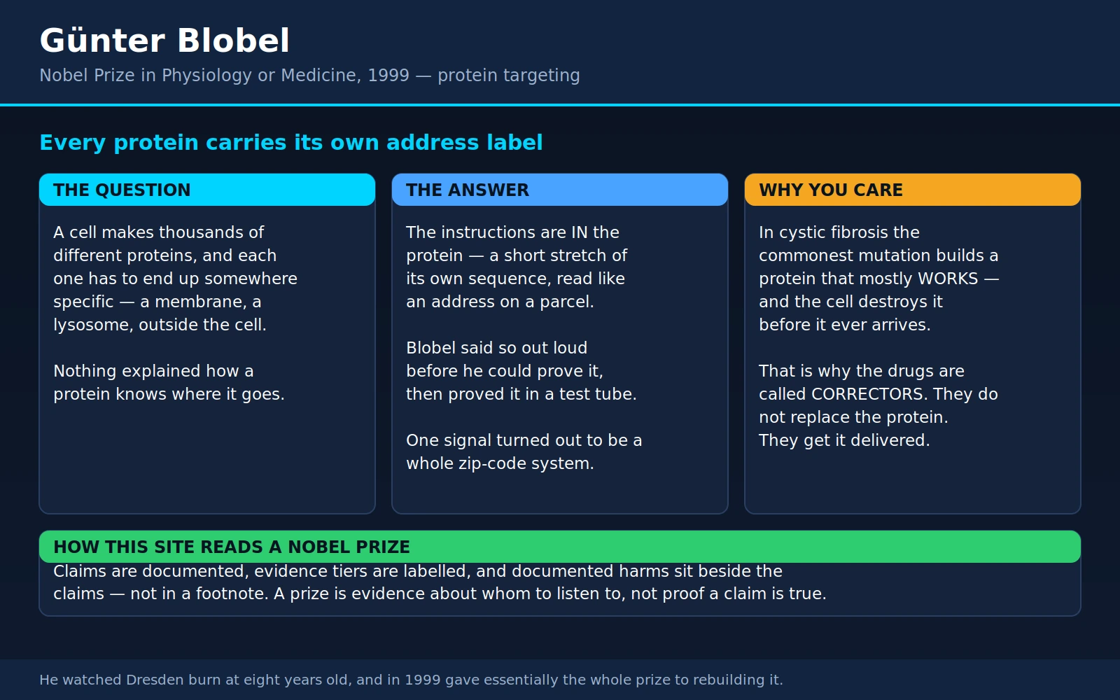
Table of Contents
- Overview
- The Question Nobody Could Answer
- The Signal Hypothesis: A Guess Made Out Loud
- 1975: Proving It in a Test Tube
- The Machinery That Reads the Label
- One Signal Becomes a Zip-Code System
- Where the Vans Come In: Schekman, Rothman and Südhof
- Cystic Fibrosis: Built Correctly, Then Destroyed
- Why the Cystic Fibrosis Drugs Are Called Correctors
- I-Cell Disease: When the Label Never Gets Printed
- Primary Hyperoxaluria Type 1: The Right Enzyme in the Wrong Room
- Familial Hypercholesterolaemia: Receptors That Never Reach the Surface
- What This Means If You or Your Child Has One of These Conditions
- Dresden: What He Did With the Prize
- Key Research Papers
- Connections
- Featured Videos
1. Overview
Günter Blobel (1936–2018) was a German-American cell biologist at Rockefeller University in New York who won the 1999 Nobel Prize in Physiology or Medicine, unshared, "for the discovery that proteins have intrinsic signals that govern their transport and localization in the cell." The official record is at nobelprize.org — Günter Blobel.
That citation is a mouthful, so here is the whole idea in one sentence: every protein your body makes carries its own shipping label, written into the protein itself. A short stretch of amino acids at the front of a newly made protein says, in effect, "send me out of the cell," or "put me in the nucleus," or "I belong in a lysosome." The cell reads that label, routes the protein, and — for the ones that get exported — snips the label off on arrival. Blobel proposed this in 1971, when there was almost no evidence for it, and then spent the next quarter-century proving it and working out the machinery that does the reading.
People often call it the zip-code hypothesis, and that is a good way to hold it. But the reason this page exists on a health site is what happens when the zip code, or the delivery, goes wrong. It is not an abstract failure. It is:
- Cystic fibrosis, where the most common mutation makes a chloride channel that works — the cell just destroys it before it ever reaches the cell surface. Two of the three drugs in the modern triple therapy are called correctors for exactly this reason.
- I-cell disease (mucolipidosis II), where the tag that marks digestive enzymes for the lysosome never gets attached, so the enzymes are secreted out of the cell instead of delivered inside it.
- Primary hyperoxaluria type 1, where a perfectly good liver enzyme is misrouted to the mitochondria when its job is in the peroxisome — a fire extinguisher installed in the wrong room — and the result is kidney stones from childhood and kidney failure.
- Familial hypercholesterolaemia, where one whole class of mutations builds an LDL receptor that never leaves the factory floor.
Blobel's other legacy is not scientific at all. He watched Dresden burn from a few kilometres away when he was eight years old, and in 1999 he gave essentially the entire Nobel award to rebuilding the city's Frauenkirche and building a new synagogue there. Section 14 tells that story.
2. The Question Nobody Could Answer
A single human cell contains billions of individual protein molecules, and a large fraction of them do not work where they were made. They are all built by ribosomes floating in the cell's interior, and then they have to end up somewhere specific: inside the nucleus, inside a mitochondrion, inside a lysosome, embedded in the outer membrane facing the right way round, or shipped clean out of the cell entirely. A liver cell secreting albumin, a pancreatic cell secreting insulin, a plasma cell secreting antibodies — all of them are running a shipping operation, continuously, with no mistakes tolerated.
By the late 1960s the route was known. George Palade — who would share the 1974 Nobel with Albert Claude and Christian de Duve, and in whose Rockefeller laboratory Blobel worked — had used electron microscopy and cell fractionation to trace secretory proteins through the cell: made on ribosomes stuck to the rough endoplasmic reticulum, then to the Golgi apparatus, then into secretory granules, then out. It was one of the great pieces of descriptive biology of the twentieth century.
What nobody could explain was the decision. Ribosomes are interchangeable. The ribosome making haemoglobin, which stays inside the red blood cell, is the same kind of machine as the ribosome making an antibody, which gets exported. So why does one ribosome end up bolted to the ER membrane, threading its product into a sealed compartment, while an identical ribosome floats free and dumps its product into the cell's interior?
Two answers were plausible. Either the ribosome was somehow specialised or pre-committed — a dedicated export machine, a dedicated domestic machine — or the protein being made carried the instruction with it. This is not a small distinction. It is the difference between a post office that decides where your parcel goes and a parcel that tells the post office.
3. The Signal Hypothesis: A Guess Made Out Loud
In 1971, Blobel and David Sabatini published a short proposal: the parcel carries the label. Their signal hypothesis said that proteins destined for export begin life with an extra stretch of amino acids at their front end — a signal sequence — which is recognised as the protein emerges from the ribosome, causes the ribosome to dock onto the endoplasmic reticulum membrane, opens a passage through that membrane, threads the growing chain across, and is then cut off. The finished protein carries no trace of the label that delivered it.
The scale of the guess is worth appreciating. In 1971 nobody had seen a signal sequence, nobody had seen a receptor for one, and nobody had seen a channel through the ER membrane. Blobel proposed all three and predicted that the sequence would be removed after use. Over the following two decades every one of those components was found and purified, largely by his own laboratory.
There was one tantalising clue already in the literature. In 1972 César Milstein's group in Cambridge reported "A possible precursor of immunoglobulin light chains" — an antibody light chain made in a cell-free system that was bigger than the light chain the same cells actually secrete. Something was being trimmed off. Blobel and Dobberstein's own 1975 paper confirms that observation in its abstract, noting that the product made in a test tube "is larger than the authentic secreted light chain" and that similar results had come from several laboratories. Nobody yet knew what the extra piece was for.
4. 1975: Proving It in a Test Tube
The proof arrived in two back-to-back papers in the Journal of Cell Biology in December 1975, by Blobel and Bernhard Dobberstein. They are among the most quietly decisive experiments in modern biology, and both are worth explaining because the logic is so clean.
The first paper (J Cell Biol 1975;67(3):835–51) used a mouse myeloma tumour that makes enormous quantities of a single antibody light chain. They showed the messenger RNA for that light chain sits exclusively on membrane-bound ribosomes — never on free ones. They found ribosome-attached chains that had already been trimmed and chains that had not, which told them the trimming happens before the protein is finished, not afterwards. And they showed the trimming activity lives in the membrane of the rough microsome, not in the ribosome. Their abstract ends by naming the framework they were testing: "the signal hypothesis."
The second paper (J Cell Biol 1975;67(3):852–62) is the one that settled it. They built a working translocation system out of parts from two different species: ribosomal subunits from rabbit reticulocytes, and stripped membranes from dog pancreas. Then they fed it two different messenger RNAs. Given antibody light-chain mRNA, the system pushed the product into a sealed, protease-resistant space and cut it to exactly the right size. Given haemoglobin mRNA, it did nothing of the kind. Same ribosomes. Same membranes. Different parcels, different outcomes. Their conclusion, in their own words: the results "establish unequivocally that the information for segregation of a translation product is encoded in the mRNA itself, not in the protein-synthesizing apparatus."
The parcel tells the post office. That is the whole thing.
5. The Machinery That Reads the Label
A signal sequence is typically a couple of dozen amino acids with a greasy, water-repelling core. On its own it does nothing; something has to read it. Over the following decade Blobel's laboratory found the readers.
The signal recognition particle (SRP) is the reader. Peter Walter, working with Blobel, purified it and then discovered something nobody expected: it is not purely a protein. In 1982 they reported that the particle contains a 7S RNA molecule that is required for both its structure and its function (Nature 1982;299(5885):691–8). That finding is why the name changed from "signal recognition protein" to "signal recognition particle." SRP grabs the emerging signal sequence, temporarily pauses the ribosome mid-sentence, and walks the whole assembly to the ER membrane — where an SRP receptor hands it off.
The translocon is the door. Blobel had predicted a protein-lined channel through the membrane; his group produced electrophysiological evidence for one in "A protein-conducting channel in the endoplasmic reticulum" (Cell 1991;65(3):371–80), and the definitive biochemical proof came in 1993 when Dirk Görlich and Tom Rapoport rebuilt working translocation into artificial vesicles from purified ER membrane components (Cell 1993;75(4):615–30). A hypothetical door had become a defined set of molecules you could put in a tube.
Signal peptidase is the doorman with the scissors. Sitting on the far side of the membrane, it clips the signal sequence off the moment the protein is through. This is why you cannot find the label on a mature secreted protein: by design, it is destroyed on delivery.
The elegance of the system is that it is co-translational. The protein is not built and then shipped. It is threaded through the door while it is still being written, so a protein destined for export never really exists loose in the cell's interior at all. That detail matters enormously in section 8, because it means the cell inspects the protein while it is folding, in the compartment it was delivered into — and can decide to destroy it right there.
6. One Signal Becomes a Zip-Code System
Getting into the ER is one destination. The cell has many. In 1980 Blobel published a sweeping generalisation in the Proceedings of the National Academy of Sciences under the title "Intracellular protein topogenesis" — his own coinage for the whole problem of how proteins get to their addresses. He proposed that the information "is encoded in discrete topogenic sequences," that the repertoire of such sequences would be "relatively small," and he sorted them into four kinds:
- Signal sequences — start the crossing of a membrane.
- Stop-transfer sequences — halt the crossing partway, which is how a protein ends up embedded in a membrane rather than passing through it. This is how every receptor on every cell surface gets anchored the right way round.
- Sorting sequences — direct onward traffic after arrival.
- Insertion sequences — drive a protein into the lipid layer without help.
This was, again, mostly prediction. And again it came true, address by address, in other people's laboratories:
- The nucleus. In 1984, Kalderon and colleagues showed that a short sequence from a viral protein, moved onto the front of β-galactosidase and of pyruvate kinase — two proteins with no business in the nucleus — sent both of them into the nucleus (Cell 1984;39(3 Pt 2):499–509). A transferable, self-sufficient nuclear address.
- The peroxisome. In 1989, Gould and colleagues narrowed the peroxisomal signal down to the last three amino acids of firefly luciferase — serine-lysine-leucine — and showed that sticking those three residues onto the tail of an ordinary cytosolic bacterial enzyme was enough to import it into peroxisomes (J Cell Biol 1989;108(5):1657–64). Three letters. Remember this one; it is the mechanism behind section 11.
- The mitochondrion. An N-terminal stretch that folds into a lopsided helix — charged on one face, greasy on the other — is read as "mitochondria." Also remember this one.
- The lysosome. The odd one out: the lysosomal address is not an amino-acid sequence at all but a sugar tag, mannose-6-phosphate, added after the protein is made. Sly and colleagues pinned it down in 1977 by showing that mannose-6-phosphate blocked enzyme uptake far more powerfully than closely related sugars, and that stripping the phosphate off with alkaline phosphatase abolished an enzyme's ability to be taken up without harming its catalytic activity at all (Proc Natl Acad Sci U S A 1977;74(5):2026–30). That is section 10.
Blobel's later work moved to the biggest gate of all — the nuclear pore complex, the structure that controls what crosses into and out of the nucleus — and his group spent the 1990s cataloguing its components in yeast. But by then the general principle was no longer in dispute. The cell runs on addresses.
7. Where the Vans Come In: Schekman, Rothman and Südhof
Blobel's machinery gets a protein into the endoplasmic reticulum. It does not get it from there to the Golgi, or to the cell surface, or into a synapse. That job belongs to the vesicle system — small membrane bubbles that bud off one compartment, travel, and fuse with another — and it earned its own Nobel Prize in 2013. The two bodies of work fit together like an address system and a courier fleet, which is why this site keeps them on one page for Rothman, Schekman and Südhof.
Randy Schekman attacked it with genetics. In 1980 his laboratory reported yeast mutants that were temperature-sensitive for secretion: shift them to 37 °C and they stop secreting, swell up with undelivered cargo, and accumulate membrane-bound organelles visible under the electron microscope. From 188 mutant clones they defined 23 complementation groups — 23 separate genes required to run the pathway (Cell 1980;21(1):205–15). Those sec genes turned out to encode the vesicle machinery of every animal cell too.
James Rothman attacked it biochemically, rebuilding vesicle transport in cell-free extracts and purifying the proteins that make fusion happen. In 1993 his group isolated the SNAREs and found something that answered the specificity question directly: the proteins came in matched pairs, one type sitting on the vesicle and its partner on the target membrane, with different pairs for different destinations (Nature 1993;362(6418):318–24). A van that only fits one loading dock.
Thomas Südhof added the timing — how a nerve terminal holds vesicles loaded and waiting, and releases them within a fraction of a millisecond when calcium floods in.
Put the two prizes together and you have the complete logistics chain: Blobel wrote the addressing standard; Schekman found the fleet; Rothman found how a van knows its dock; Südhof found the dispatch signal. Every disease in the next five sections is a failure somewhere in that chain, and knowing where the failure is turns out to be the thing that determines whether a drug can help.
8. Cystic Fibrosis: Built Correctly, Then Destroyed
Cystic fibrosis is the clearest example in all of medicine of a trafficking disease, and it is the one where Blobel's principle has already changed what happens in clinic.
The gene was identified in 1989 (Kerem et al., Science 1989;245(4922):1073–80). It encodes CFTR, a chloride channel that sits in the surface membrane of cells lining the airways, pancreas, gut and sweat glands. Without working CFTR at the surface, the fluid layer on those surfaces goes wrong: mucus turns thick and sticky, the lungs cannot clear bacteria, the pancreas cannot deliver its digestive enzymes, and sweat comes out salty — which is why the sweat chloride test exists.
The overwhelmingly common mutation is F508del (also written Phe508del): a single missing amino acid. Nearly 90% of people with cystic fibrosis carry at least one copy of it, a figure stated in the 2019 New England Journal of Medicine trial we discuss in the next section.
Here is the part that matters. In 1990, Seng Cheng and colleagues at Genzyme worked out what F508del actually does — and it is not that the channel doesn't work. They found that F508del CFTR never gets its mature sugar coating, which is the signature of a protein that has never left the endoplasmic reticulum. Their interpretation, in their abstract: the mutant protein "is recognized as abnormal and remains incompletely processed in the endoplasmic reticulum where it is subsequently degraded." They calculated that mutations behaving this way accounted for at least 70% of known CF chromosomes, and concluded that "the molecular basis of most cystic fibrosis is the absence of mature CFTR at the correct cellular location" (Cell 1990;63(4):827–34). Read that last phrase again: not the absence of CFTR. The absence of CFTR at the correct cellular location.
Two follow-up experiments nailed it down.
The protein is actively destroyed, not merely lost. In 1995, Kopito's laboratory showed that immature CFTR is tagged with ubiquitin and shredded by the proteasome — the cell's demolition service. Block the proteasome with an inhibitor and polyubiquitinated immature CFTR piles up instead of disappearing (Cell 1995;83(1):121–7). This is quality control doing its job too zealously: a slightly-misfolded but perfectly serviceable channel gets condemned at inspection.
The protein is fine. This is the finding that made modern CF drugs conceivable. In 1992, Denning and colleagues in Michael Welsh's laboratory simply lowered the temperature of cells expressing F508del CFTR. As the temperature dropped, the protein's processing "reverts towards that of wild-type" — and, in their words, "when the processing defect is corrected, cAMP-regulated Cl− channels appear in the plasma membrane" (Nature 1992;358(6389):761–4). Give the mutant protein an easier folding environment and it makes it to the surface and works.
So the analogy is precise. The factory builds a functioning machine. The inspector on the loading dock decides it looks slightly odd and sends it to the shredder. Nothing is wrong with the machine. Everything is wrong with the delivery.
9. Why the Cystic Fibrosis Drugs Are Called Correctors
Once you know the defect is trafficking, the drug you need is not a replacement channel. It is something that helps the protein survive inspection. That distinction is baked into the vocabulary of CF medicine, and it is worth understanding because it explains which drugs work for which mutations.
A potentiator works on a channel that has already reached the cell surface but will not open properly. Ivacaftor is the potentiator. It was developed for gating mutations such as G551D, where the protein traffics normally but the gate barely opens. In the 2011 pivotal trial, ivacaftor raised percent-predicted FEV1 by 10.6 percentage points over placebo through week 24, made pulmonary exacerbations 55% less likely through week 48, added 2.7 kg of body weight, and dropped sweat chloride by 48.1 mmol/L (N Engl J Med 2011;365(18):1663–72). For the small group of patients with those mutations it was transformative.
For F508del it was useless on its own — because there is nothing at the surface to potentiate. You cannot hold open a door that was never installed.
A corrector works upstream, in the endoplasmic reticulum, stabilising the folding protein so it passes quality control and gets delivered. Tezacaftor and elexacaftor are correctors, and the combination of both plus ivacaftor is the modern triple therapy. Two trials published within weeks of each other in late 2019 established it:
- One copy of F508del. 403 patients aged 12 and over, each with one Phe508del allele and one mutation that produces essentially nothing, randomised to triple therapy or placebo for 24 weeks. Percent-predicted FEV1 was 13.8 points higher at week 4 and 14.3 points higher through 24 weeks; the pulmonary exacerbation rate was 63% lower; the respiratory quality-of-life score was 20.2 points higher on a scale whose minimum clinically important difference is 4 points; and sweat chloride fell by 41.8 mmol/L. All comparisons P<0.001. Only 1% stopped the drug because of side effects (N Engl J Med 2019;381(19):1809–19).
- Two copies of F508del. A harder test: everyone first spent four weeks on tezacaftor plus ivacaftor, then 107 patients were randomised either to add elexacaftor or to continue the two-drug regimen. Adding the third drug gained a further 10.0 percentage points of FEV1 (95% CI 7.4 to 12.6), dropped sweat chloride by a further 45.1 mmol/L (95% CI −50.1 to −40.1), and raised the respiratory score by 17.4 points (95% CI 11.8 to 23.0) — on top of a regimen that was already working. Nobody discontinued (Lancet 2019;394(10212):1940–8).
Sweat chloride is the number to watch, because it is a direct readout of how much CFTR is actually working at the cell surface. A 40–45 mmol/L fall is a large amount of channel that was not there before — channel that the cell was building all along and throwing away.
10. I-Cell Disease: When the Label Never Gets Printed
I-cell disease, formally mucolipidosis II, is a severe inherited condition that shows itself in infancy: coarsened facial features, stiff restricted joints, skeletal abnormalities, enlarged organs, poor growth and developmental delay. Its name comes from the dense inclusions that fill the cells of affected children — lysosomes stuffed with material they cannot digest.
The mechanism is the purest possible illustration of Blobel's principle, because in this disease the enzymes are entirely normal. In 1972, Susan Hickman and Elizabeth Neufeld published a hypothesis whose title says the whole thing: "A hypothesis for I-cell disease: defective hydrolases that do not enter lysosomes". The digestive enzymes are made. They work. They simply never get delivered to the compartment where the work is, and are released outside the cell instead.
The reason they are not delivered is that the lysosomal address label never gets written. As section 6 described, that label is a sugar modification — mannose-6-phosphate — stamped onto the enzyme's carbohydrate chains after it is made, and read by receptors that route the enzyme to the lysosome. In 1977, Sly's group established that this phosphate group is what makes an enzyme deliverable: mannose-6-phosphate blocked enzyme uptake far more strongly than mannose-1-phosphate or related sugars, and removing the phosphate with alkaline phosphatase destroyed the enzyme's deliverability while leaving its catalytic activity untouched (Proc Natl Acad Sci U S A 1977;74(5):2026–30).
In 1981, Reitman, Varki and Kornfeld found the broken step. The enzyme that writes the first half of the tag — GlcNAc-1-phosphotransferase — is missing. Fibroblasts from six normal people transferred between 0.67 and 1.46 picomoles per milligram of protein per hour; fibroblasts from five I-cell patients and five patients with the milder related disease transferred less than 0.02 (J Clin Invest 1981;67(5):1574–9). The gene responsible was identified in 2005 (Nat Med 2005;11(10):1109–12).
The laboratory picture that results is unmistakable and follows straight from the mechanism: cells are deficient in lysosomal enzymes while the blood shows elevated activity of multiple lysosomal enzymes at once, because everything that should have gone into lysosomes was secreted instead. That combination is characteristic enough that other conditions producing it are described in the literature as "mimicking a biochemical phenotype of mucolipidosis" (Am J Med Genet A 2017;173(2):501–9).
It is a cruel disease with no corrector-style fix, because the failure is not in the cargo but in the labelling machine itself. But it makes the general point better than anything else: a protein in the wrong place is as useless as a protein that was never made. Compare this with the more familiar lysosomal storage diseases such as Fabry, Pompe or Tay-Sachs, where one specific enzyme is genuinely defective. I-cell disease is what happens when the enzymes are all fine and the postal service has failed.
11. Primary Hyperoxaluria Type 1: The Right Enzyme in the Wrong Room
If cystic fibrosis is the trafficking disease where the protein gets destroyed, primary hyperoxaluria type 1 is the one where the protein arrives safely at the wrong address — and that turns out to be just as bad.
The liver has to dispose of a reactive small molecule called glyoxylate. The enzyme for the job is alanine:glyoxylate aminotransferase (AGT), and in humans it belongs in the peroxisome — which is exactly where glyoxylate is produced. In 1986, Christopher Danpure and Patricia Jennings showed that peroxisomal AGT activity was completely absent from the liver of a patient with this disease, and proposed that primary hyperoxaluria type 1 "should be added to the rather select list of peroxisomal disorders" (FEBS Lett 1986;201(1):20–4).
Four years later came the twist. In a subset of patients the enzyme is not absent and not broken. It is in the mitochondria. Purdue, Takada and Danpure sequenced AGT from such a patient and found three point mutations; screening a wider group, they found that all eight patients with mitochondrial AGT carried at least one allele bearing the same three changes. The mechanism they proposed is beautiful and awful at once: a proline-to-leucine substitution at residue 11 creates, out of nowhere, "an amphiphilic alpha-helix with characteristics similar to recognized mitochondrial targeting sequences" — a brand-new address label at the front of the protein — and its full effect depends on a second change at residue 170 that appears to impair peroxisomal import (J Cell Biol 1990;111(6 Pt 1):2341–51). One of the contributing variants is common: the residue 11 and residue 340 pair occurs in the general population at an allelic frequency of 5–10%, and on its own reroutes only a small proportion of the enzyme.
The consequence is entirely a consequence of geography. In the mitochondria the enzyme is catalytically fine but has no substrate to act on; in the peroxisome, glyoxylate accumulates unprocessed and gets oxidised to oxalate instead. Oxalate binds calcium, and calcium oxalate is the stuff of kidney stones. Children with this disease get recurrent stones, calcium deposits throughout the kidney tissue, and progressive kidney failure — and once the kidneys can no longer excrete oxalate, it deposits in bone, heart, retina, skin and blood vessels, a state called systemic oxalosis.
Two treatment strands follow directly, and both are instructive about what you can and cannot do to a mislabelled protein.
Vitamin B6 as a chemical chaperone. Pyridoxine is AGT's cofactor, and in some patients it appears to help the enzyme fold and route correctly. A Mayo Clinic series reported that approximately half of patients with this disease improve on pyridoxine, and that the response is associated specifically with the G170R and F152I mutations — both of them mistargeting mutations. The authors were careful about the mechanism, writing that whether the benefit "is specific to the peroxisomal-to-mitochondrial mistargeting caused by these changes or due to another mechanism remains to be determined" (Am J Nephrol 2005;25(2):183–8). Genotype predicts who responds. See our page on Vitamin B6 for the vitamin itself.
Turning down the tap instead. The modern drug does not attempt to fix the address at all. Lumasiran is an RNA-interference therapy that silences an upstream enzyme so the liver simply makes less glyoxylate in the first place. In the phase 3 ILLUMINATE-A trial, 39 patients aged 6 and over were randomised 2:1 to lumasiran or placebo for six months. Twenty-four-hour urinary oxalate fell by 65.4% in the lumasiran group, a difference of 53.5 percentage points versus placebo (P<0.001); plasma oxalate differed by 39.5 percentage points; and 84% of lumasiran patients reached a urinary oxalate no higher than 1.5 times the upper limit of normal at month 6, compared with 0% on placebo. Mild, transient injection-site reactions occurred in 38% (N Engl J Med 2021;384(13):1216–26).
That is the honest state of play for a mistargeting disease: you can sometimes coax the protein back to the right room with its own cofactor, and if you cannot, you can reduce the mess it was supposed to clean up.
12. Familial Hypercholesterolaemia: Receptors That Never Reach the Surface
Michael Brown and Joseph Goldstein won the 1985 Nobel Prize for the LDL receptor — the protein on the surface of liver cells that pulls cholesterol-carrying LDL particles out of the bloodstream. People who inherit a defective copy have high LDL from birth and premature coronary artery disease: familial hypercholesterolaemia.
What makes this relevant to Blobel is how Goldstein and Brown's group catalogued the mutations. Rather than listing them by position, they sorted them by what goes wrong in the protein's life cycle. By 1992, reviewing 150 characterised mutations, Hobbs, Brown and Goldstein described how the accumulated mutations had illuminated "the structure/function relationship of the receptor protein and the clinical manifestations" of the disease (Hum Mutat 1992;1(6):445–66). The classes are essentially a tour of the trafficking pathway:
- Receptors that are never synthesised at all.
- Receptors that are synthesised but cannot leave the endoplasmic reticulum — a pure trafficking failure, structurally the same problem as F508del CFTR.
- Receptors that reach the surface but cannot grip an LDL particle.
- Receptors that grip LDL but cannot be pulled into the cell — a defect in a short sequence in the receptor's tail whose only job is to say "gather me into a coated pit." Another address label, this time for internalisation.
- Receptors that get in but cannot recycle back out for another trip.
Class 2 is Blobel's world exactly: a receptor that would work, made in normal quantity, held and destroyed before it ever reaches the surface. Class 4 is arguably even more on-point, because the mutations that defined it are mutations in a targeting signal — the discovery of an address code by way of the patients in whom it was misspelled.
13. What This Means If You or Your Child Has One of These Conditions
Blobel's discovery sounds like pure cell biology, and for twenty-five years it was. Its practical consequence, now, is a single question worth carrying into a specialist appointment: not just "which gene," but "what does my mutation actually do to the protein?"
Because the answers point to different treatments:
- The protein is never made. Nothing to fold, nothing to reroute. Treatment aims elsewhere — replacing the protein's product, replacing the enzyme, gene therapy, or managing consequences.
- The protein is made, works, and is destroyed in the ER. This is the class that correctors were invented for. It is the most tractable target in the whole field, precisely because the hard part — a functioning protein — is already done.
- The protein is made, works, and is delivered to the wrong compartment. Sometimes a cofactor acts as a chaperone and improves routing, as pyridoxine appears to do for particular hyperoxaluria genotypes. Where it does not, the strategy shifts to reducing the substrate.
- The protein reaches the right place but malfunctions there. This is where potentiators and similar drugs work.
Two practical notes. First, genetic testing in these conditions is no longer just about family counselling — in cystic fibrosis it determines drug eligibility outright, and in primary hyperoxaluria it predicts who is likely to respond to pyridoxine. Second, recurrent calcium-oxalate kidney stones beginning in childhood, or nephrocalcinosis found on imaging in a young person, are worth asking a nephrologist about specifically, because primary hyperoxaluria is treatable and its damage is cumulative.
14. Dresden: What He Did With the Prize
Günter Blobel was born on 21 May 1936 in Waltersdorf, a village in Silesia that is now part of Poland. In January 1945, with the Red Army advancing, his family fled west and went to stay with relatives near Dresden. He was eight years old.
In February 1945 he watched the city burn. As he described it to an interviewer years later: "I saw the firebombing destruction of Dresden from very near, only a few kilometers away; for an 8½-year-old, this was all very impressive. The bombing was so bright that you could read the newspaper by the red sky." His older sister Ruth was killed in a wartime air raid.
He went on to take a medical degree at Tübingen in 1960 and a PhD at the University of Wisconsin–Madison in 1967, then joined Palade's laboratory at Rockefeller, where he stayed for the rest of his career. He received the Albert Lasker Basic Medical Research Award in 1993 and the Nobel Prize in 1999.
In the mid-1990s, long before the prize, he founded a non-profit in the United States called Friends of Dresden to support the reconstruction of the city's destroyed architecture. When the Nobel money arrived in 1999 he gave essentially all of it away, to two buildings.
The first was the Frauenkirche, the baroque church whose dome collapsed two days after the raid and whose rubble sat in a heap in the city centre for half a century. It was rebuilt using the original stones wherever they could be identified and reconsecrated in 2005.
The second was a new synagogue. Dresden's main synagogue, designed by Gottfried Semper, was burned by the Nazis on Kristallnacht in November 1938 — six years before the firestorm, and by the city's own government rather than by bombers. The replacement was built on the same ground and consecrated on 9 November 2001, sixty-three years to the day after the building it replaced was destroyed. Rockefeller University's obituary records that both gifts were made in memory of his sister.
Blobel died in New York on 18 February 2018, aged 81. The university's account of his life and work is at rockefeller.edu — Günter Blobel, a Nobel laureate who redefined cell biology, has died, and the account of his donation and the quotation above is at The Times of Israel.
There is a symmetry in it that he never claimed and that we will not overstate. But it is there. The scientist who spent his life proving that things have to arrive at the right address spent his prize putting two buildings back where they belonged.
15. Key Research Papers
Every citation below was checked directly against the PubMed record before it was written, and re-checked against the finished page. Where a paper carries no abstract in PubMed, we say so above rather than describing findings we could not read.
- Milstein C, Brownlee GG, Harrison TM, Mathews MB. A possible precursor of immunoglobulin light chains. Nat New Biol 1972;239(91):117-20 — the oversized light chain. No abstract in PubMed.
- Blobel G, Dobberstein B. Transfer of proteins across membranes. I. Presence of proteolytically processed and unprocessed nascent immunoglobulin light chains on membrane-bound ribosomes of murine myeloma. J Cell Biol 1975;67(3):835-51
- Blobel G, Dobberstein B. Transfer of proteins across membranes. II. Reconstitution of functional rough microsomes from heterologous components. J Cell Biol 1975;67(3):852-62 — the two-species reconstitution that settled the question.
- Blobel G. Intracellular protein topogenesis. Proc Natl Acad Sci U S A 1980;77(3):1496-500 — the four classes of topogenic sequence.
- Walter P, Blobel G. Signal recognition particle contains a 7S RNA essential for protein translocation across the endoplasmic reticulum. Nature 1982;299(5885):691-8
- Görlich D, Rapoport TA. Protein translocation into proteoliposomes reconstituted from purified components of the endoplasmic reticulum membrane. Cell 1993;75(4):615-30
- Blobel G. Protein targeting (Nobel lecture). Chembiochem 2000;1(2):86-102 — his own account. No abstract in PubMed.
- Kalderon D, Roberts BL, Richardson WD, Smith AE. A short amino acid sequence able to specify nuclear location. Cell 1984;39(3 Pt 2):499-509
- Gould SJ, Keller GA, Hosken N, Wilkinson J, Subramani S. A conserved tripeptide sorts proteins to peroxisomes. J Cell Biol 1989;108(5):1657-64
- Cheng SH, Gregory RJ, Marshall J, et al. Defective intracellular transport and processing of CFTR is the molecular basis of most cystic fibrosis. Cell 1990;63(4):827-34
- Denning GM, Anderson MP, Amara JF, Marshall J, Smith AE, Welsh MJ. Processing of mutant cystic fibrosis transmembrane conductance regulator is temperature-sensitive. Nature 1992;358(6389):761-4
- Ward CL, Omura S, Kopito RR. Degradation of CFTR by the ubiquitin-proteasome pathway. Cell 1995;83(1):121-7
- Middleton PG, Mall MA, Dřevínek P, et al. Elexacaftor-Tezacaftor-Ivacaftor for Cystic Fibrosis with a Single Phe508del Allele. N Engl J Med 2019;381(19):1809-1819
- Heijerman HGM, McKone EF, Downey DG, et al. Efficacy and safety of the elexacaftor plus tezacaftor plus ivacaftor combination regimen in people with cystic fibrosis homozygous for the F508del mutation: a double-blind, randomised, phase 3 trial. Lancet 2019;394(10212):1940-1948
- Reitman ML, Varki A, Kornfeld S. Fibroblasts from patients with I-cell disease and pseudo-Hurler polydystrophy are deficient in uridine 5'-diphosphate-N-acetylglucosamine: glycoprotein N-acetylglucosaminylphosphotransferase activity. J Clin Invest 1981;67(5):1574-9
- Purdue PE, Takada Y, Danpure CJ. Identification of mutations associated with peroxisome-to-mitochondrion mistargeting of alanine/glyoxylate aminotransferase in primary hyperoxaluria type 1. J Cell Biol 1990;111(6 Pt 1):2341-51
- Monico CG, Olson JB, Milliner DS. Implications of genotype and enzyme phenotype in pyridoxine response of patients with type I primary hyperoxaluria. Am J Nephrol 2005;25(2):183-8
- Garrelfs SF, Frishberg Y, Hulton SA, et al. Lumasiran, an RNAi Therapeutic for Primary Hyperoxaluria Type 1. N Engl J Med 2021;384(13):1216-1226
Live PubMed Searches
- Signal hypothesis and protein translocation
- CFTR correctors and F508del trafficking
- Mannose-6-phosphate lysosomal targeting
- Primary hyperoxaluria and AGT mistargeting
- LDL receptor mutation classes
- ER quality control and human disease
Connections
- All Notable Doctors
- Nobel Prize in Physiology or Medicine — every laureate, and where 1999 sits in the sequence
- Rothman, Schekman & Südhof — the 2013 prize for vesicle traffic: the couriers that Blobel's addressing standard feeds
- Claude, de Duve & Palade — the 1974 prize; Palade mapped the secretory route and was Blobel's mentor, and de Duve discovered the lysosome
- Goldstein & Brown — the LDL receptor, and the mutation classes that include a pure trafficking failure
- Cajal & Golgi — Camillo Golgi found the sorting station every exported protein passes through
- Yoshinori Ohsumi — autophagy: the other half of cellular quality control, disposal rather than delivery
- Stanley Prusiner — prions, and protein misfolding as a disease mechanism in its own right
- Cystic Fibrosis — the disease, its diagnosis and its modern treatment
- History of Cystic Fibrosis — from a fatal childhood illness to a treatable chronic one
- Primary Hyperoxaluria — the mistargeted enzyme, in full clinical detail
- Kidney Stones — including calcium-oxalate stones and what raises oxalate
- Cholesterol Management — LDL receptors, statins, and familial hypercholesterolaemia
- Vitamin B6 — the cofactor that acts as a chemical chaperone in some hyperoxaluria genotypes
- Genetic Diseases — the wider category these conditions belong to
- Gaucher Disease — a lysosomal storage disease where one enzyme, not the delivery system, is at fault
- Fabry Disease — another single-enzyme lysosomal disorder, for contrast with I-cell disease
- Pompe Disease — lysosomal glycogen storage, and what enzyme replacement can and cannot do
- Protein Folding & Chaperones — an interactive animation of the folding and quality-control step this page describes