Arber, Nathans and Smith: The Molecular Scissors That Made Genetic Engineering Possible
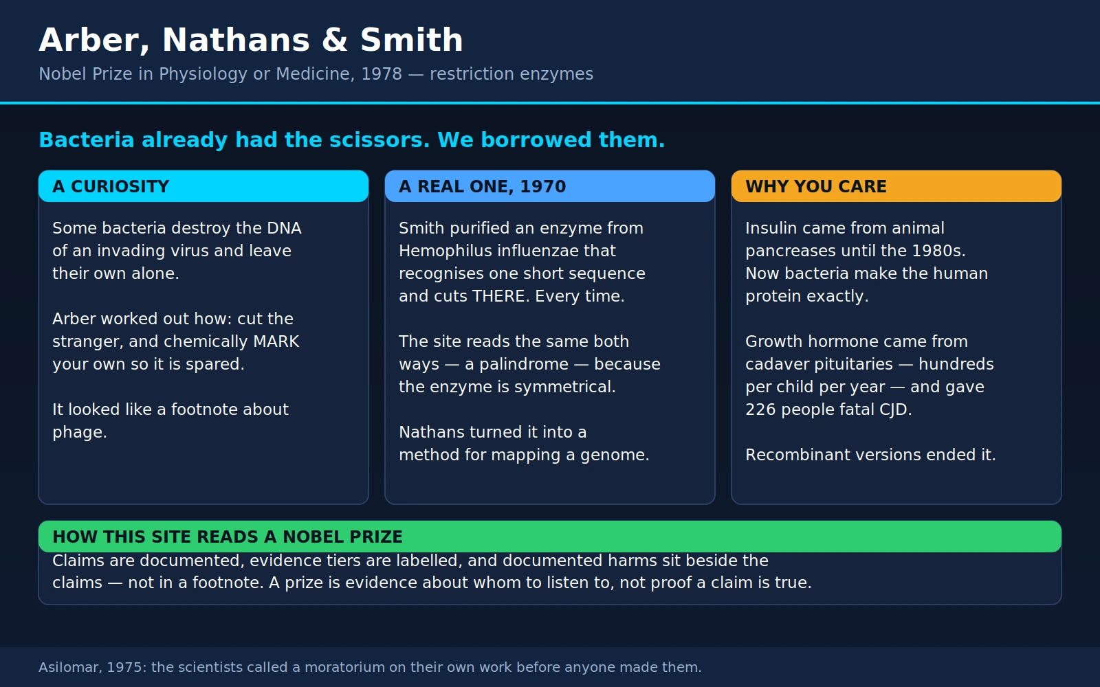
In 1978 the Nobel Prize in Physiology or Medicine went to three men “for the discovery of restriction enzymes and their application to problems of molecular genetics.” The phrase is dry even by Nobel standards, and it hides what is probably the single largest return on investment in the history of publicly funded biology. Werner Arber had been trying to understand why a particular virus grew well on one strain of bacteria and badly on another. Hamilton Smith had been studying how a bacterium takes up DNA. Daniel Nathans had been trying to find a way to take a virus genome apart. None of the three set out to invent a technology.
What came out of their work is the reason a person with type 1 diabetes today injects human insulin made by bacteria instead of insulin scraped from pig pancreases; the reason a child with growth hormone deficiency is not injected with an extract of pituitary glands taken from cadavers; the reason a person with haemophilia is not treated with a product pooled from the plasma of thousands of donors; and the reason the hepatitis B vaccine is not manufactured from the blood of people who are infected with hepatitis B. Those four changes, taken together, removed a set of very specific and very lethal infection risks from routine medical care. All four rest on a bacterial enzyme that cuts DNA at a sequence it recognises.
Table of Contents
- A Curiosity About Bacteria
- Arber's Answer: Cut the Stranger, Mark Your Own
- Hamilton Smith Finds a Real One
- What a “Recognition Site” Is, and Why It Makes a Tool
- Daniel Nathans Makes It a Method
- Sticky Ends and Recombinant DNA
- What This Gave Patients
- Asilomar, 1975
- Restriction Enzymes in the Clinic and the Lab Today
- The Bacterial Immune System, Continued
- The Three Men
- What a Reader Should Take From This
- Where Mainstream Medicine Agrees — and What Remains Debated
- Key Research Papers
- Connections
- Featured Videos
1. A Curiosity About Bacteria
In the early 1950s, bacteriophage researchers noticed something that looked like a nuisance. A phage — a virus that infects bacteria — grown on one strain of Escherichia coli would plate beautifully on that same strain, producing thousands of plaques. Take the identical phage stock and put it on a slightly different strain, and the count could fall by a factor of ten thousand. The virus had not changed. The bacterium had.
Stranger still, the effect was not permanent. Of the very few phage particles that did manage to grow on the new strain, the descendants plated perfectly well on it from then on — and had now lost their ability to grow on the original strain. Whatever the host was doing, it was doing it to the virus's own material, and the change was inherited for as long as the virus stayed in that host.
Salvador Luria and Mary Human described this in 1952; Giuseppe Bertani and Jean Weigle described it independently in 1953. They called it host-controlled variation, and it was, at the time, mostly an irritation — something you had to control for when you did phage genetics. Luria's own work on bacteria and viruses would be recognised with the 1969 Nobel Prize, shared with Max Delbrück and Alfred Hershey. This page is the direct sequel to that one: Luria saw the phenomenon, and Arber explained it.
It is worth pausing on how unpromising this looked. A graduate student in 1955 who announced that they intended to spend several years on why a phage stock plates poorly after a change of host would not have been describing a medical project. There was no patient in the room. There was no disease. There was an odd number on a plate.
2. Arber's Answer: Cut the Stranger, Mark Your Own
Werner Arber, working in Geneva with his graduate student Daisy Dussoix, took the odd number seriously. In two companion papers in the Journal of Molecular Biology in 1962, they showed that the host's effect was exerted on the DNA of the incoming phage — not on its protein coat, not on some later step of infection. DNA that arrived carrying the wrong host's signature was destroyed. DNA carrying the right signature was let through.
From this Arber built a model that turned out to be correct. A bacterium, he proposed, carries two paired activities:
- A restriction activity — an enzyme that recognises a particular short sequence of DNA letters and, finding it unprotected, cuts the DNA. Chopped DNA cannot make a virus.
- A modification activity — an enzyme that chemically marks that same sequence in the cell's own chromosome, by attaching a methyl group to one of the bases. Marked DNA is invisible to the restriction enzyme.
Put together, the pair is a self/non-self discrimination system. The cell marks everything it owns and destroys everything unmarked that comes through the door. The rare phage that survives gets marked on its way past, which is exactly why its descendants inherit the new host range. This is, in the plainest sense, an immune system — a primitive, hard-coded one, but an immune system: a mechanism for telling self from foreign and destroying the foreign.
That framing matters for everything that follows. Restriction enzymes were not invented as laboratory reagents. They are bacterial anti-virus weapons that human beings borrowed. Nobody designing a gene-editing tool from first principles would have thought to look inside E. coli for one.
There was, however, a catch, and it is the reason Arber alone could not have started a biotechnology industry. The systems Arber was studying — now classified as Type I restriction systems — do recognise a specific sequence, but then travel along the DNA and cut it at some unpredictable distance away. As a defence mechanism that is perfectly adequate: cut anywhere and the virus is dead. As a tool it is useless. If the cut lands in a different place every time, you get a smear of random fragments, and a smear tells you nothing.
3. Hamilton Smith Finds a Real One
Hamilton O. Smith, at the Johns Hopkins University School of Medicine in Baltimore, was not looking for a restriction enzyme. He was studying how Haemophilus influenzae strain Rd takes up DNA from its environment — natural transformation. In the course of that work he and his research fellow Kent Wilcox noticed that when they gave the bacterium DNA from a phage, the foreign DNA was degraded, while the bacterium's own DNA sat there untouched.
That is Arber's prediction, showing up uninvited in someone else's experiment. Smith recognised it, dropped what he was doing, and purified the responsible activity. The 1970 paper — A restriction enzyme from Hemophilus influenzae. I. Purification and general properties, by Smith and Wilcox — reports a purified enzyme, then called endonuclease R and now known as HindII, that made a limited and reproducible number of double-strand breaks in foreign DNA.
The word doing the work in that sentence is limited. Not a smear. A countable number of cuts, in the same places every time.
The companion paper, published back-to-back in the same issue — A restriction enzyme from Hemophilus influenzae. II. Base sequence of the recognition site, by Thomas Kelly Jr. and Smith — worked out what the enzyme was actually reading. It recognises the six-letter sequence GTPy↓PuAC: G, T, then either pyrimidine (C or T), then either purine (A or G), then A, then C. And it cuts in the middle, between the fourth and fifth letters, on both strands.
4. What a “Recognition Site” Is, and Why It Makes a Tool
DNA is a two-stranded molecule, and the two strands run in opposite directions. Each strand is read in a fixed direction, conventionally written 5′ to 3′. Because the bases pair only one way — A with T, G with C — knowing one strand tells you the other.
Now consider the sequence recognised by EcoRI, a restriction enzyme found a little later in E. coli, and probably the most-used enzyme in the history of molecular biology. Written out, both strands, with the direction marked:
- Top strand, 5′→3′: G A A T T C
- Bottom strand, 3′→5′: C T T A A G
Read the bottom strand in its own 5′→3′ direction — that is, backwards from how it is written above — and you get G A A T T C. The same six letters. A sequence like this is called a palindrome, and most restriction enzymes recognise one. It is not a coincidence: these enzymes generally work as a pair of identical protein subunits, one sitting on each strand, and a symmetrical protein needs a symmetrical target. HindII's site, GTPyPuAC, is a palindrome too, just a slightly fuzzy one — the middle two positions can vary as long as they stay complementary.
Here is why that specificity is the whole story. Imagine two machines you could point at a length of DNA:
- A shredder cuts wherever it happens to land. Feed it the same DNA sample twice and you get two different sets of pieces. The information in the original molecule — what was next to what — is destroyed.
- A restriction enzyme cuts only where its sequence appears. Feed it the same DNA twice and you get exactly the same pieces, of exactly the same lengths, in exactly the same quantities. Run those pieces out on a gel and you see a ladder of discrete bands, not a smear.
Reproducibility is what converts destruction into measurement. A shredder tells you nothing. A restriction enzyme turns an enormous, unreadable molecule into a small set of defined, countable, separable objects that you can look at one at a time — and, crucially, put back together. That is the difference between breaking something and taking it apart.
5. Daniel Nathans Makes It a Method
Daniel Nathans worked one floor away from Smith at Johns Hopkins. He was studying SV40, a small monkey virus with a circular DNA genome of about 5,200 base pairs that could transform cells in culture and was, at the time, one of the few animal genomes small enough to contemplate studying whole. His problem was that he had no way to get at any particular part of it. A circular genome with no landmarks is a closed loop of five thousand indistinguishable letters.
Nathans got some of Smith's enzyme and put it on SV40 DNA. The 1971 paper by Kathleen Danna and Nathans in the Proceedings of the National Academy of Sciences reports the result: digestion gave eleven fragments that could be separated by gel electrophoresis, eight of them present in the same molar amount as the starting DNA. The genome had been cut into a specific, repeatable set of pieces.
Two years later Danna, George Sack Jr. and Nathans published the next step, which is the one that mattered: they worked out the order the eleven fragments occupy around the circle. This was the first restriction map of any genome.
What a restriction map actually is
Think of a circular road with no signposts. You know its total length and nothing else. Someone hands you a machine that will cut the road at every point where the surface is a particular colour. You run it, and you get eleven segments of known lengths — but you have no idea what order they went in, because the machine cut them all at once and they fell into a heap.
So you cheat. You run the machine again but stop it early, so that most stretches of road get cut once or twice rather than everywhere. Now you have longer pieces that each contain two or three adjacent segments still joined together. Measure those, and each one tells you which segments are neighbours. Do this enough times and the overlaps force a single consistent ordering. Then you bring in a second machine that cuts on a different colour, and where its cuts fall inside your segments gives you a second, independent set of landmarks to check the first against.
That is a restriction map: a diagram of a genome showing where each enzyme cuts, in order, with distances. Once you have one, an unreadable molecule becomes an addressable one. You can say the mutation is in fragment C, or transcription starts near the boundary between A and H, and then go and purify fragment C in isolation and work on it. Nathans and his group promptly did exactly that: they used the map to locate the SV40 origin of replication and to place its genes, years before anyone could sequence DNA.
This is the step the Nobel citation means by “their application to problems of molecular genetics.” Arber found the phenomenon. Smith found a usable enzyme. Nathans turned a usable enzyme into a general method for taking any genome apart into knowable, orderable pieces — and every genetic map since, including the human one, is a descendant of that idea.
6. Sticky Ends and Recombinant DNA
HindII cuts straight across the middle of its site, leaving what are called blunt ends — two flat, flush ends. Blunt ends can be joined, but reluctantly; there is nothing holding two of them together while the joining enzyme works.
EcoRI turned out to behave differently, and the difference changed everything. It cuts its palindrome off-centre — between the G and the first A on each strand. Because the cuts on the two strands are staggered, each fragment comes away with a short single-stranded tail hanging off it:
- Left fragment ends: …G on the top strand, with AATT unpaired and dangling from the bottom.
- Right fragment begins: AATTC…, with AATT unpaired and dangling from the top.
Those two tails are complementary. Bring them close and they will pair up spontaneously, the same way the two strands of DNA pair anywhere else. This is why they are called cohesive ends or, universally, sticky ends.
Janet Mertz and Ronald Davis spelled out the consequence in 1972. Because every EcoRI cut produces the same four-letter overhang, every EcoRI end in the world is complementary to every other EcoRI end in the world. It does not matter whether one fragment came from a virus, a bacterium, a mouse or a person. Cut two DNA molecules from any two organisms with the same enzyme, mix them, let the sticky ends find each other, and seal the joins with DNA ligase — and you have a single hybrid molecule that never existed before.
The other half of the problem was getting such a molecule to survive and multiply. Paul Berg, at Stanford, had built the first hybrid DNA molecule in 1972, joining SV40 DNA to DNA from a bacterial virus and the E. coli galactose operon — but by a laborious chemical route, and he stopped short of putting it into a living cell. The practical solution came in 1973 from Stanley Cohen, Annie Chang, Herbert Boyer and Robert Helling, who used plasmids as the vehicle.
A plasmid is a small circle of DNA that lives inside a bacterium alongside the main chromosome and copies itself independently. Bacteria swap them routinely; it is how antibiotic resistance spreads. For an experimenter a plasmid is close to ideal: cut it open with a restriction enzyme, drop in a fragment of foreign DNA with matching sticky ends, seal it, and put the resulting circle back into a bacterium. The bacterium copies the plasmid — and therefore your insert — every time it divides, and it divides every twenty minutes. Overnight, one molecule becomes billions. If the inserted fragment happens to be a gene, and it is placed behind a signal the bacterium's machinery recognises, the bacterium will not only copy it but read it, and make the protein.
A restriction enzyme, a ligase and a plasmid: that is the whole of the technique, and that is the technical basis of the biotechnology industry. Boyer and the venture capitalist Robert Swanson founded Genentech in 1976 on precisely this. Every recombinant protein drug on the market — and there are hundreds — traces back through this three-step procedure to a bacterial defence enzyme.
7. What This Gave Patients
This is the section that justifies the page. The recombinant proteins that reached patients first were not, for the most part, better than what they replaced in the sense of working harder. They were better because of where they came from. Each one removed a human or animal body from the supply chain, and with it a specific, documented, sometimes fatal infection risk.
Insulin: out of the slaughterhouse
From 1922 until the early 1980s, every unit of injected insulin came from an animal pancreas. Frederick Banting and Charles Best's discovery had been made with dog and then cattle pancreas extract, and the industry that grew from it depended on collecting pancreases from abattoirs — on the order of tens of thousands of animals to supply one patient for a lifetime.
Animal insulin worked, and it saved millions of lives. But pig insulin differs from human insulin by one amino acid and cattle insulin by three, and the immune system notices. A substantial fraction of long-term users developed anti-insulin antibodies; a minority developed antibody-mediated insulin resistance requiring escalating doses; injection-site fat loss (lipoatrophy) and local allergic reactions were routine clinical problems, especially with the less-purified preparations of earlier decades. And the supply was tied to meat production, which was a genuine long-term worry as diabetes prevalence rose.
In 1979 David Goeddel and colleagues at Genentech expressed chemically synthesised genes for the two chains of human insulin in E. coli. In 1980 Harry Keen and colleagues in London published the first controlled comparison in people: recombinant human insulin against highly purified pig insulin in healthy men, by intracutaneous, subcutaneous and intravenous routes. No local reactions over 48 hours; the fall in blood glucose was closely similar, with only small differences in dose–response. Their conclusion was that genetically synthesised human insulin appeared safe and effective in humans. Recombinant human insulin (Humulin) was approved in the United States in 1982 — the first medicine made by recombinant DNA ever licensed for human use.
What changed for patients: a molecule identical to the one their own pancreas would have made, in unlimited supply, with markedly less immunogenicity and none of the batch-to-batch variability of an animal-tissue extract. See also type 1 diabetes.
Growth hormone: the hardest case
This one is not a story about convenience. Children with growth hormone deficiency were treated, from 1958 to 1985, with human growth hormone extracted from pituitary glands collected at autopsy. There was no alternative: growth hormone is species-specific, so unlike insulin it could not be taken from an animal. A single year of treatment for one child required the pituitaries of hundreds of cadavers, and the glands were pooled in large batches.
Some of those donors had died of undiagnosed Creutzfeldt-Jakob disease. The infectious agent is a prion — a misfolded protein, discovered and characterised by Stanley Prusiner, which is not destroyed by the treatments that inactivate bacteria and viruses. It survived the extraction process, and it was distributed, in pooled batches, to children.
The first case was reported in 1985: a young adult with hypopituitarism who had received cadaveric growth hormone and died of Creutzfeldt-Jakob disease. Distribution through the United States National Hormone and Pituitary Program was suspended on 19 April 1985. By 1991, Judith Fradkin and colleagues reported seven neuropathologically confirmed cases among 6,284 American recipients — all seven among the roughly 700 who had begun treatment before 1970, and with a median treatment duration more than twice that of the cohort as a whole. The incubation period averaged around fifteen years, so most of the exposed population had not yet been at risk long enough to know.
The final worldwide tally, published by Paul Brown and colleagues in 2012, was 226 cases of Creutzfeldt-Jakob disease caused by contaminated growth hormone, alongside 228 caused by cadaveric dura mater grafts. Every one of those was a person who received a medical treatment and got a fatal, untreatable brain disease from it.
Recombinant methionyl human growth hormone was tested in growth-hormone-deficient children and reported by Selna Kaplan and colleagues in 1986: growth rate rose from 3.2 to 10.5 cm per year, essentially the same as pituitary-derived hormone, with somatomedin C responses to match. It was approved in the United States in October 1985, six months after cadaveric distribution stopped. A protein made in a bacterium cannot carry a human prion, because it never passed through a human body. The risk did not become smaller; it became structurally impossible. See also hypopituitarism.
Clotting factors: the pooled-plasma catastrophe
Haemophilia A is caused by a deficiency of clotting factor VIII. From the late 1960s it was treated with factor VIII concentrate — a genuine advance, because it let people treat bleeds at home instead of in hospital. But concentrate was manufactured by pooling plasma from thousands to tens of thousands of donors per lot. A single infected donor contaminated the entire lot, and a person with severe haemophilia was exposed to every lot they used, several times a month, for years.
When HIV entered the blood supply at the end of the 1970s, that manufacturing design turned a rare infection into a near-universal one within this population. James Goedert and colleagues followed a multicentre cohort of 1,219 people with haemophilia and related disorders and analysed 319 with documented HIV-1 seroconversion dates, reporting AIDS incidence rising with age at seroconversion — the cohort exists because so many people were infected at once and at a known time.
Hepatitis C was, if anything, more complete. Victor Blanchette and colleagues tested stored sera from Canadian children with haemophilia between 1987 and 1989. Among those regularly treated with unheated or dry-heat-treated concentrates, 95% (21 of 22) were hepatitis C antibody positive. Among untransfused children with haemophilia: 0 of 11. Among those treated only with cryoprecipitate or single-donor products: 0 of 9. Among those given vapour-heated concentrate: 0 of 9. The concentrate was the exposure.
Recombinant factor VIII was tested through the late 1980s and licensed in 1992. The trial of previously untreated children published by Jeanne Lusher and colleagues in 1993 states the rationale in its own opening lines: viral attenuation of plasma-derived concentrates had improved, but the possibility of transmitting blood-borne viruses remained, and recombinant factor VIII is virus-free. Ninety-five children received it exclusively, some for more than three years, across 3,315 infusions, with no treatment failures and three minor adverse reactions. See also HIV/AIDS and hepatitis C.
Hepatitis B vaccine: a vaccine that was made of blood
Baruch Blumberg discovered the hepatitis B surface antigen — the “Australia antigen” — and with Irving Millman devised the first vaccine against it. That vaccine, licensed in 1981, was made by purifying surface antigen from the plasma of chronic hepatitis B carriers. The purification was rigorous and the product was effective, but the raw material was the blood of infected people, and it reached the market at precisely the moment the public was learning that the blood supply could transmit a fatal virus. Uptake suffered.
William McAleer, Maurice Hilleman and colleagues at Merck, working with vectors developed by William Rutter's and Benjamin Hall's groups, expressed the surface antigen in recombinant baker's yeast. Their 1984 paper in Nature reports that the yeast-derived antigen raised antibody in mice, monkeys and chimpanzees, and that vaccinated chimpanzees were totally protected against intravenous challenge with hepatitis B virus of both the matching and non-matching subtypes. They noted, correctly, that this was the first vaccine made from recombinant cells shown to be effective against a human viral infection. The recombinant vaccine was licensed in 1986 and is what is given today, worldwide, including the birth dose. See also hepatitis B.
The pattern
Four products, four different diseases, one shared logic. In each case the older product came out of a body — a pig's pancreas, a cadaver's pituitary, thousands of donors' plasma, an infected person's blood — and carried whatever that body carried. The recombinant version is assembled from a written gene sequence by an organism that has never been near a human being. That is not a marginal improvement in purity. It is the removal of an entire category of risk, and it is the reason this particular piece of basic science shows up in the life of almost every patient who takes a biological medicine.
8. Asilomar, 1975
The part of this history that is least known outside science is that the people who built the technology stopped it, voluntarily, before anyone told them to.
By 1973 the recombinant plasmid method was working, and it was obvious to its inventors that it would work on anything. At the 1973 Gordon Research Conference on Nucleic Acids, participants voted to write to the National Academy of Sciences asking for the safety questions to be studied. The Academy convened a committee chaired by Paul Berg.
In July 1974 that committee published a short letter in Science — also carried in Nature and the Proceedings of the National Academy of Sciences — titled “Potential biohazards of recombinant DNA molecules.” It is a letter, not a research paper, and it is listed as such below. Its signatories included Berg, David Baltimore, Herbert Boyer, Stanley Cohen, Ronald Davis, Daniel Nathans and Sherman Weissman, among others — that is, the people who had personally done the experiments. It asked scientists worldwide to voluntarily defer:
- construction of new bacterial plasmids that would introduce antibiotic resistance or bacterial toxin genes into strains that did not already carry them; and
- linking DNA from cancer-causing or other animal viruses to plasmids or other autonomously replicating DNA.
It further asked that experiments linking animal DNA to bacterial plasmids or phage be weighed carefully; that the National Institutes of Health establish an advisory committee to assess the hazards and develop procedures; and that an international meeting be convened early in 1975. The moratorium was honoured, essentially worldwide, on nothing but the authority of the people who signed it.
The meeting took place from 24 to 27 February 1975 at the Asilomar Conference Grounds in Pacific Grove, California. About 140 people attended — mostly molecular biologists, but deliberately also lawyers, physicians and journalists, who were present for the discussions and not excluded from them. Berg chaired; the organising committee included Baltimore, Sydney Brenner, Richard Roblin and Maxine Singer.
The summary statement, published in the Proceedings of the National Academy of Sciences in June 1975 and simultaneously in Science, concluded these things:
- The work should go forward. The moratorium should be lifted. The conference explicitly judged that stopping the research was not the right answer.
- Containment should be matched to estimated risk. Experiments were sorted into categories, and each category assigned a level of containment. This graded principle, rather than any single rule, is Asilomar's real product.
- Two kinds of containment, used together. Physical containment — safety cabinets, controlled airflow, laboratory practice — graded from minimal to high. And biological containment: the deliberate use of host organisms and vectors so enfeebled that they cannot survive outside the specific conditions of a laboratory. The safe-vector programme that followed produced strains such as E. coli χ1776, engineered with nutritional requirements it could not meet in the human gut or the environment.
- Some experiments should not be done at all yet. A small number of categories — among them cloning DNA from highly pathogenic organisms and genes for potent toxins — were judged to carry risks too serious to permit with the containment then available.
- The rules should be revisited as evidence accumulated, in either direction, along with training and monitoring of laboratory workers.
The National Institutes of Health established the Recombinant DNA Advisory Committee, which issued formal guidelines in June 1976. Those guidelines were progressively relaxed over the following years as the feared hazards failed to materialise.
Asilomar is invoked, repeatedly and by name, whenever a new technology raises questions of the same shape — in debates over human germline editing, over gain-of-function research on pathogens, and more recently over artificial intelligence. Whether the analogy holds in any given case is argued about by people who study these things; what is not in dispute is that the precedent is real and that it is cited. It happened because a small number of researchers decided that the right time to work out the rules was before, rather than after.
9. Restriction Enzymes in the Clinic and the Lab Today
Honesty is required here, because a lot of what restriction enzymes were once used for in medicine is now genuinely obsolete.
What they did: RFLP and the first DNA diagnostics
If a stretch of DNA differs between two people at a single letter, and that letter happens to sit inside a restriction enzyme's recognition site, then the enzyme will cut one person's DNA there and not the other's. Run both digests on a gel and the two people give different band patterns. This is a restriction fragment length polymorphism, or RFLP — and for about fifteen years it was the only practical way to read a difference in human DNA.
Yuet Wai Kan and Andrée Dozy made the first clinical use of it in 1978. They found a restriction-site polymorphism next to the beta-globin gene that travelled with the sickle cell mutation in the families they studied, and used it, in a companion paper the same year, to make an antenatal diagnosis of sickle cell anaemia from amniotic fluid cells. That was the first prenatal diagnosis of a genetic disease made by reading DNA rather than by measuring a protein.
Two years later David Botstein, Ray White, Mark Skolnick and Ronald Davis proposed using RFLPs systematically to build a genetic linkage map of the entire human genome. That proposal is the direct intellectual ancestor of positional cloning, of the disease-gene hunts of the 1980s and 1990s, and of the Human Genome Project itself. RFLP analysis was also the original basis of DNA fingerprinting in forensic and paternity casework.
Why sequencing replaced it
RFLP has severe limits, and they are all structural rather than fixable. It can only see a variant if that variant happens to create or destroy a restriction site — the great majority do not. It reads one locus at a time. It needs micrograms of intact high-molecular-weight DNA, which means a decent blood sample, not a cheek swab. And a Southern blot takes the better part of a week.
Sequencing has none of those constraints. It reads the actual letters, at any position, whether or not an enzyme happens to care about them; modern instruments read millions of positions at once from nanograms of material. Clinical genetics moved to targeted gene panels, then exomes, then whole genomes, and RFLP as a diagnostic method is now essentially a historical technique. If a clinician today wants to know whether a patient carries a particular variant, nobody digests their DNA with an enzyme and looks at a gel.
Where restriction digestion is still genuinely used
- Verifying that a construct is what you think it is. A “diagnostic digest” — cutting a plasmid with one or two enzymes and checking that the band sizes match the predicted map — remains the fastest, cheapest quality-control check in molecular biology, and is run daily in laboratories that also make clinical-grade material.
- Assembling DNA. Type IIS enzymes, which cut at a fixed distance outside their recognition site and therefore leave a programmable overhang, are the basis of Golden Gate assembly, used to build multi-part constructs including engineered cell therapies.
- Reading DNA methylation. Some enzymes cut a site only when it is unmethylated; their methylation-blind partners cut it either way. Comparing the two digests reads the methylation state directly, which is used in epigenetic work and in some cancer assays.
- Reduced-representation sequencing. Several sequencing library methods use a restriction digest deliberately, to sample a consistent, reproducible subset of a large genome rather than sequencing all of it.
- Cheap single-variant testing. PCR followed by a restriction digest is still occasionally the most economical way to test one known variant in many samples where sequencing capacity is limited.
There is also a nice closing of the circle. In 1995 the first complete genome sequence of any free-living organism was published: Haemophilus influenzae Rd, sequenced by whole-genome shotgun assembly at The Institute for Genomic Research. The organism was chosen partly because Hamilton Smith, who was on the team, had been working on it since the 1960s — it is the bacterium his restriction enzyme came from.
10. The Bacterial Immune System, Continued
Restriction-modification was the first bacterial defence against viruses ever described. It was not the last, and the second one to be found repeated the pattern almost exactly.
In 1987 Japanese researchers sequencing a gene in E. coli noticed a strange array of short repeated sequences separated by spacers of unrelated DNA. Nobody knew what it was for. Similar arrays turned up in many bacteria and archaea and acquired the name CRISPR — clustered regularly interspaced short palindromic repeats. In the mid-2000s several groups noticed that the spacer sequences matched fragments of phage genomes.
The function was demonstrated experimentally in 2007 by Rodolphe Barrangou and colleagues, working at a food company on Streptococcus thermophilus — the bacterium used to make yoghurt and mozzarella, which was being studied because phage infections ruin industrial fermentations. They showed that when the bacterium survived a phage attack, it inserted a piece of the phage's own DNA into its CRISPR array, and that this gave it specific, heritable resistance to that phage. Change the spacers and you change which phages it resists.
That is an adaptive immune system in a bacterium: it acquires a memory of a specific pathogen from experience, stores it, and passes it to its descendants. Where restriction-modification is a blunt innate defence — destroy anything unmarked — CRISPR is a targeted one, guided by a stored record of past infections.
The tool followed from the mechanism. Because the CRISPR system's cutting enzyme, Cas9, is aimed by a short guide RNA rather than by the shape of the protein itself, it can be reprogrammed simply by supplying a different guide. Work published in 2012 by Jennifer Doudna, Emmanuelle Charpentier and colleagues showed Cas9 could be directed by a single engineered guide RNA to cut any chosen sequence; groups led by Feng Zhang and George Church demonstrated it working in human cells in 2013. Doudna and Charpentier received the 2020 Nobel Prize in Chemistry for it.
The first CRISPR-based therapy to be approved is exagamglogene autotemcel (brand name Casgevy). The United Kingdom's medicines regulator authorised it in November 2023. The United States Food and Drug Administration approved it for sickle cell disease on 8 December 2023, and for transfusion-dependent beta-thalassaemia on 16 January 2024, in both cases for patients aged 12 and over.
It is worth being precise about what it does, because it is commonly described inaccurately. It does not correct the sickle mutation. Doctors collect the patient's own blood stem cells, use CRISPR-Cas9 in the laboratory to disrupt an enhancer that controls the gene BCL11A, and return the edited cells. BCL11A is the switch that shuts off fetal haemoglobin production after birth; disabling the enhancer turns fetal haemoglobin back on, and fetal haemoglobin does not sickle. The first-in-human report, published by Haydar Frangoul and colleagues in 2021, described two patients — one with beta-thalassaemia, one with sickle cell disease — both transfusion-independent more than a year later. The phase 3 trial, published in 2024, treated 44 patients with severe sickle cell disease: of the 30 with enough follow-up to assess, 29 (97%) had no severe vaso-occlusive crisis for at least twelve consecutive months, and all 30 avoided hospitalisation for one.
Two honest caveats. The procedure requires myeloablative chemotherapy — busulfan conditioning to clear the marrow before the edited cells go back — which carries real toxicity including infertility, and is why this is a hospital-based transplant procedure rather than an injection. And the follow-up, at a median of about nineteen months in the phase 3 trial, is short for a therapy meant to last a lifetime.
The parallel with 1970 is exact and worth stating plainly. Both restriction-modification and CRISPR are bacterial defences against viruses. Both were found by people studying bacteria for their own sake — one a phage geneticist puzzled by plating efficiency, the other a food-industry laboratory trying to keep phages out of yoghurt vats. Neither was found by anyone looking for a way to edit genes. In both cases the immune system turned out to be a programmable, sequence-specific DNA-cutting machine, and in both cases that is the property that made it the standard tool of a generation.
11. The Three Men
Werner Arber (born 1929, Gränichen, Switzerland) trained as a chemist and physicist before moving into phage genetics at the University of Geneva, where the 1962 work with Daisy Dussoix was done. He was professor of molecular microbiology at the University of Basel from 1971 until 1996 and remains professor emeritus there. He was the first Swiss scientist to receive the Nobel Prize in Physiology or Medicine.
Daniel Nathans (1928–1999) was born in Wilmington, Delaware, the youngest of nine children of Russian-Jewish immigrants; his father lost his small business in the Depression. He trained in medicine at Washington University in St. Louis, practised as a physician, and joined Johns Hopkins in 1962, where he spent the rest of his career and served as interim president of the university. He was awarded the National Medal of Science in 1993.
Hamilton O. Smith (1931–2025) was born in New York City and raised in Illinois, took his medical degree at Johns Hopkins, and served in the United States Navy before returning to research. He joined the Johns Hopkins faculty in 1967, where the restriction enzyme work was done in the laboratory next door to Nathans's. In the 1990s and 2000s he worked with J. Craig Venter on whole-genome shotgun sequencing — including the first sequenced free-living organism, his own Haemophilus influenzae — and later on synthetic genomes. He died in October 2025, aged 94.
It is a small detail, but a telling one, that two of the three laureates worked in adjacent laboratories in the same building and that the crucial step happened because one of them walked down the corridor to borrow the other's enzyme.
12. What a Reader Should Take From This
Set out the chain in order, with the dates:
- 1952–53. Luria and Human, and Bertani and Weigle, notice that a phage plates poorly after a change of host. It is an annoyance in an experiment.
- 1962. Arber and Dussoix show the effect is exerted on the phage's DNA, and Arber proposes cutting-plus-marking.
- 1970. Smith and Wilcox purify an enzyme that makes a limited number of cuts; Kelly and Smith determine the sequence it reads.
- 1971–73. Danna and Nathans cut a viral genome into eleven defined pieces and then put them in order — the first restriction map.
- 1972. Mertz and Davis show that identical sticky ends make any two DNAs joinable.
- 1973. Cohen, Chang, Boyer and Helling get a recombinant plasmid replicating in a living bacterium.
- 1975. Asilomar sets the safety framework that lets the work proceed.
- 1982. Human insulin made by bacteria is licensed.
- 1985–86. Recombinant growth hormone and the recombinant hepatitis B vaccine follow.
- 1992. Recombinant factor VIII removes the pooled-plasma risk from haemophilia care.
Thirty years separate the odd number on a plate from the insulin in the pharmacy. Nobody along that chain could have written down the destination at the start of it. In 1960 there was no proposal anyone could have submitted that said: fund my work on phage plating efficiency and in twenty-two years people with diabetes will stop injecting pig hormone. The connection is real, it is direct, and it was invisible in advance.
The honest version of the lesson is narrower than the one usually offered, and stronger for being narrower. It is not that all curiosity-driven research pays off — most individual projects do not produce anything like this, and that is the normal outcome. It is not an argument against targeted research; targeted research produced the trials that proved these products safe. The claim is this: the projects that turn out to matter most cannot be identified in advance from the question they are asking, because the payoff runs through a mechanism nobody knows exists yet. A funding system that only supports work with a stated application would have declined all three of these men at the point where their work was most valuable.
And this particular lineage has an unusually good record. The same corner of biology — bacteria defending themselves against viruses — has now produced, twice, the defining laboratory tool of an era. Someone was studying yoghurt cultures.
13. Where Mainstream Medicine Agrees — and What Remains Debated
Agreed, and not seriously disputed anywhere
- Restriction enzymes and their specificity are settled biochemistry. Thousands are characterised, catalogued and sold; their recognition sequences are experimentally verified reagent specifications, not inferences.
- Recombinant DNA is the manufacturing basis of modern biological medicine. Insulin, growth hormone, erythropoietin, interferons, clotting factors, monoclonal antibodies, several vaccines and most enzyme replacement therapies are made this way. No mainstream body disputes this and no clinical guideline treats it as controversial.
- Replacing human- and animal-sourced biologics removed real infection risks. The prion transmissions through cadaveric growth hormone and the HIV and hepatitis C transmissions through pooled plasma are documented in the epidemiological record with case counts, and the recombinant products cannot transmit those agents because they never contact the source tissue.
- Asilomar's graded-containment framework worked well enough to proceed on. The specific hazards feared in 1974 — a laboratory-engineered organism escaping and causing disease or ecological harm — have not materialised from recombinant DNA research of the kind the moratorium covered, and the guidelines were relaxed as that became clear.
Genuinely open or contested
- Recombinant is not automatically better on every endpoint. The SIPPET trial, published in 2016, randomised 251 previously untreated boys with severe haemophilia A to plasma-derived factor VIII containing von Willebrand factor or to recombinant factor VIII, and found more inhibitor formation with the recombinant product: cumulative incidence 44.5% versus 26.8%, hazard ratio 1.87 (95% CI 1.17–2.96). Inhibitors are neutralising antibodies that make replacement therapy stop working, and they are the most serious complication of haemophilia care. The result has been argued over ever since — on trial population, on which specific products were used, on how it applies where safe plasma supply is not guaranteed — and practice varies between countries. It is an important corrective to a simple “recombinant is safer, full stop” reading of this page's section 7, and it does not overturn the infection-risk argument, which is about a different endpoint entirely.
- How long gene-edited therapies last is not yet known. Follow-up on exagamglogene autotemcel is measured in a few years. Durability, late effects of busulfan conditioning, and any long-term consequence of the edit itself will only be answered by time.
- Cost and access. Exagamglogene autotemcel carries a United States list price of about $2.2 million per patient, before the cost of the transplant admission. Sickle cell disease is most common in populations with the least access to that kind of expenditure, and how these therapies reach the people who need them — including in sub-Saharan Africa and India, where most patients live — is unresolved. The same tension applied, in a milder form, to recombinant clotting factor for decades.
- Whether Asilomar is the right template for later technologies. Historians and policy scholars note that Asilomar addressed a narrow, technical, biosafety question among a small group of specialists who all shared a research goal, and that questions about germline editing or artificial intelligence involve contested values and a far wider set of stakeholders. Whether the precedent transfers is an active scholarly argument; that it is repeatedly cited is not.
14. Key Research Papers
Every citation below was verified in PubMed against journal, year, volume and pages, and its publication type checked. Where a paper is a letter rather than a research report, it is labelled as one.
- Arber W, Dussoix D. Host specificity of DNA produced by Escherichia coli. I. Host controlled modification of bacteriophage lambda. Journal of Molecular Biology. 1962;5:18–36. PMID 13862047. — The foundational paper, showing that host-controlled restriction acts on the phage's DNA. Its companion, Dussoix D, Arber W, part II (J Mol Biol. 1962;5:37–49; PMID 13888713), covers acceptance of DNA from the infecting phage. Neither carries an abstract in PubMed.
- Smith HO, Wilcox KW. A restriction enzyme from Hemophilus influenzae. I. Purification and general properties. Journal of Molecular Biology. 1970;51(2):379–391. PMID 5312500. — The first purified sequence-specific restriction endonuclease. Cite this part for the enzyme itself, not for its recognition sequence.
- Kelly TJ Jr, Smith HO. A restriction enzyme from Hemophilus influenzae. II. Base sequence of the recognition site. Journal of Molecular Biology. 1970;51(2):393–409. PMID 5312501. — The recognition sequence itself, GTPy↓PuAC, cut in the middle. PubMed truncates the subtitle to “II.”; the full title is as given here.
- Danna K, Nathans D. Specific cleavage of simian virus 40 DNA by restriction endonuclease of Hemophilus influenzae. Proceedings of the National Academy of Sciences USA. 1971;68(12):2913–2917. PMID 4332003. — Eleven resolvable fragments from the SV40 genome, eight of them equimolar with the starting DNA. The first application of a restriction enzyme to an animal virus genome.
- Jackson DA, Symons RH, Berg P. Biochemical method for inserting new genetic information into DNA of Simian Virus 40: circular SV40 DNA molecules containing lambda phage genes and the galactose operon of Escherichia coli. Proceedings of the National Academy of Sciences USA. 1972;69(10):2904–2909. PMID 4342968. — The first recombinant DNA molecule, built by a chemical tailing route rather than with sticky ends. Berg's caution about this construction is what led to the 1974 letter.
- Mertz JE, Davis RW. Cleavage of DNA by R1 restriction endonuclease generates cohesive ends. Proceedings of the National Academy of Sciences USA. 1972;69(11):3370–3374. PMID 4343968. — Sticky ends. Shows that all EcoRI-generated ends are identical and complementary, so any two DNA molecules with EcoRI sites can be recombined with ligase.
- Danna KJ, Sack GH Jr, Nathans D. Studies of simian virus 40 DNA. VII. A cleavage map of the SV40 genome. Journal of Molecular Biology. 1973;78(2):363–376. PMID 4355833. — The first restriction map of any genome: the eleven fragments placed in order around the circle.
- Cohen SN, Chang ACY, Boyer HW, Helling RB. Construction of biologically functional bacterial plasmids in vitro. Proceedings of the National Academy of Sciences USA. 1973;70(11):3240–3244. PMID 4594039. — Recombinant plasmids that replicate in living bacteria. The practical birth of genetic engineering.
- Berg P, Baltimore D, Boyer HW, Cohen SN, Davis RW, et al. [Letter] Potential biohazards of recombinant DNA molecules. Science. 1974;185(4148):303. PMID 4600381. — The self-imposed moratorium. This is a letter, not a research paper. Note that PubMed also holds a duplicate record for the same page attributed to a single author; PMID 4600381 is the one carrying the full signatory list.
- Berg P, Baltimore D, Brenner S, Roblin RO, Singer MF. Summary statement of the Asilomar conference on recombinant DNA molecules. Proceedings of the National Academy of Sciences USA. 1975;72(6):1981–1984. PMID 806076. — The conference's own conclusions: lift the moratorium, match containment to risk, use physical and biological containment together, defer a small number of experiment classes entirely. Published simultaneously in Science (1975;188(4192):991–994; PMID 1056638).
- Kan YW, Dozy AM. Polymorphism of DNA sequence adjacent to human beta-globin structural gene: relationship to sickle mutation. Proceedings of the National Academy of Sciences USA. 1978;75(11):5631–5635. PMID 281713. — The restriction-site polymorphism linked to the sickle mutation. The companion clinical paper (Lancet. 1978;2(8096):910–912; PMID 81926) reports the first antenatal diagnosis of sickle cell anaemia by DNA analysis of amniotic-fluid cells.
- Botstein D, White RL, Skolnick M, Davis RW. Construction of a genetic linkage map in man using restriction fragment length polymorphisms. American Journal of Human Genetics. 1980;32(3):314–331. PMID 6247908. — The proposal that made human disease-gene mapping possible and set the stage for the Human Genome Project.
- Keen H, Glynne A, Pickup JC, Viberti GC, Bilous RW, Jarrett RJ, Marsden R. Human insulin produced by recombinant DNA technology: safety and hypoglycaemic potency in healthy men. Lancet. 1980;2(8191):398–401. PMID 6105520. — First controlled human comparison of recombinant human insulin against highly purified porcine insulin: no local reactions, closely similar glycaemic response, small dose–response differences.
- McAleer WJ, Buynak EB, Maigetter RZ, Wampler DE, Miller WJ, Hilleman MR. Human hepatitis B vaccine from recombinant yeast. Nature. 1984;307(5947):178–180. PMID 6318124. — Hepatitis B surface antigen made in yeast; vaccinated chimpanzees fully protected against challenge with both matching and non-matching virus subtypes. The first recombinant-cell vaccine effective against a human viral infection.
- Kaplan SL, Underwood LE, August GP, Bell JJ, Blethen SL, Blizzard RM, et al. Clinical studies with recombinant-DNA-derived methionyl human growth hormone in growth hormone deficient children. Lancet. 1986;1(8483):697–700. PMID 2870221. — Thirty-six children treated up to 48 months; growth rate rose from 3.2 to 10.5 cm/year, comparable to pituitary-derived hormone. Antibody formation was more frequent than with pituitary hormone but affected growth in only one patient.
- Blanchette VS, Vorstman E, Shore A, Wang E, Petric M, Jett BW, Alter HJ. Hepatitis C infection in children with hemophilia A and B. Blood. 1991;78(2):285–289. PMID 1712646. — 95% (21/22) of children regularly treated with unheated or dry-heat-treated concentrate were anti-HCV positive, against 0% of untransfused children and 0% of those given vapour-heated concentrate.
- Lusher JM, Arkin S, Abildgaard CF, Schwartz RS; Kogenate Previously Untreated Patient Study Group. Recombinant factor VIII for the treatment of previously untreated patients with hemophilia A. Safety, efficacy, and development of inhibitors. New England Journal of Medicine. 1993;328(7):453–459. PMID 8421474. — Ninety-five previously untreated children, 3,315 infusions, no treatment failures. The paper states the rationale explicitly: plasma-derived concentrates may still transmit blood-borne viruses; recombinant factor VIII is virus-free.
- Barrangou R, Fremaux C, Deveau H, Richards M, Boyaval P, Moineau S, Romero DA, Horvath P. CRISPR provides acquired resistance against viruses in prokaryotes. Science. 2007;315(5819):1709–1712. PMID 17379808. — Experimental proof that CRISPR is a bacterial adaptive immune system: spacers acquired from a phage confer heritable, sequence-specific resistance to it.
- Brown P, Brandel JP, Sato T, Nakamura Y, MacKenzie J, Will RG, Ladogana A, Pocchiari M, Leschek EW, Schonberger LB. Iatrogenic Creutzfeldt-Jakob disease, final assessment. Emerging Infectious Diseases. 2012;18(6):901–907. PMID 22607808. — The final worldwide count: 226 cases from contaminated cadaveric growth hormone and 228 from cadaveric dura mater grafts. This is the number that recombinant growth hormone made impossible to repeat.
- Peyvandi F, Mannucci PM, Garagiola I, El-Beshlawy A, Elalfy M, Ramanan V, et al.; SIPPET Study Group. A randomized trial of factor VIII and neutralizing antibodies in hemophilia A. New England Journal of Medicine. 2016;374(21):2054–2064. PMID 27223147. — The counter-evidence. In 251 previously untreated boys, inhibitor incidence was 44.5% with recombinant factor VIII against 26.8% with plasma-derived factor VIII containing von Willebrand factor (hazard ratio 1.87; 95% CI 1.17–2.96).
- Frangoul H, Locatelli F, Sharma A, Bhatia M, Mapara M, Molinari L, et al.; CLIMB SCD-121 Study Group. Exagamglogene autotemcel for severe sickle cell disease. New England Journal of Medicine. 2024;390(18):1649–1662. PMID 38661449. — Phase 3, 44 patients infused; of 30 evaluable, 29 (97%) free of severe vaso-occlusive crises for at least 12 consecutive months and all 30 free of hospitalisation for them. The first-in-human report of two patients is Frangoul H, et al. N Engl J Med. 2021;384(3):252–260; PMID 33283989. Note that PubMed also lists an authors' Reply letter under a near-identical title (PMID 34107197); it is a comment, not the trial.
Live PubMed Searches
- Restriction endonucleases — history and molecular biology
- Recombinant human insulin versus animal insulin — immunogenicity
- Iatrogenic Creutzfeldt-Jakob disease from cadaveric growth hormone
- Recombinant factor VIII — inhibitors in previously untreated patients
- CRISPR gene editing for sickle cell disease and beta-thalassaemia
15. Connections
- Notable Doctors — the full index of physicians and scientists profiled on this site
- The Nobel Prize in Physiology or Medicine — every laureate, by year, with what the prize was actually for
- Delbrück, Hershey and Luria — the phage group; Luria observed the host-controlled variation that Arber went on to explain
- Watson, Crick and Wilkins — the double helix, without which a sequence-reading enzyme makes no sense
- Frederick Banting — insulin from animal pancreas in 1922; recombinant human insulin replaced it sixty years later
- Stanley Prusiner — prions, the agent that cadaveric growth hormone carried into 226 patients
- Baruch Blumberg — the hepatitis B surface antigen and the first vaccine, which was made from human plasma until yeast replaced it
- Brenner, Horvitz and Sulston — Sydney Brenner was on the Asilomar organising committee and co-signed the summary statement
- Type 1 Diabetes — the first disease treated with a recombinant medicine
- Hypopituitarism — growth hormone deficiency, and why its treatment had to change in 1985
- Haemophilia — pooled-plasma concentrate, the infections it carried, and recombinant factor VIII
- Sickle Cell Disease — first DNA-based prenatal diagnosis in 1978, first approved CRISPR therapy in 2023
- Thalassemia — the second indication approved for exagamglogene autotemcel
- Hepatitis B — the vaccine that stopped being made from infected human plasma
- HIV/AIDS — how the blood supply transmitted it to people with haemophilia
- Genetic Conditions — the diagnostic field that restriction enzymes opened and sequencing inherited
- Covered in depth on its own page: Hepatitis