IBD Diagnostic Workup: Calprotectin, Colonoscopy, and MR Enterography
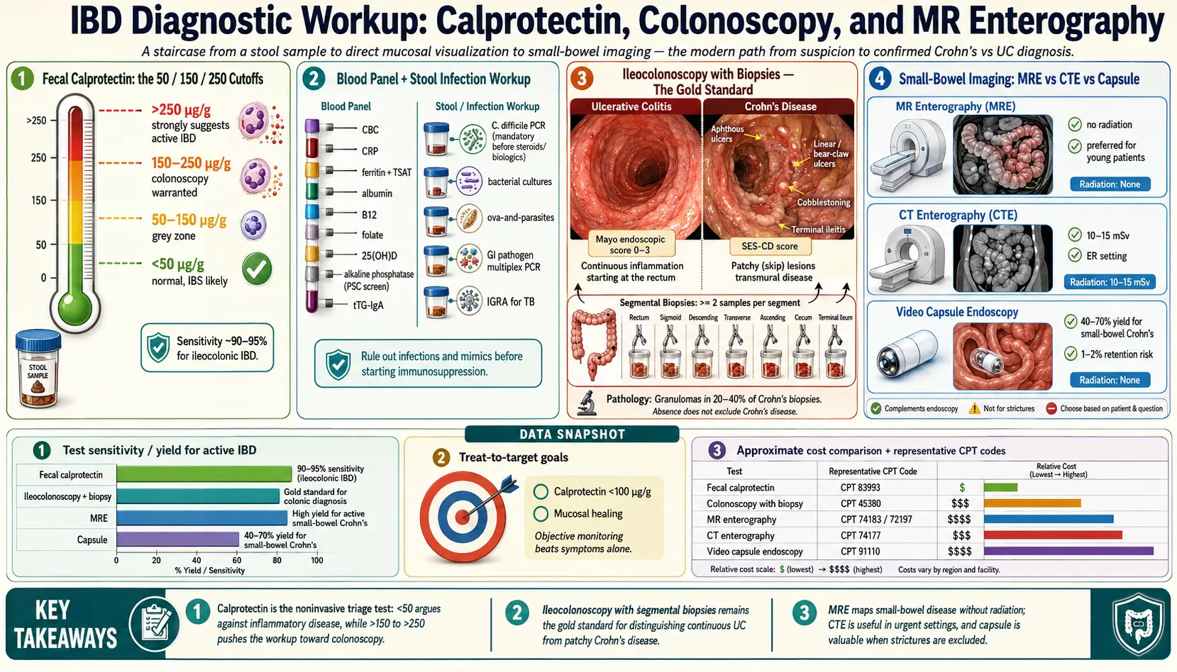
Table of Contents
- Why the Workup Matters
- Fecal Calprotectin — What the Cutoffs Mean
- Calprotectin: IBD vs IBS
- Blood Panels — CBC, CRP, Iron, Albumin
- Stool Testing to Rule Out Infection
- Ileocolonoscopy with Biopsy — The Gold Standard
- CT Enterography vs MR Enterography
- Small-Bowel Capsule Endoscopy
- Upper Endoscopy (EGD) for Crohn's
- Serology — ASCA, pANCA, anti-OmpC Limits
- Preparation and What Happens
- Insurance and Cost
- Key Research Papers
- Connections
- Featured Videos
Why the Workup Matters
Crohn's disease and ulcerative colitis are chronic inflammatory conditions, and their symptoms — diarrhea, cramping, blood in the stool, weight loss, fatigue — overlap with other conditions. The 2025 American College of Gastroenterology (ACG) Crohn's guideline lists conditions that can produce symptoms indistinguishable from active Crohn's disease, including bile salt diarrhea, intestinal infection, small intestinal bacterial overgrowth, dietary intolerances, disorders of gut-brain interaction, medication side effects and mimics such as endometriosis and tuberculosis (Lichtenstein 2025).
The same guideline states that there is no single laboratory test that can make an unequivocal diagnosis of Crohn's disease: the clinician integrates history and examination, laboratory tests, endoscopy, pathology and imaging, and the sequence of testing depends on the presenting features (Lichtenstein 2025). The tests described below are the pieces the ACG Crohn's and ulcerative colitis guidelines discuss: stool and blood tests for inflammation and infection, ileocolonoscopy with biopsies, cross-sectional imaging of the small bowel such as MR enterography, CT enterography and intestinal ultrasound, and in selected cases capsule endoscopy, upper endoscopy and serology.
This article describes what each test measures, what the published cutoffs and accuracy figures are, and what the guidelines say about when each is used.
Fecal Calprotectin — What the Cutoffs Mean
Calprotectin is a calcium-binding protein derived from neutrophils, a type of white blood cell. The ACG Crohn's guideline describes it as a sensitive marker of intestinal inflammation that is released into the stool from an inflamed bowel (Lichtenstein 2025). The ACG ulcerative colitis guideline describes it as a nonspecific neutrophil marker: it is raised in infectious as well as inflammatory colitis, but not in noninflammatory causes of diarrhea such as irritable bowel syndrome (Rubin 2025). Results are reported in micrograms per gram of stool (µg/g).
The published cutoffs differ with the question being asked:
- Separating inflammatory from noninflammatory disease (diagnosis). The ACG Crohn's guideline recommends a calprotectin cutoff of more than 50–100 µg/g to differentiate inflammatory from noninflammatory disease of the colon (strong recommendation, moderate level of evidence) (Lichtenstein 2025). In the meta-analysis it cites, a level of 50 µg/g had a sensitivity of 88% and a specificity of 72% for distinguishing IBD from functional gut disorders; the difference between 50 and 100 µg/g cutoffs was not statistically significant (An 2019). The ACG ulcerative colitis guideline notes that studies have used cutoffs ranging from 6 to 280 µg/g (Rubin 2025).
- Monitoring people already diagnosed. The 2023 American Gastroenterological Association (AGA) biomarker guidelines address established disease, not first diagnosis. For people with Crohn's disease or ulcerative colitis in symptomatic remission, the panels suggest that a calprotectin below 150 µg/g with a normal CRP (Crohn's disease), or a calprotectin below 150 µg/g, a normal fecal lactoferrin and/or a normal CRP (ulcerative colitis), rules out active inflammation, so endoscopy is not needed to assess activity; elevated values in that setting merit endoscopic confirmation before treatment is changed. In people with mild symptoms, the Crohn's panel found that neither normal nor elevated biomarkers alone were accurate enough to determine endoscopic activity, and the ulcerative colitis panel suggests endoscopic assessment (Ananthakrishnan 2023; Singh 2023).
The ACG Crohn's guideline adds that calprotectin levels can depend on disease location, extent and severity, and that biomarkers should not serve exclusively as a treatment endpoint, because a biomarker can normalize while mucosal inflammation or ulceration is still present (Lichtenstein 2025). The ulcerative colitis guideline reports that calprotectin levels correlate with clinical remission, endoscopic remission and histologic healing on treatment (Rubin 2025).
Calprotectin: IBD vs IBS
The most studied use of calprotectin is separating inflammatory gut disease from functional gut disorders such as irritable bowel syndrome, which produce similar symptoms without mucosal inflammation.
- In a 2010 BMJ meta-analysis of people with suspected IBD, calprotectin had a pooled sensitivity of 0.93 and specificity of 0.96 in adult studies, and 0.92 and 0.76 in studies of children and teenagers. The authors estimated that screening with calprotectin would reduce the number of adults needing endoscopy by 67%, at the cost of a delayed diagnosis in 6% of adults because of a false-negative result (van Rheenen 2010).
- In a 2015 meta-analysis of adults with IBS-type symptoms, a calprotectin of 40 µg/g or less, or a CRP of 0.5 or less, corresponded to a probability of IBD of 1% or less; none of the markers reliably distinguished IBS from healthy controls (Menees 2015).
- For ulcerative colitis, the ACG guideline gives a pooled sensitivity of 0.88 and specificity of 0.79 for an elevated calprotectin, with a positive likelihood ratio of 4.2 and a negative likelihood ratio of 0.15 (Rubin 2025).
Calprotectin is less sensitive in Crohn's disease than in ulcerative colitis: in a meta-analysis of symptomatic patients, it was more sensitive for endoscopic activity in ulcerative colitis than in Crohn's disease (Mosli 2015), and the ACG Crohn's guideline notes that levels depend on disease location (Lichtenstein 2025). The workup for IBS is covered on the Irritable Bowel Syndrome page.
Blood Panels — CBC, CRP, Iron, Albumin
The ACG Crohn's guideline states that the initial laboratory investigation should include evaluation for inflammation, anemia, dehydration and malnutrition (Lichtenstein 2025). What the guidelines and reviews report about the main tests:
- Complete blood count (CBC). Anemia and an elevated platelet count are the most common blood-count changes in suspected Crohn's disease (Lichtenstein 2025).
- C-reactive protein (CRP). An acute-phase protein made by the liver. It is elevated in a subset of people with Crohn's disease; its short half-life of 19 hours makes it useful for detecting and monitoring inflammation. Up to 40% of people with IBD who have mild inflammation may have a normal CRP and ESR (Lichtenstein 2025). In ulcerative colitis, up to a quarter of patients with endoscopically active disease may have a normal CRP (Rubin 2025). In a meta-analysis of symptomatic patients, CRP had a pooled sensitivity of 0.49 and specificity of 0.92 for endoscopic activity, against 0.88 and 0.73 for calprotectin and 0.82 and 0.79 for stool lactoferrin (Mosli 2015).
- Erythrocyte sedimentation rate (ESR). The ACG Crohn's guideline describes it as possibly useful in an individual patient, but not predictive of IBD and not able to discriminate IBD from IBS or healthy controls (Lichtenstein 2025).
- Iron studies. Iron deficiency and anemia in IBD are the subject of a dedicated European consensus (Dignass 2015).
- Albumin. In ulcerative colitis, a low serum albumin is associated with a greater risk of hospitalization and surgery and a lower likelihood of response to medical therapy (Rubin 2025).
- Vitamin B12. A 2014 systematic review of 42 studies found that Crohn's disease without ileal resection did not increase the risk of B12 deficiency, regardless of ileal involvement; ileal resections longer than 30 cm were associated with deficiency, and ulcerative colitis did not predispose to it (Battat 2014).
- Associated conditions. The ACG Crohn's guideline lists celiac disease among the immune-mediated diseases associated with Crohn's disease, and identifies primary sclerosing cholangitis, a bile-duct disease, as a factor that raises colorectal cancer risk and prompts surveillance colonoscopy from the time of its diagnosis (Lichtenstein 2025). Celiac testing is covered on the celiac disease page.
Stool Testing to Rule Out Infection
Infections can mimic IBD and can complicate it. The ACG ulcerative colitis guideline recommends stool testing to rule out Clostridioides difficile in people suspected of having ulcerative colitis (strong recommendation, very low quality of evidence) and states that infectious causes should be excluded at the time of diagnosis (Rubin 2025). It reports that the prevalence of C. difficile infection among newly diagnosed or relapsing IBD patients ranges from 5% to 47%, and that concurrent infection is associated with worse outcomes, including higher mortality (Rubin 2025). The ACG Crohn's guideline states that the risk of C. difficile infection may be up to five-fold higher in people with IBD (Lichtenstein 2025).
What the guidelines describe:
- C. difficile testing, typically by polymerase chain reaction (PCR) or enzyme-linked immunosorbent assay (Rubin 2025).
- Other enteric pathogens that can mimic ulcerative colitis, including Escherichia coli O157:H7, Salmonella, Shigella, Yersinia and Campylobacter, and parasitic infections such as amebiasis in the right clinical setting (Rubin 2025).
- Multiplex PCR pathogen panels, which several institutions use; the guideline notes that the prevalence and impact of non-C. difficile pathogens found this way remain to be robustly established (Rubin 2025).
Tuberculosis is listed among the conditions that can mimic Crohn's disease, and the ACG Crohn's guideline states that testing for latent and active tuberculosis should be undertaken before anti-TNF therapy, with preventive antituberculous treatment for several weeks first if latent infection is found (Lichtenstein 2025).
Ileocolonoscopy with Biopsy — The Gold Standard
An ileocolonoscopy is a colonoscopy that also enters the terminal ileum, the last part of the small bowel. The ACG Crohn's guideline states that ileocolonoscopy with biopsies should be performed in the assessment of suspected Crohn's disease, with biopsies of both involved and uninvolved mucosa; it reports that over 80% of people with IBD have mucosal involvement within reach of the colonoscope, and that the ileum can be entered in 80%–97% of patients in whom the cecum is reached (Lichtenstein 2025). The ulcerative colitis guideline states that colonoscopy with intubation of the ileum and biopsies of affected and unaffected areas should be obtained to confirm the diagnosis, interpreted by a pathologist (Rubin 2025).
Features the guidelines describe:
- Ulcerative colitis. Most often a continuously inflamed segment starting in the distal rectum and extending upward, with loss of vascular markings, granularity and friability of the lining, erosions, and in severe disease deep ulcers and spontaneous bleeding. Biopsies are taken from normal-looking colon as well, because inflammation seen under the microscope can extend beyond what is visible. In severe disease, a complete colonoscopy may carry a greater risk of perforation, and a sigmoidoscopy with biopsies is described as sufficient (Rubin 2025).
- Crohn's disease. Clues that favor Crohn's over ulcerative colitis include discontinuous involvement with skip areas, sparing of the rectum, deep, linear or serpiginous ulcers, strictures, fistulas and granulomatous inflammation. Classical granulomas are seen in a minority of patients (up to 33%) and are helpful but not required for the diagnosis (Lichtenstein 2025).
A side-by-side comparison is on the Crohn's vs Ulcerative Colitis: Key Differences page.
Endoscopic scores. The Simple Endoscopic Score for Crohn's Disease (SES-CD) sums scores for ulcer size, ulcerated surface, affected surface and narrowing; in its validation study it correlated strongly with the older Crohn's Disease Endoscopic Index of Severity (r = 0.920), with excellent agreement between observers (Daperno 2004). The ACG Crohn's guideline describes SES-CD categories of 0–2 for endoscopic remission, 3–6 mild, 7–15 moderate and above 15 severe (Lichtenstein 2025). For ulcerative colitis, the guideline states that endoscopic severity should be reported with a validated scale such as the Mayo Endoscopic Score or the UC Endoscopic Index of Severity, and it defines endoscopic remission as a Mayo score of 0 or 1 (Rubin 2025).
Mucosal healing as a target. The STRIDE-II consensus set clinical remission and endoscopic healing as long-term treatment targets, with symptom relief and normalization of CRP and calprotectin as short-term targets (Turner 2021). The ACG Crohn's guideline notes that few studies have examined the long-term impact of endoscopic healing, but reports that in early-stage Crohn's disease complete endoscopic healing after two years of therapy predicted sustained steroid-free remission three and four years out, and that mucosal healing has been associated with lower rates of surgery and hospitalization (Lichtenstein 2025).
CT Enterography vs MR Enterography
Much of the small bowel lies beyond the reach of the colonoscope. The ACG Crohn's guideline reports that in up to 50% of people with active small-bowel disease, inflammation may skip the terminal ileum or lie within the bowel wall and not be detected by ileocolonoscopy, and that strictures and fistulas are best identified with small-bowel imaging (Lichtenstein 2025). A multisociety consensus recommends that cross-sectional enterography be performed at the diagnosis of Crohn's disease and considered for monitoring small-bowel disease (Bruining 2018).
Accuracy. The ACG Crohn's guideline states that CT enterography (CTE) is sensitive for small-bowel disease and comparable with MR enterography (MRE) (Lichtenstein 2025). In a meta-analysis of six studies (290 patients), the pooled sensitivity and specificity for detecting active small-bowel Crohn's disease were 87.9% and 81.2% for MRE and 85.8% and 83.6% for CTE, with no statistically significant difference in yield (Qiu 2014).
Radiation. CTE uses ionizing radiation; MRE does not. The ACG guideline states that repeated CTE studies lead to levels of diagnostic radiation that theoretically might increase cancer risk, and that because of the absence of radiation, MRE should be used preferentially in people younger than 35 and in those likely to need serial examinations (Lichtenstein 2025). In a meta-analysis of six studies (1,704 participants), 8.8% of IBD patients had received potentially harmful cumulative doses, defined as 50 mSv or more (described by the authors as equivalent to five CT abdomen scans) — 11.1% in Crohn's disease and 2% in ulcerative colitis; IBD-related surgery and corticosteroid use were associated with higher exposure (Chatu 2012). In a single-centre study of 399 people with Crohn's disease, CT accounted for 77.2% of diagnostic radiation, and 15.5% had a cumulative dose above 75 mSv (Desmond 2008).
Perianal disease. The ACG Crohn's guideline states that MRI of the pelvis and/or endoscopic ultrasound may be used to further characterize perianal Crohn's disease and perirectal abscesses (Lichtenstein 2025).
Intestinal ultrasound. The ACG Crohn's guideline describes intestinal ultrasound (IUS) as a noninvasive, radiation-free way to assess the bowel wall, mesentery and nearby structures and as an adjunct to diagnosis and to monitoring response to therapy, noting that it has been used in Europe for over a decade and that interest and training are growing in the United States (Lichtenstein 2025). In a 2011 systematic review, ultrasonography had a sensitivity of 0.84 and specificity of 0.92 for diagnosing suspected Crohn's disease and assessing activity, with lower accuracy for disease above the terminal ileum; ultrasound, CT and MRI all had sensitivities and specificities above 0.80 for fistulas, abscesses and stenosis, and the authors favored ultrasound or MRI over CT because of radiation, particularly in young patients (Panés 2011). The ulcerative colitis guideline lists intestinal ultrasound among the tools for monitoring response to therapy (Rubin 2025).
Small-Bowel Capsule Endoscopy
Small-bowel capsule endoscopy, in which a camera capsule is swallowed, allows direct visualization of the lining of the small intestine (Lichtenstein 2025). The ACG Crohn's guideline describes it as a useful adjunct in diagnosing small-bowel Crohn's disease when the index of suspicion is high. It reports that isolated small-bowel involvement may be seen in up to 30% of people with Crohn's disease; that in meta-analyses capsule endoscopy had an incremental diagnostic yield of 32% over small-bowel barium studies, 47% over CTE and 22% over ileocolonoscopy in suspected Crohn's disease; and that it has a negative predictive value of 96%. It also notes that some studies have questioned the specificity of capsule findings and that there is no consensus on exactly which findings constitute a diagnosis of Crohn's disease (Lichtenstein 2025).
The main risk is capsule retention, the capsule failing to pass. In a 2020 meta-analysis, the retention rate was 3.32% across all Crohn's patients: 2.35% in suspected and 4.63% in established Crohn's disease, and 3.49% in adults against 1.64% in children. In established disease, retention rates were lower after a patency capsule (2.88%) or MR/CT enterography (2.32%) (Pasha 2020). The ACG guideline states that people with obstructive symptoms should have small-bowel imaging and/or a patency capsule evaluation before video capsule endoscopy to decrease the risk of retention (Lichtenstein 2025).
Upper Endoscopy (EGD) for Crohn's
Crohn's disease can involve the esophagus, stomach and duodenum. The ACG Crohn's guideline reports that most adult studies put upper-tract Crohn's disease at 0.3%–5%, but that prospective studies, performed mainly in children, found endoscopic and histologic changes in up to 16% of patients, only 37% of whom had upper GI symptoms; the clinical significance of these mild changes appears limited (Lichtenstein 2025).
- Adults with Crohn's disease: the guideline states that upper endoscopy should be performed in patients with upper GI complaints (Lichtenstein 2025).
- Adults with ulcerative colitis: routine upper endoscopy is not required and is restricted to those with upper GI symptoms (Rubin 2025).
- Children: routine upper endoscopy is performed at diagnosis of colitis, after which up to 8% of children with ulcerative colitis may have their diagnosis changed to Crohn's disease (Rubin 2025).
Serology — ASCA, pANCA, anti-OmpC Limits
Antibody tests sometimes discussed in IBD evaluation include anti-Saccharomyces cerevisiae antibodies (ASCA), perinuclear anti-neutrophil cytoplasmic antibodies (pANCA) and antibodies to bacterial antigens such as anti-OmpC.
In a meta-analysis of 60 studies (3,841 people with ulcerative colitis and 4,019 with Crohn's disease), the combination ASCA-positive / pANCA-negative had a sensitivity of 54.6% and specificity of 92.8% for Crohn's disease, and a positive pANCA had a sensitivity of 55.3% and specificity of 88.5% for ulcerative colitis. The authors concluded that the tests are specific but not sensitive, and may be particularly useful for telling Crohn's disease from ulcerative colitis in children (Reese 2006).
The ACG Crohn's guideline states that routine use of serologic markers to establish the diagnosis of Crohn's disease is not indicated: anti-glycan antibodies are more common in Crohn's disease than in ulcerative colitis but have low sensitivity, and combination panels of serologic, genetic and inflammatory markers have not improved their usefulness as a screening tool (Lichtenstein 2025). The ACG ulcerative colitis guideline recommends against serologic antibody testing to establish or rule out the diagnosis, or to determine prognosis (strong recommendation, very low quality of evidence) (Rubin 2025).
Preparation and What Happens
Calprotectin. Collection and handling instructions for the stool sample vary by laboratory and assay.
Colonoscopy preparation. The 2025 US Multi-Society Task Force on Colorectal Cancer consensus recommends a split-dose bowel preparation regimen and suggests that a 2-liter regimen may be sufficient. It describes a same-day regimen as an acceptable alternative for afternoon colonoscopy but an inferior one for morning colonoscopy, recommends limiting dietary restrictions to the day before the colonoscopy (clear liquids, or low-fiber/low-residue foods for the early and midday meals), and suggests adding oral simethicone (Jacobson 2025).
MR enterography, CT enterography and capsule endoscopy. Protocols for fasting, oral contrast, intravenous contrast and timing differ between centres; the imaging centre or endoscopy unit gives the instructions that apply. Decisions about sedation and anti-anxiety medication for scans belong with the clinician and the imaging team.
Insurance and Cost
Prices, coverage rules, billing codes and prior-authorization requirements for these tests vary by country, insurer, facility and the reason a test is ordered, and change over time. The figures that apply to an individual case come from the insurer and the facility's billing office.
Key Research Papers
- Mosli MH, Zou G, Garg SK, Feagan SG, MacDonald JK, Chande N, Sandborn WJ, Feagan BG. C-reactive protein, fecal calprotectin, and stool lactoferrin for detection of endoscopic activity in symptomatic inflammatory bowel disease patients: a systematic review and meta-analysis. Am J Gastroenterol. 2015;110(6):802-819. — doi:10.1038/ajg.2015.120 (PubMed PMID: 25964225)
- Rubin DT, Ananthakrishnan AN, Siegel CA, Sauer BG, Long MD. ACG Clinical Guideline: Ulcerative Colitis in Adults. Am J Gastroenterol. 2019;114(3):384-413. Updated by the 2025 guideline below. — doi:10.14309/ajg.0000000000000152 (PubMed PMID: 30840605)
- Maaser C, Sturm A, Vavricka SR, et al. ECCO-ESGAR Guideline for Diagnostic Assessment in IBD Part 1: Initial diagnosis, monitoring of known IBD, detection of complications. J Crohns Colitis. 2019;13(2):144-164. — doi:10.1093/ecco-jcc/jjy113 (PubMed PMID: 30137275)
- van Rheenen PF, Van de Vijver E, Fidler V. Faecal calprotectin for screening of patients with suspected inflammatory bowel disease: diagnostic meta-analysis. BMJ. 2010;341:c3369. — doi:10.1136/bmj.c3369 (PubMed PMID: 20634346)
- Turner D, Ricciuto A, Lewis A, et al. STRIDE-II: An Update on the Selecting Therapeutic Targets in Inflammatory Bowel Disease (STRIDE) Initiative of the International Organization for the Study of IBD (IOIBD): Determining Therapeutic Goals for Treat-to-Target strategies in IBD. Gastroenterology. 2021;160(5):1570-1583. — doi:10.1053/j.gastro.2020.12.031 (PubMed PMID: 33359090)
- Bruining DH, Zimmermann EM, Loftus EV Jr, Sandborn WJ, Sauer CG, Strong SA; Society of Abdominal Radiology Crohn's Disease-Focused Panel. Consensus Recommendations for Evaluation, Interpretation, and Utilization of Computed Tomography and Magnetic Resonance Enterography in Patients With Small Bowel Crohn's Disease. Radiology. 2018;286(3):776-799. — doi:10.1148/radiol.2018171737 (PubMed PMID: 29319414)
- Lichtenstein GR, Loftus EV, Afzali A, Long MD, Barnes EL, Isaacs KL, Ha CY. ACG Clinical Guideline: Management of Crohn's Disease in Adults. Am J Gastroenterol. 2025;120(6):1225-1264. — doi:10.14309/ajg.0000000000003465 (PubMed PMID: 40701562)
- Rubin DT, Ananthakrishnan AN, Siegel CA, Barnes EL, Long MD. ACG Clinical Guideline Update: Ulcerative Colitis in Adults. Am J Gastroenterol. 2025;120(6):1187-1224. — doi:10.14309/ajg.0000000000003463 (PubMed PMID: 40701556) (correction published 2026: PubMed PMID: 41384833)
- Ananthakrishnan AN, Adler J, Chachu KA, et al. AGA Clinical Practice Guideline on the Role of Biomarkers for the Management of Crohn's Disease. Gastroenterology. 2023;165(6):1367-1399. — doi:10.1053/j.gastro.2023.09.029 (PubMed PMID: 37981354)
- Singh S, Ananthakrishnan AN, Nguyen NH, et al. AGA Clinical Practice Guideline on the Role of Biomarkers for the Management of Ulcerative Colitis. Gastroenterology. 2023;164(3):344-372. — doi:10.1053/j.gastro.2022.12.007 (PubMed PMID: 36822736)
- An YK, Prince D, Gardiner F, et al. Faecal calprotectin testing for identifying patients with organic gastrointestinal disease: systematic review and meta-analysis. Med J Aust. 2019;211(10):461-467. — doi:10.5694/mja2.50384 (PubMed PMID: 31680263)
- Menees SB, Powell C, Kurlander J, Goel A, Chey WD. A meta-analysis of the utility of C-reactive protein, erythrocyte sedimentation rate, fecal calprotectin, and fecal lactoferrin to exclude inflammatory bowel disease in adults with IBS. Am J Gastroenterol. 2015;110(3):444-454. — doi:10.1038/ajg.2015.6 (PubMed PMID: 25732419)
- Battat R, Kopylov U, Szilagyi A, et al. Vitamin B12 deficiency in inflammatory bowel disease: prevalence, risk factors, evaluation, and management. Inflamm Bowel Dis. 2014;20(6):1120-1128. — doi:10.1097/MIB.0000000000000024 (PubMed PMID: 24739632)
- Dignass AU, Gasche C, Bettenworth D, et al. European consensus on the diagnosis and management of iron deficiency and anaemia in inflammatory bowel diseases. J Crohns Colitis. 2015;9(3):211-222. — doi:10.1093/ecco-jcc/jju009 (PubMed PMID: 25518052)
- Daperno M, D'Haens G, Van Assche G, et al. Development and validation of a new, simplified endoscopic activity score for Crohn's disease: the SES-CD. Gastrointest Endosc. 2004;60(4):505-512. — doi:10.1016/s0016-5107(04)01878-4 (PubMed PMID: 15472670)
- Qiu Y, Mao R, Chen BL, et al. Systematic review with meta-analysis: magnetic resonance enterography vs. computed tomography enterography for evaluating disease activity in small bowel Crohn's disease. Aliment Pharmacol Ther. 2014;40(2):134-146. — doi:10.1111/apt.12815 (PubMed PMID: 24912799)
- Chatu S, Subramanian V, Pollok RC. Meta-analysis: diagnostic medical radiation exposure in inflammatory bowel disease. Aliment Pharmacol Ther. 2012;35(5):529-539. — doi:10.1111/j.1365-2036.2011.04975.x (PubMed PMID: 22239831)
- Desmond AN, O'Regan K, Curran C, et al. Crohn's disease: factors associated with exposure to high levels of diagnostic radiation. Gut. 2008;57(11):1524-1529. — doi:10.1136/gut.2008.151415 (PubMed PMID: 18443021)
- Panés J, Bouzas R, Chaparro M, et al. Systematic review: the use of ultrasonography, computed tomography and magnetic resonance imaging for the diagnosis, assessment of activity and abdominal complications of Crohn's disease. Aliment Pharmacol Ther. 2011;34(2):125-145. — doi:10.1111/j.1365-2036.2011.04710.x (PubMed PMID: 21615440)
- Pasha SF, Pennazio M, Rondonotti E, et al. Capsule retention in Crohn's disease: a meta-analysis. Inflamm Bowel Dis. 2020;26(1):33-42. — doi:10.1093/ibd/izz083 (PubMed PMID: 31050736)
- Reese GE, Constantinides VA, Simillis C, et al. Diagnostic precision of anti-Saccharomyces cerevisiae antibodies and perinuclear antineutrophil cytoplasmic antibodies in inflammatory bowel disease. Am J Gastroenterol. 2006;101(10):2410-2422. — doi:10.1111/j.1572-0241.2006.00840.x (PubMed PMID: 16952282)
- Jacobson BC, Anderson JC, Burke CA, et al. Optimizing Bowel Preparation Quality for Colonoscopy: Consensus Recommendations by the US Multi-Society Task Force on Colorectal Cancer. Gastroenterology. 2025;168(4):798-829. — doi:10.1053/j.gastro.2025.02.002 (PubMed PMID: 40047732)
PubMed Topic Searches
For further reading, the following PubMed topic searches return current peer-reviewed work on each component of the IBD diagnostic workup:
- PubMed: Fecal calprotectin in IBD diagnosis and monitoring
- PubMed: Calprotectin differentiating IBS from IBD
- PubMed: Ileocolonoscopy and biopsy protocols for IBD
- PubMed: MR enterography in Crohn's disease
- PubMed: CT enterography for small-bowel evaluation
- PubMed: Capsule endoscopy in suspected small-bowel Crohn's
- PubMed: ASCA, pANCA, and IBD serology
- PubMed: Mucosal healing as a treatment target in IBD
Connections
- Gastroenterology
- Inflammatory Bowel Disease Overview
- Crohn's vs Ulcerative Colitis: Key Differences
- Biologics: TNF, IL-23, and Integrin Inhibitors
- Diet for IBD: SCD and Mediterranean
- IBD Surgery Decisions
- Extraintestinal Manifestations: Joints, Skin, Eyes
- IBD Fatigue, Anemia, and Nutrient Deficiencies
- Pregnancy and IBD
- JAK Inhibitors and S1P Modulators
- Crohn's Disease
- Ulcerative Colitis
- Endoscopy, Biopsy, and Marsh Classification
- Full-Body MRI Screening
- Ankylosing Spondylitis