Aortic Aneurysm
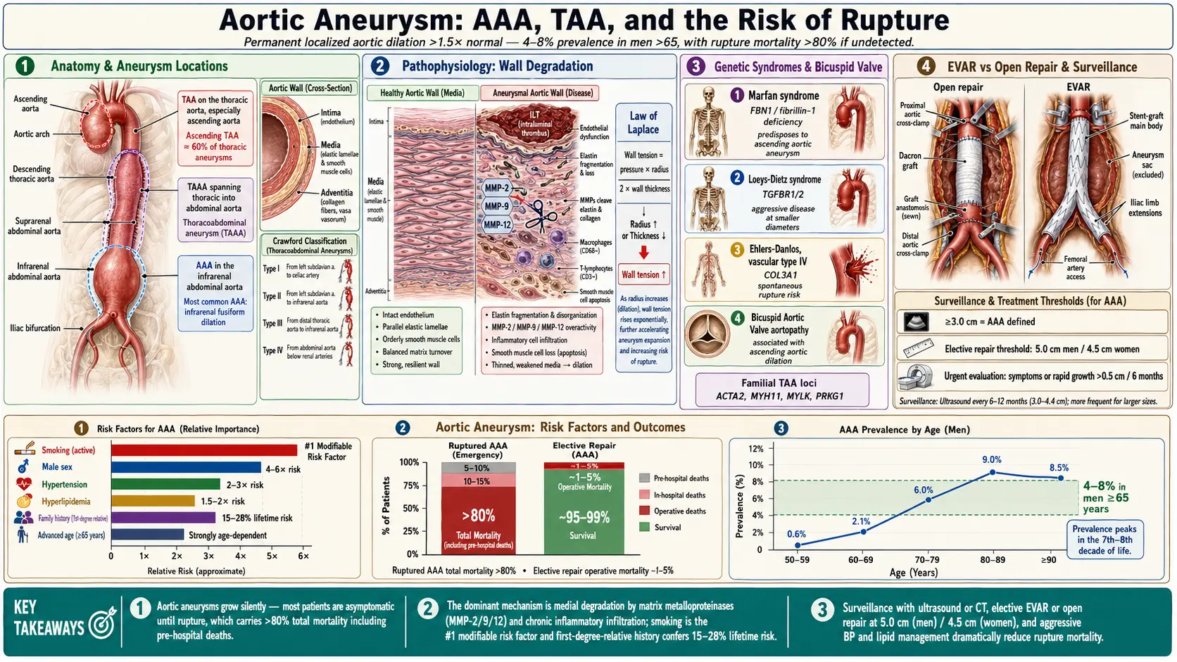
Table of Contents
- Overview
- Epidemiology
- Pathophysiology
- Etiology and Risk Factors
- Clinical Presentation
- Diagnosis
- Classification (Crawford and Other Systems)
- Treatment and Surgical Indications
- EVAR vs. Open Repair
- Surveillance Guidelines
- Complications
- Prognosis
- Prevention
- Recent Research
- Key Research Papers
- Connections
- Featured Videos
1. Overview
An aortic aneurysm is a permanent, localized widening of the aorta. The conventional definition of an arterial aneurysm is an artery dilated to at least 1.5 times its expected normal diameter; the 2022 ACC/AHA aortic disease guideline notes that this definition works well for the abdominal and descending thoracic aorta but not for the aortic root and ascending aorta, where it treats a diameter of 4.0 cm or more as "dilated" and 4.5 cm or more as an "aneurysm" in adults of average body size (Isselbacher 2022). An abdominal aortic aneurysm (AAA) is typically defined as an abdominal aorta 3.0 cm or larger in diameter (USPSTF 2019).
Aneurysms are grouped by location: thoracic aortic aneurysms (TAA) of the root, ascending aorta, arch or descending aorta; abdominal aortic aneurysms (AAA); and thoracoabdominal aortic aneurysms (TAAA), descending thoracic aneurysms that extend into the abdominal aorta (Isselbacher 2022). Most AAAs cause no symptoms until they rupture (USPSTF 2019), which is why screening, surveillance imaging and diameter-based repair thresholds dominate the guidelines summarized on this page.
2. Epidemiology
The USPSTF reports that the prevalence of AAA has declined over the past two decades among screened men 65 or older in several European countries, and that current US prevalence is unclear because few people are screened (USPSTF 2019). A US trial report describes AAA as affecting more than 3% of older adults (Baxter 2020). The 2022 guideline gives a lifetime risk of AAA of 8.2% in men and 10.5% in current smokers (Isselbacher 2022).
Thoracic aortic aneurysms occur in 5 to 10 per 100,000 person-years. Aneurysms of the aortic root, ascending aorta or both are the most common (about 60%), followed by the descending aorta (about 30%) and the arch (under 10%) (Isselbacher 2022).
Key epidemiological features include:
- Age: the guideline lists older age among the strong risk factors for AAA. Root and ascending aneurysms are diagnosed at younger ages than descending thoracic aneurysms (mean 60 versus 72 years in the series it cites) (Isselbacher 2022).
- Sex: male sex is a strong, independent risk factor for AAA. The guideline notes that female sex has not been proven an independent risk factor and that prevalence is lower in women (Isselbacher 2022). Once an aneurysm is present, outcomes differ: in an individual-patient meta-analysis of 15,475 people with small AAAs, rupture rates were almost fourfold higher in women than in men (Sweeting 2012), and registry data reviewed for the USPSTF showed higher surgical complications and postoperative mortality in women (Guirguis-Blake 2019).
- Race: the guideline lists White race among the additional risk factors for AAA (Isselbacher 2022).
- Mortality: the guideline estimates the mortality of ruptured AAA at 80% to 90%, with most patients never reaching the hospital (Isselbacher 2022); the USPSTF gives a risk of death with rupture as high as 81% (USPSTF 2019).
- Younger patients: in a US National Inpatient Sample study cited by the guideline, 24% of patients with ruptured AAA were under 65, below the usual screening age (Isselbacher 2022).
3. Pathophysiology
The aortic wall has three layers: a thin inner intima, a thicker media of smooth muscle cells, elastic fibers and collagen arranged in more than 50 elastic lamellae, and an outer adventitia carrying the vasa vasorum. The media provides the wall's strength and distensibility (Isselbacher 2022).
Medial Degeneration
Although AAA shares risk factors with atherosclerosis, the guideline describes it as histopathologically distinct, characterized by degeneration of the aortic media (Isselbacher 2022).
Intraluminal Thrombus
Most AAAs develop an intraluminal thrombus, which the guideline describes as contributing to ongoing wall degradation through oxidative stress, smooth muscle cell apoptosis, proteolysis of the extracellular matrix and adventitial inflammation (Isselbacher 2022).
Diameter, Growth and Risk
The risk of aortic dissection or rupture correlates with aneurysm diameter, as does the rate of growth (Isselbacher 2022). AAA growth is nonlinear and faster in larger aneurysms: in the RESCAN individual-patient meta-analysis, each 0.5-cm increase in diameter raised the growth rate by an average of 0.59 mm per year and the rupture rate by a factor of 1.91 (Bown 2013).
Genetic and Connective Tissue Disorders
About 20% of thoracic aortic aneurysms are related to a heritable condition (heritable thoracic aortic disease, HTAD) (Isselbacher 2022). The guideline's tables list:
- Marfan syndrome (FBN1).
- Loeys-Dietz syndrome (TGFBR1, TGFBR2, SMAD3, TGFB2, TGFB3). Dissection can occur at smaller diameters than in Marfan syndrome when the cause is a TGFBR1, TGFBR2 or SMAD3 variant; TGFB2 and TGFB3 disease may be less aggressive, based on limited data (Isselbacher 2022). The Loeys-Dietz Syndrome page covers this in detail.
- Vascular Ehlers-Danlos syndrome (COL3A1).
- Nonsyndromic HTAD — variants in ACTA2, MYH11, PRKG1, MYLK and other genes, and familial thoracic aneurysm without an identified variant.
- Congenital conditions — bicuspid aortic valve, Turner syndrome, coarctation of the aorta.
Genome-wide association studies of sporadic AAA have identified multiple risk loci; a 2017 meta-analysis of 10,204 cases and 107,766 controls added four, including one near MMP9, and found that they were not associated with coronary artery disease, blood pressure, lipids or diabetes (Jones 2017).
4. Etiology and Risk Factors
The guideline's table of TAA causes lists heritable disorders, congenital conditions, hypertension, atherosclerosis, degenerative disease, previous aortic dissection, inflammatory aortitis, infectious aortitis and previous traumatic aortic injury (Isselbacher 2022).
Risk Factors for AAA
The 2022 guideline lists as strong risk factors smoking history, older age, male sex, family history of AAA and atherosclerotic cardiovascular disease, and as additional risk factors hypertension, hyperlipidemia, White race and inherited vascular connective tissue disorders (Isselbacher 2022).
- Tobacco smoking — the guideline calls cigarette smoking the largest modifiable risk factor for AAA. A smoking history is defined as lifetime use of 100 or more cigarettes, and risk is lower with fewer pack-years. In one study it cites, smoking was associated with a 15-fold increased risk of AAA in women (relative risk 15.0) versus 7-fold in men (7.3) (Isselbacher 2022). Among people with small AAAs, growth was 0.35 mm/year faster in smokers and rupture rates were double in current smokers (Sweeting 2012).
- Family history — at least 10% to 25% of patients with AAA have a family member with the condition, and the lifetime prevalence of AAA is estimated at 32% in brothers of people with AAA (Isselbacher 2022).
- Hypertension — listed as an additional risk factor; in small AAAs, higher blood pressure was associated with higher rupture rates, while mean arterial pressure had no effect on growth (Sweeting 2012).
- Thoracic aneurysms — hypertension, smoking, hypercholesterolemia and heritable genetic variants are listed as risk factors for TAA disease (Isselbacher 2022).
Diabetes: An Inverse Association
Diabetes is not on the guideline's list of AAA risk factors. A 2016 meta-analysis found a strongly negative association between diabetes and AAA prevalence in population-based screening studies (adjusted odds ratio 0.66) and in prospective studies (0.52), and slower aneurysm growth in people with diabetes (mean −0.60 mm/year) (Xiong 2016). The 15,475-patient individual-data meta-analysis likewise found growth reduced by 0.51 mm/year in patients with diabetes (Sweeting 2012). The guideline's surveillance text nonetheless mentions diabetes, together with smoking, as a reason shorter scan intervals are often used (Isselbacher 2022).
Inflammatory Aortitis
The guideline describes inflammatory aortitis as a rare cause of AAA; its list of inflammatory causes of TAA includes giant cell arteritis, Takayasu arteritis, Behçet disease and IgG4-related disease (Isselbacher 2022).
Infectious Aortitis ("Mycotic" Aneurysms)
The guideline notes that "infectious aortitis" has replaced the older term "mycotic aneurysm," which implies a fungal cause. Infection reaches the aortic wall by contiguous spread or by septic emboli and bloodstream spread to a vulnerable plaque or existing aneurysm; Staphylococcus aureus, Pneumococcus, Escherichia coli and Salmonella are the pathogens identified in most reports. In a single-institution series of 2,520 patients operated on for infectious aortic aneurysms, 24% had already ruptured at presentation (Isselbacher 2022).
Post-Dissection Aneurysm
After acute aortic dissection or intramural hematoma, residual aortic disease can enlarge; for chronic residual disease with a total diameter of 5.5 cm or more, the guideline recommends elective repair (Isselbacher 2022).
Connective Tissue and Genetic Causes
Root and ascending aneurysms tend to have a heritable influence and present at younger ages, whereas descending thoracic aneurysms tend to be degenerative and present at older ages (Isselbacher 2022). Genetic conditions carry their own repair thresholds (see the treatment section below).
Traumatic Aortic Injury
The aortic isthmus is the most common site of blunt traumatic thoracic aortic injury. For injuries that meet indications for repair, the guideline recommends TEVAR over open repair when the anatomy is appropriate (Isselbacher 2022).
5. Clinical Presentation
Most AAAs are asymptomatic until they rupture (USPSTF 2019).
Symptomatic (Unruptured) Aneurysm
The guideline reports that about 6% to 22% of treated AAAs are symptomatic but unruptured. It describes these symptoms as high risk for impending rupture:
- Pain in the back, abdomen or flank attributable to the aneurysm, sometimes radiating to the groin.
and these as warranting expedited, though not necessarily urgent, repair:
- Tenderness over the aneurysm in the abdomen, back or flank.
- Embolism from the aneurysm (for example, blue toe syndrome).
- Compressive symptoms (for example, obstructive uropathy).
Patients treated for symptomatic aneurysms have higher mortality and morbidity than those treated electively (Isselbacher 2022). For the aortic root and ascending aorta, symptoms attributable to the aneurysm are an indication for surgery at any size (Isselbacher 2022).
Ruptured Aortic Aneurysm
Ruptured AAA has an estimated mortality of 80% to 90%, most patients never reaching the hospital. Patients who do arrive often maintain adequate blood pressure, in part because of catecholamine responses, and can collapse once general anesthesia removes that response (Isselbacher 2022).
6. Diagnosis
The guideline recommends that aortic diameters be measured at reproducible anatomic landmarks perpendicular to the axis of blood flow, reporting the longest diameter and its perpendicular when the contour is oval, and that ionizing radiation doses be kept as low as feasible (Isselbacher 2022).
Ultrasound
Ultrasound has been the standard for surveillance imaging of the abdominal aorta and is widely used; it is the screening test in the USPSTF and ACC/AHA recommendations (USPSTF 2019; Isselbacher 2022).
Computed Tomography (CT)
CT gives superior visualization of the abdominal aorta and its branches, is the alternative when ultrasound is inadequate, and is recommended for planning once an AAA meets repair criteria. For the thoracic aorta, CT and MRI are described as the gold standard for all segments, and electrocardiographic gating minimizes motion artifact for root and ascending measurements (Isselbacher 2022).
Magnetic Resonance Imaging (MRI)
MRI is a reasonable alternative to CT; it avoids ionizing radiation and iodinated contrast but has higher cost, longer acquisition times, lower resolution and limited visualization of metallic stent-graft components. Non-contrast MRI techniques have also been useful for defining AAAs (Isselbacher 2022).
Echocardiography
For a dilated thoracic aorta, a transthoracic echocardiogram (TTE) at diagnosis is recommended to assess aortic valve anatomy and function and thoracic aortic diameters; transesophageal echocardiography is an alternative when imaging windows are poor (Isselbacher 2022).
7. Classification (Crawford and Other Systems)
Several classification systems describe aneurysm extent and related aortic disease.
Crawford Classification of Thoracoabdominal Aortic Aneurysms
The Crawford classification of TAAA, later modified by Safi and colleagues (who added extent V), describes how much of the thoracic and abdominal aorta an aneurysm involves and may predict the morbidity and mortality of repair (Isselbacher 2022). In the guideline's discussion of spinal cord protection, extent I and II repairs are the high-risk group in which cerebrospinal fluid drainage was shown in a randomized trial to reduce spinal cord injury (Isselbacher 2022).
Stanford and DeBakey Classifications (Aortic Dissection)
These systems classify dissection, which is relevant to post-dissection aneurysm (Isselbacher 2022):
- Stanford type A: any dissection involving the ascending aorta, wherever the tear starts. The guideline recommends emergency surgical consultation and immediate surgical intervention for acute type A dissection.
- Stanford type B: dissections that do not involve the ascending aorta. Uncomplicated acute type B dissection has a 30-day mortality of 10%; with complications such as malperfusion or rupture, mortality rises to 20% by day 2 and 25% by day 30.
- DeBakey type I: tear in the ascending aorta, extending to the arch and typically the descending aorta.
- DeBakey type II: confined to the ascending aorta.
- DeBakey type III: tear in the descending thoracic aorta (IIIa confined to it; IIIb extending below the diaphragm).
Anatomic Location Classification
The guideline divides the aorta into five segments (Isselbacher 2022):
- Aortic root: aortic valve annulus to the sinotubular junction.
- Ascending aorta: sinotubular junction to the innominate artery.
- Aortic arch: innominate artery to the left subclavian artery.
- Descending thoracic aorta: left subclavian artery to the diaphragm.
- Abdominal aorta: diaphragm to the aortic bifurcation.
Morphology
- Fusiform: the usual shape of AAA.
- Saccular: a focal outpouching. In a Dutch registry of 7,659 treated AAAs, 6.1% were saccular; saccular aneurysms were more common in women and more often symptomatic at smaller sizes, and 25% of acutely presenting saccular aneurysms measured under 5.5 cm, against 8.1% of fusiform ones (Isselbacher 2022).
8. Treatment and Surgical Indications
Management combines medical therapy, surveillance for aneurysms below repair thresholds, and endovascular or open repair when the risk of rupture or dissection outweighs the risk of the procedure. The thresholds below are those of the 2022 ACC/AHA aortic disease guideline (Isselbacher 2022).
Medical Management
- Smoking cessation: the guideline recommends smoking cessation efforts in patients with AAA or TAA who smoke. No randomized trial has tested the effect of quitting on aneurysm outcomes; the recommendation rests on smoking being the largest modifiable risk factor (Isselbacher 2022).
- Blood pressure: antihypertensive medication is recommended when average systolic pressure is 130 mm Hg or higher or diastolic 80 mm Hg or higher, to reduce cardiovascular events. For AAA, the guideline states that in prospective human trials no specific agent has been proven to inhibit growth; for TAA, a beta blocker is reasonable to reach blood pressure targets and an ARB is a reasonable adjunct (Isselbacher 2022).
- Statins: moderate- or high-intensity statin therapy is recommended in AAA with evidence of aortic atherosclerosis and may be considered without it. A meta-analysis cited by the guideline associated statins with slower growth, fewer ruptures and lower 30-day mortality after repair, while one prospective cohort found no slowing of growth (Isselbacher 2022).
- Antiplatelet therapy: low-dose aspirin may be considered in AAA with concomitant atheroma or penetrating ulcer; aortic-specific outcome data are limited (Isselbacher 2022).
- Doxycycline: two randomized trials found no reduction in small-AAA growth (Meijer 2013; Baxter 2020); see Recent Research.
Indications for Repair: Abdominal Aortic Aneurysm
- Diameter 5.5 cm or more in men, 5.0 cm or more in women: repair recommended (class 1, level A). Trials of early repair at 4.0–5.4 cm (UKSAT, ADAM, CAESAR, PIVOTAL) found no survival benefit; in UKSAT, mean diameter at rupture was 5.0 cm in women and 6.0 cm in men.
- Symptoms attributable to the aneurysm: repair recommended to reduce the risk of rupture.
- Growth of 0.5 cm or more in 6 months: repair may be reasonable; the guideline's text treats 0.5 cm in 6 months or 1 cm in 1 year as rapid growth.
- Saccular aneurysm: intervention may be reasonable.
- Rupture: in stable patients CT is recommended to judge suitability for endovascular repair, and endovascular repair is recommended over open repair when the anatomy is suitable.
- Infectious aortitis with aneurysm: open surgical repair is recommended, and the guideline describes prolonged antimicrobial therapy as warranted.
Indications for Repair: Thoracic Aortic Aneurysm
- Sporadic root or ascending aneurysm, 5.5 cm or more: surgery indicated.
- Sporadic root or ascending aneurysm, 5.0 cm or more: surgery reasonable when performed by experienced surgeons in a multidisciplinary aortic team.
- Growth: for root or ascending aneurysms under 5.5 cm, surgery is indicated when growth confirmed by tomographic imaging is 0.3 cm/year or more in 2 consecutive years, or 0.5 cm or more in 1 year.
- Concomitant cardiac surgery: ascending replacement is reasonable at 4.5 cm or more during repair or replacement of a tricuspid aortic valve when performed by experienced surgeons in a multidisciplinary aortic team, and may be reasonable at 5.0 cm or more during other cardiac surgery.
- Marfan syndrome: root and ascending replacement recommended at 5.0 cm or more; reasonable at 4.5 cm or more with features linked to dissection (family history of dissection, growth of 0.3 cm/year or more, diffuse root and ascending dilation, marked vertebral arterial tortuosity), or when the root cross-sectional area to height ratio is 10 cm²/m or more.
- Loeys-Dietz syndrome (gene-specific, guideline Table 11): TGFBR1 or TGFBR2 without high-risk features, 4.5 cm or more; TGFBR1 or TGFBR2 with high-risk features, 4.0 cm or more; SMAD3, 4.5 cm or more; TGFB2, 4.5 cm or more; TGFB3, 5.0 cm or more. The guideline states that the threshold is informed by the specific variant, diameter, growth rate, features outside the aorta, family history, age, sex and patient and physician preferences.
- Bicuspid aortic valve: surgery recommended at 5.5 cm or more; reasonable at 5.0–5.4 cm with an additional risk factor for dissection; may be reasonable at 5.0–5.4 cm without risk factors in patients at low surgical risk, by experienced surgeons.
- Aortic arch: open replacement is reasonable at 5.5 cm or more in asymptomatic patients at low operative risk; recommended for symptomatic arch aneurysms at low or intermediate risk.
- Descending thoracic aorta: repair recommended at 5.5 cm or more; may be considered below 5.5 cm with risk factors for rupture (growth of 0.5 cm/year or more, symptoms, a heritable aortic syndrome). For patients without Marfan, Loeys-Dietz or vascular Ehlers-Danlos syndrome whose anatomy is suitable, TEVAR is recommended over open surgery.
- Distal aorta in heritable disease: in Marfan syndrome, replacement of a nondissected arch, descending or abdominal aneurysm is reasonable at 5.0 cm or more; in Loeys-Dietz syndrome due to TGFBR1, TGFBR2 or SMAD3, surgery on the intact arch, descending or abdominal aorta may be considered at 4.5 cm or more.
Indications for Repair: Thoracoabdominal Aortic Aneurysm
- Intact degenerative TAAA, 6.0 cm or more: repair recommended.
- 5.5 cm or more: repair reasonable when performed by experienced surgeons in a multidisciplinary aortic team.
- Below 5.5 cm with features linked to rupture (for example, confirmed growth of 0.5 cm/year or more, symptoms): repair reasonable.
- Marfan, Loeys-Dietz or vascular Ehlers-Danlos syndrome: open repair recommended over endovascular repair.
9. EVAR vs. Open Repair
For nonruptured AAA in patients at low to moderate operative risk whose anatomy suits either method, the 2022 guideline recommends shared decision-making weighing the risks and benefits of each; for high perioperative risk, EVAR is reasonable to reduce 30-day morbidity and mortality (Isselbacher 2022).
Endovascular Aortic Repair (EVAR / TEVAR)
EVAR places a stent-graft inside the aneurysm to exclude the sac from arterial pressure; TEVAR applies the same approach in the thoracic aorta. The guideline lists neck diameter, length and angulation, iliac seal diameter and length, and vessel access as anatomic factors, and recommends adherence to the manufacturer's instructions for use: observational studies link treatment outside them to more sac enlargement, graft-related events and mortality (Isselbacher 2022).
The Randomized Trials
- EVAR trial 1 (UK; 1,082 patients, aneurysms 5.5 cm or more): 30-day mortality was 1.7% with EVAR versus 4.7% with open repair; secondary interventions were more common after EVAR (9.8% vs 5.8%) (Greenhalgh 2004). At up to 15 years (1,252 patients, mean follow-up 12.7 years), overall mortality did not differ significantly (adjusted HR 1.11); EVAR had lower mortality in the first 6 months, but beyond 8 years open repair had significantly lower total and aneurysm-related mortality, the latter mainly from secondary aneurysm sac rupture (13 deaths after EVAR vs 2 after open repair) (Patel 2016).
- DREAM (Netherlands; 351 patients, aneurysms 5 cm or more): at 6 years, cumulative survival was 69.9% after open repair and 68.9% after EVAR (P=0.97); freedom from secondary interventions was 81.9% versus 70.4% (P=0.03) (De Bruin 2010).
- OVER (US Veterans Affairs; 881 patients): at up to 9 years, all-cause mortality was the same (146 deaths in each group); the perioperative survival advantage of EVAR lasted through about 3 years, six ruptures occurred after EVAR versus none after open repair, and EVAR improved survival in patients under 70 but not in older patients (Lederle 2012). At up to 14 years, deaths were 68.0% after EVAR and 70.0% after open repair (HR 0.96), with no significant trend in any period; more EVAR patients underwent secondary procedures. The authors note that this did not match the worse long-term EVAR results of the two European trials (Lederle 2019).
- Pooled trials: in pooled data from 7 trials cited by the guideline, perioperative mortality was much lower with EVAR (OR 0.36), survival became equivalent after about 6 months, and after 8 years EVAR carried higher risks of aneurysm-related death, secondary intervention and rupture (Isselbacher 2022).
- EVAR trial 2 (patients unfit for open repair): EVAR at first did not improve survival compared with no intervention, but more than a decade later aneurysm-related mortality was lower with EVAR (HR 0.55) (Isselbacher 2022).
Endoleak and Late Failure after EVAR
An endoleak is persistent blood flow outside the graft and inside the aneurysm sac; the guideline describes five types and states that patients with endografts need lifelong surveillance imaging for them. Endoleaks are present after 10% to 17% of EVARs at 30 days; late aortic rupture after EVAR exceeds 5% through 8 years of follow-up, with endoleak plus sac enlargement as significant risk factors; stent-graft fracture and migration occur in 3% to 4% by 4 years (Isselbacher 2022).
Complex Endovascular Repair
For juxtarenal aneurysms in patients at moderate to high risk with suitable anatomy, endovascular repair with an FDA-approved fenestrated device is reasonable over open repair. Observational data show fewer immediate complications after fenestrated EVAR than after complex open repair, but higher rates of late reintervention and persistent renal impairment and higher 3-year mortality excluding perioperative deaths (Isselbacher 2022). For intact degenerative TAAA, fenestrated or branched endovascular repair may be considered in experienced centers; as of November 2022 no FDA-approved device for endovascular TAAA repair existed (Isselbacher 2022).
Open Surgical Repair
The guideline describes open repair of the thoracic aorta as durable, with treatment failure requiring reintervention of 1% to 7% at 10 years. After open AAA repair, para-anastomotic aneurysms occur in an estimated 1%, 6% and 27% to 35% of patients at 5, 10 and 15 years, so surveillance imaging continues after open repair as well (Isselbacher 2022). In a Medicare comparison cited by the guideline, myocardial infarction, pneumonia, acute renal failure and need for dialysis were all significantly less likely after infrarenal EVAR than after open repair (Isselbacher 2022).
Descending Thoracic Aneurysms
No randomized trial compares TEVAR with open repair of descending TAA. Device trials showed lower perioperative morbidity and aneurysm-related mortality with TEVAR, but reintervention after TEVAR is substantial, and in Medicare claims the perioperative advantage was lost within a year, with 5-year survival of 79% after TEVAR versus 89% after open repair. Open repair is reasonable for suitable patients with a life expectancy of at least 10 years (Isselbacher 2022).
Open Repair of TAAA
Open repair remains the preferred therapy for TAAA requiring intervention. In the largest series cited by the guideline, operative mortality was 7.5% in 3,309 patients; other high-volume centers reported 5.6% and 6.8%. Spinal cord injury occurs in 2% to 15% of thoracoabdominal repairs depending on extent, cause, comorbidity, urgency and center experience. Cerebrospinal fluid drainage is recommended for patients at high risk and is the only technique proven to reduce perioperative spinal cord injury; measures listed for delayed spinal cord dysfunction include drain insertion, raising mean arterial pressure above 100 mm Hg, transfusion to hemoglobin above 10 g/dL, volume resuscitation and cardioversion of tachyarrhythmias (Isselbacher 2022).
10. Surveillance Guidelines
Guidelines set imaging intervals for screening, for known aneurysms below repair thresholds, and after repair.
Screening
- USPSTF 2019: recommends one-time ultrasound screening for AAA in men aged 65 to 75 who have ever smoked (grade B); recommends that clinicians selectively offer screening to men 65 to 75 who have never smoked (grade C); recommends against routine screening in women who have never smoked and have no family history of AAA (grade D); and finds the evidence insufficient for women 65 to 75 who have ever smoked or have a family history (I statement) (USPSTF 2019).
- ACC/AHA 2022: recommends ultrasound screening in men 65 or older who have ever smoked, and in men or women 65 or older who are first-degree relatives of patients with AAA; considers it reasonable in women 65 or older who have ever smoked; says it may be considered under 65 with multiple risk factors or a first-degree relative with AAA; and does not recommend repeat screening after a negative scan in asymptomatic people over 75 (Isselbacher 2022).
- Evidence: in a meta-analysis of 4 population-based trials in men 65 or older, a screening invitation was associated with lower AAA-related mortality (OR 0.65) and fewer ruptures (OR 0.62) over 12 to 15 years, but not with lower all-cause mortality (relative risk 0.99), and with more elective operations (Guirguis-Blake 2019).
Surveillance of Known AAA (Below Repair Threshold)
ACC/AHA 2022 surveillance ultrasound intervals (Isselbacher 2022):
- 3.0–3.9 cm: every 3 years.
- 4.0–4.9 cm in men, 4.0–4.4 cm in women: annually.
- 5.0 cm or more in men, 4.5 cm or more in women: every 6 months.
- Inadequate ultrasound: CT recommended, or MRI when CT is contraindicated or to limit radiation; CT recommended for planning once repair criteria are met.
The RESCAN meta-analysis of 15,471 patients concluded that surveillance intervals of several years may be acceptable for most small AAAs: keeping the risk of rupture below 1% in men required intervals of about 8.5 years for a 3.0-cm aneurysm and 17 months for a 5.0-cm aneurysm (Bown 2013).
Surveillance of Known TAA (Below Repair Threshold)
- TTE at diagnosis; CT or MRI at diagnosis is reasonable to assess thoracic anatomy and diameters.
- Follow-up imaging in 6 to 12 months is reasonable to establish the growth rate; if stable, surveillance every 6 to 24 months depending on diameter (Isselbacher 2022).
- Marfan syndrome: TTE at diagnosis and 6 months later, then annually if stable (Isselbacher 2022).
- Loeys-Dietz syndrome: TTE at baseline and 6 months later, then annually if stable; baseline head-to-pelvis MRI or CT; annual MRI or CT of affected segments when the aorta or branches are dilated or dissected; chest-to-pelvis imaging every 2 years is reasonable otherwise (Isselbacher 2022).
Post-EVAR Surveillance
- CT at 1 month; if there is no endoleak or sac enlargement, duplex ultrasound at 12 months and annually thereafter (Isselbacher 2022).
- With annual duplex ultrasound, additional CT or MRI every 5 years is reasonable; abnormal duplex findings (sac enlargement, any endoleak, stent-graft fracture, migration or separation) prompt cross-sectional imaging (Isselbacher 2022).
- Duplex ultrasound is described as 95% accurate for sac diameter and 100% specific for type I and III endoleaks, but insufficient for type II endoleaks or for graft migration or failure (Isselbacher 2022).
Post-TEVAR Surveillance
- CT at 1 and 12 months, then annually if stable; MRI is a reasonable alternative to reduce radiation or avoid iodinated contrast (Isselbacher 2022).
Post-Open Repair Surveillance
- After open AAA repair or open thoracic repair without residual disease, CT or MRI within 1 year and then every 5 years is reasonable; annual imaging is reasonable with residual aortopathy or abnormal findings (Isselbacher 2022).
11. Complications
Complications may arise from the aneurysm itself or from its repair.
Aneurysm-Related Complications
- Rupture: estimated mortality 80% to 90%, most deaths occurring before hospital arrival; historical mortality for open repair of rupture was about 50% (Isselbacher 2022).
- Aortic dissection: risk rises with diameter; in heritable conditions it may occur at smaller diameters (Isselbacher 2022).
- Embolism: for example, blue toe syndrome (Isselbacher 2022).
- Compression of neighboring structures (for example, obstructive uropathy) (Isselbacher 2022).
Complications of Repair
- Open repair: myocardial infarction, pneumonia, acute renal failure and dialysis were more frequent than after infrarenal EVAR in a Medicare comparison; late complications include graft infection and anastomotic pseudoaneurysm (Isselbacher 2022).
- Thoracoabdominal repair: spinal cord injury in 2% to 15%, including delayed injury up to 2 weeks after surgery (Isselbacher 2022).
- EVAR: endoleak, sac enlargement, stent-graft migration, fracture or separation, and late rupture (Isselbacher 2022; Patel 2016).
- TEVAR: endoleak, retrograde type A dissection, stent-graft migration, fracture or collapse, and growth of the aorta; reintervention rates of 7% to 23% are reported (Isselbacher 2022).
12. Prognosis
Prognosis depends on aneurysm size, rupture status, comorbidities and type of repair.
- Rupture risk by AAA diameter: the guideline cites historical estimates of 0.5% to 5% for aneurysms under 5 cm, 3% to 15% for 5–6.9 cm, and 30% or more for 8 cm or larger (Isselbacher 2022). In 198 veterans with AAAs of 5.5 cm or more who did not have elective repair, the 1-year incidence of probable rupture was 9.4% at 5.5–5.9 cm, 10.2% at 6.0–6.9 cm (19.1% at 6.5–6.9 cm) and 32.5% at 7.0 cm or more (Lederle 2002).
- AAA growth: mean growth of 2.6 mm/year for AAAs under 5.0 cm; pooled surveillance data give 1.5–2 mm/year at 3.0–3.9 cm and 3.3–5.7 mm/year at 4.0–5.9 cm (Isselbacher 2022). The individual-patient meta-analysis found a mean of 2.21 mm/year, independent of age and sex (Sweeting 2012).
- Thoracic growth: reported mean rates are 0.26 cm/year for the aortic root in Marfan syndrome (faster above 6.0 cm), 0.03–0.06 cm/year in bicuspid aortic valve aortopathy, as low as 0.01 cm/year for sporadic ascending dilation with a tricuspid valve, 0.25 cm/year for the arch and 0.19 cm/year for descending and thoracoabdominal aneurysms (Isselbacher 2022).
- Descending TAA: 1-year rates of definite aortic events (dissection or rupture) by baseline diameter were 5.5% at 5.0 cm, 7.2% at 5.5 cm, 9.3% at 6.0 cm and 15.4% at 7.0 cm in the series in the guideline's Table 16 (Isselbacher 2022).
- After repair: long-term survival is similar after EVAR and open repair in the OVER trial (Lederle 2019); EVAR trial 1 found inferior late survival after EVAR beyond 8 years (Patel 2016).
- Marfan syndrome: elective root and ascending replacement before dissection improves survival; a 1995 report documented a marked improvement in lifespan compared with historical controls (Isselbacher 2022).
Atherosclerotic AAA is considered a coronary artery disease equivalent, with a greater than 20% risk of a cardiovascular event within 10 years (Isselbacher 2022).
13. Prevention
Findings and guideline statements on preventing aneurysm formation, growth and rupture:
- Smoking: the guideline calls smoking the largest modifiable risk factor for AAA and a risk factor for TAA expansion; no randomized trial has measured the effect of quitting on aneurysm outcomes (Isselbacher 2022).
- Blood pressure: uncontrolled hypertension is described as a known risk factor for aortic rupture and dissection (Isselbacher 2022).
- Lipids: statins are recommended in AAA with atherosclerosis; evidence on aneurysm-specific benefit is mixed (Isselbacher 2022).
- Screening: one-time screening of men 65 or older was associated with fewer AAA-related deaths and ruptures (Guirguis-Blake 2019); see the Screening section.
- Genetic testing and family screening: for root or ascending aneurysm or dissection with risk factors for heritable disease, the guideline recommends genetic testing, genetic counseling when a pathogenic variant is found, and testing and imaging of at-risk relatives (Isselbacher 2022).
- Fluoroquinolone antibiotics: in a Swedish cohort of 360,088 matched treatment episodes, fluoroquinolone use was associated with a higher 60-day risk of aortic aneurysm or dissection than amoxicillin (HR 1.66; an estimated 82 extra cases per million treatment episodes), driven mainly by aneurysm (Pasternak 2018). The FDA issued a warning in December 2018, aimed at older adults and patients with conditions such as hypertension, Marfan syndrome, Ehlers-Danlos syndrome, atherosclerosis, peripheral vascular disease and a history of aneurysm (Rizk 2024). The current ciprofloxacin label states that epidemiologic studies report an increased rate of aortic aneurysm and dissection within two months of fluoroquinolone use, particularly in elderly patients, that the cause has not been identified, and that in patients with a known aortic aneurysm or at greater risk the drug is to be reserved for when no alternative antibacterial treatment is available (FDA ciprofloxacin label). The guideline notes the association and that its mechanism is unknown (Isselbacher 2022).
- Physical activity: the guideline recommends education about avoiding intense isometric exercise (such as heavy weightlifting or activities requiring the Valsalva maneuver), burst exertion and collision sports for patients with significant aortic disease, and considers 30 to 60 minutes of mild-to-moderate aerobic activity at least 3 to 4 days a week reasonable when blood pressure is controlled (Isselbacher 2022).
Decisions about screening, medicines and surgery belong with a clinician.
14. Recent Research
- Drugs to slow AAA growth: doxycycline did not reduce growth in a Dutch trial of 286 patients (growth was 4.1 mm with doxycycline vs 3.3 mm with placebo at 18 months) (Meijer 2013) or in a US trial of 261 patients over 2 years (Baxter 2020). The AARDVARK trial of 224 patients with small AAAs found no effect of the ACE inhibitor perindopril or of amlodipine on growth compared with placebo, despite greater blood pressure lowering (Bicknell 2016). Metformin is being tested in randomized trials; the MetAAA trial (54 patients) reported a limited improvement in quality of life with metformin, and that abstract does not report aneurysm growth (Klopf 2025).
- ARBs and a retracted trial: in a meta-analysis of 1,510 randomized patients with Marfan syndrome, ARBs slowed aortic root growth compared with placebo, did not differ from beta blockers, and slowed growth further when combined with them. The Jikei Heart Study, which had supported ARB use in the 2010 guideline, was later retracted, and the level of evidence for ARBs was downgraded (Isselbacher 2022).
- Indexing aneurysm size: aortic size index (diameter divided by body surface area) has been shown to predict rupture better than diameter alone in women; the guideline states that further research is needed before it replaces diameter thresholds (Isselbacher 2022).
- Genetics: genome-wide association studies continue to identify AAA risk loci; the four added in 2017 were near SMYD2, LINC00540, PCIF1/MMP9/ZNF335 and ERG (Jones 2017).
- Long-term EVAR outcomes: the trials disagree on late survival: EVAR trial 1 found higher mortality after EVAR beyond 8 years (Patel 2016), while OVER found similar survival through 14 years (Lederle 2019).
- Fluoroquinolone safety: a study of prescribing claims found no change in fluoroquinolone prescribing rates in the targeted population after the FDA's 2018 warning (Rizk 2024).
Key Research Papers
- Isselbacher EM, Preventza O, Hamilton Black J, et al. 2022 ACC/AHA Guideline for the Diagnosis and Management of Aortic Disease: A Report of the American Heart Association/American College of Cardiology Joint Committee on Clinical Practice Guidelines. Circulation. 2022;146(24):e334–e482. — PubMed PMID: 36322642 — Also published in J Am Coll Cardiol 2022;80(24):e223–e393 (PMID 36334952); facts on this page were checked against that copy's full text.
- US Preventive Services Task Force; Owens DK, Davidson KW, Krist AH, et al. Screening for Abdominal Aortic Aneurysm: US Preventive Services Task Force Recommendation Statement. JAMA. 2019;322(22):2211–2218. — PubMed PMID: 31821437
- Guirguis-Blake JM, Beil TL, Senger CA, Coppola EL. Primary Care Screening for Abdominal Aortic Aneurysm: Updated Evidence Report and Systematic Review for the US Preventive Services Task Force. JAMA. 2019;322(22):2219–2238. — PubMed PMID: 31821436
- Chaikof EL, Dalman RL, Eskandari MK, et al. The Society for Vascular Surgery practice guidelines on the care of patients with an abdominal aortic aneurysm. Journal of Vascular Surgery. 2018;67(1):2–77.e2. — PubMed PMID: 29268916 — The full text was not accessible for this review; no figure on this page is taken from it.
- Greenhalgh RM, Brown LC, Kwong GP, Powell JT, Thompson SG; EVAR trial participants. Comparison of endovascular aneurysm repair with open repair in patients with abdominal aortic aneurysm (EVAR trial 1), 30-day operative mortality results: randomised controlled trial. Lancet. 2004;364(9437):843–848. — PubMed PMID: 15351191
- Patel R, Sweeting MJ, Powell JT, Greenhalgh RM; EVAR trial investigators. Endovascular versus open repair of abdominal aortic aneurysm in 15-years' follow-up of the UK endovascular aneurysm repair trial 1 (EVAR trial 1): a randomised controlled trial. Lancet. 2016;388(10058):2366–2374. — PubMed PMID: 27743617
- De Bruin JL, Baas AF, Buth J, et al.; DREAM Study Group. Long-term outcome of open or endovascular repair of abdominal aortic aneurysm. New England Journal of Medicine. 2010;362(20):1881–1889. — PubMed PMID: 20484396
- Lederle FA, Freischlag JA, Kyriakides TC, et al.; OVER Veterans Affairs Cooperative Study Group. Long-term comparison of endovascular and open repair of abdominal aortic aneurysm. New England Journal of Medicine. 2012;367(21):1988–1997. — PubMed PMID: 23171095
- Lederle FA, Kyriakides TC, Stroupe KT, et al.; OVER Veterans Affairs Cooperative Study Group. Open versus Endovascular Repair of Abdominal Aortic Aneurysm. New England Journal of Medicine. 2019;380(22):2126–2135. — PubMed PMID: 31141634
- Lederle FA, Johnson GR, Wilson SE, et al. Rupture rate of large abdominal aortic aneurysms in patients refusing or unfit for elective repair. JAMA. 2002;287(22):2968–2972. — PubMed PMID: 12052126
- Sweeting MJ, Thompson SG, Brown LC, Powell JT; RESCAN collaborators. Meta-analysis of individual patient data to examine factors affecting growth and rupture of small abdominal aortic aneurysms. British Journal of Surgery. 2012;99(5):655–665. — PubMed PMID: 22389113
- RESCAN Collaborators; Bown MJ, Sweeting MJ, Brown LC, Powell JT, Thompson SG. Surveillance intervals for small abdominal aortic aneurysms: a meta-analysis. JAMA. 2013;309(8):806–813. — PubMed PMID: 23443444
- Xiong J, Wu Z, Chen C, Wei Y, Guo W. Association between diabetes and prevalence and growth rate of abdominal aortic aneurysms: A meta-analysis. International Journal of Cardiology. 2016;221:484–495. — PubMed PMID: 27414727
- Pasternak B, Inghammar M, Svanström H. Fluoroquinolone use and risk of aortic aneurysm and dissection: nationwide cohort study. BMJ. 2018;360:k678. — PubMed PMID: 29519881
- Rizk JG, Slejko JF, Heil EL, Seo D, Qato DM. Impact of the US Food and Drug Administration warning regarding increased risk of aortic aneurysms or aortic dissections on fluoroquinolone prescribing trends. BMJ Open Quality. 2024;13(3):e002925. — PubMed PMID: 39053916
- US Food and Drug Administration. Ciprofloxacin tablets prescribing information, section 5.9 Risk of Aortic Aneurysm and Dissection (label version effective 2025-03-06, via the openFDA drug label database). — openFDA label record
- Baxter BT, Matsumura J, Curci JA, et al. Effect of Doxycycline on Aneurysm Growth Among Patients With Small Infrarenal Abdominal Aortic Aneurysms: A Randomized Clinical Trial. JAMA. 2020;323(20):2029–2038. — PubMed PMID: 32453369
- Meijer CA, Stijnen T, Wasser MN, et al. Doxycycline for stabilization of abdominal aortic aneurysms: a randomized trial. Annals of Internal Medicine. 2013;159(12):815–823. — PubMed PMID: 24490266
- Bicknell CD, Kiru G, Falaschetti E, Powell JT, Poulter NR; AARDVARK Collaborators. An evaluation of the effect of an angiotensin-converting enzyme inhibitor on the growth rate of small abdominal aortic aneurysms: a randomized placebo-controlled trial (AARDVARK). European Heart Journal. 2016;37(42):3213–3221. — PubMed PMID: 27371719
- Klopf J, Willixhofer R, Ahmadi-Fazel D, et al. MetAAA Trial Patients Receiving Metformin Therapy Show Limited Improvement in Quality of Life Compared to AAA Patients with Placebo Intake—A Double-Blind, Randomized, and Placebo-Controlled Trial. Medical Sciences (Basel). 2025;13(4):273. — PubMed PMID: 41283274
- Jones GT, Tromp G, Kuivaniemi H, et al. Meta-Analysis of Genome-Wide Association Studies for Abdominal Aortic Aneurysm Identifies Four New Disease-Specific Risk Loci. Circulation Research. 2017;120(2):341–353. — PubMed PMID: 27899403
PubMed Topic Searches
- PubMed: Abdominal aortic aneurysm
- PubMed: Thoracic aortic aneurysm
- PubMed: Aortic aneurysm screening
- PubMed: Endovascular aneurysm repair (EVAR)
- PubMed: Open surgical repair aortic aneurysm
- PubMed: Aortic aneurysm rupture
- PubMed: Marfan syndrome aortic aneurysm
- PubMed: Aortic dissection
- PubMed: Aortic aneurysm risk factors
- PubMed: Aortic aneurysm guidelines
- PubMed: Aortic aneurysm CT imaging
- PubMed: Aortic aneurysm mortality
Connections
- Cardiology
- Hypertension
- Atherosclerosis
- Coronary Artery Disease
- Peripheral Artery Disease
- Cardiovascular Disease
- Stroke
- Valvular Heart Disease
- Lipid Panel
- Full Body MRI
- Deep Vein Thrombosis
- Omega-3 Fatty Acids
- Anti-Inflammatory Diet
- Chest Pain
- Oxidative Stress
- Garlic
- Ehlers-Danlos Syndrome
- Polycystic Kidney Disease
- Magnesium