Loeys-Dietz Syndrome
- Overview and Genetics
- LDS Types and Genes
- Vascular Manifestations
- Craniofacial and Skeletal Features
- Skin and Joint Findings
- Diagnosis and Genetic Testing
- Surveillance and Imaging
- Medical and Surgical Treatment
- Prognosis and Natural History
- Key Research Papers
- Connections
- Featured Videos
Overview and Genetics
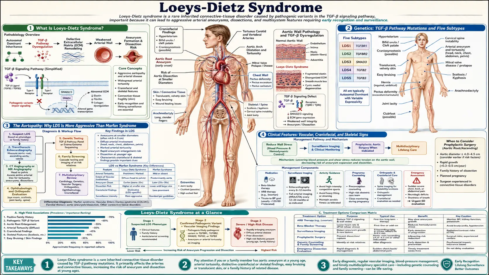
Loeys-Dietz syndrome (LDS) is a connective tissue disorder first described in 2005 by Loeys, Dietz and colleagues, who reported heterozygous mutations in the genes for the type I or type II transforming growth factor beta (TGF-β) receptor in ten families with a newly recognized pattern of cardiovascular, craniofacial, neurocognitive and skeletal findings (Loeys 2005). The 2006 follow-up study of 52 affected families described the syndrome by the triad of arterial tortuosity and aneurysms, hypertelorism (widely spaced eyes), and bifid uvula or cleft palate (Loeys 2006).
GeneReviews describes LDS as vascular findings (cerebral, thoracic and abdominal arterial aneurysms and/or dissections), skeletal findings, craniofacial features and skin findings, with a predisposition to widespread and aggressive arterial aneurysms and to pregnancy-related complications including uterine rupture and death. It also reports a strong tendency to allergic and inflammatory disease (asthma, eczema, food or environmental allergy) and an increased incidence of gastrointestinal inflammation such as eosinophilic esophagitis, gastritis or inflammatory bowel disease (Loeys and Dietz, GeneReviews). Features vary widely in distribution and severity, even among relatives who carry the same variant.
The TGF-β paradox. Some of the mutated receptors cannot carry the TGF-β signal in laboratory tests, yet tissue from affected people showed increased collagen and connective tissue growth factor and nuclear enrichment of phosphorylated Smad2 — signs of increased TGF-β signaling (Loeys 2005). In knock-in mice carrying LDS mutations in Tgfbr1 or Tgfbr2, the aortic wall showed progressive upregulation of Smad2 phosphorylation and TGF-β1 ligand expression that paralleled worsening aneurysm; mice that were simply haploinsufficient (one working copy) did not reproduce the LDS phenotype, and the authors interpreted the steady-state findings as a chronic compensation (Gallo 2014). The TGF-β2 discovery paper likewise found upregulated TGF-β signaling in aortic tissue and proposed that compensatory autocrine and/or paracrine events contribute to disease (Lindsay 2012).
Inheritance. LDS caused by variants in SMAD2, SMAD3, TGFB2, TGFB3, TGFBR1 or TGFBR2 is autosomal dominant: GeneReviews states that about 75% of probands have a de novo variant and about 25% have an affected parent, and that each child of an affected person has a 50% chance of inheriting the variant. A recessive form caused by two variants in IPO8 is also listed. No population prevalence figure was found in the sources checked for this page.
LDS Types and Genes
The first reports split patients with TGFBR1 or TGFBR2 mutations into two clinical types: type I, with typical craniofacial features, and type II, with more prominent skin features resembling vascular Ehlers-Danlos syndrome (Loeys 2006). The 2014 primer proposed instead that a mutation in TGFBR1, TGFBR2, SMAD3 or TGFB2 together with a documented aneurysm or dissection is enough for the diagnosis, whatever the craniofacial findings (MacCarrick 2014). The numbered types now designate the gene (Schepers 2018):
- LDS type 1 — TGFBR1 (TGF-β receptor 1; chromosome 9q22.33).
- LDS type 2 — TGFBR2 (TGF-β receptor 2; chromosome 3p24.1).
- LDS type 3 — SMAD3 (chromosome 15q22.33). First described as aneurysms-osteoarthritis syndrome: aneurysms, dissections and tortuosity throughout the arterial tree with mild craniofacial features, skeletal and skin findings, and early-onset osteoarthritis in most affected individuals (van de Laar 2011).
- LDS type 4 — TGFB2 (the TGF-β2 ligand; chromosome 1q41). Two 2012 papers reported it: one as familial thoracic aortic aneurysm and dissection with mild systemic features of Marfan syndrome (Boileau 2012), the other as a syndromic presentation within the LDS spectrum (Lindsay 2012).
- LDS type 5 — TGFB3 (the TGF-β3 ligand; chromosome 14q24.3). The discovery study reported 43 patients from 11 families with thoracic/abdominal aortic aneurysm and dissection and mitral valve disease, plus features overlapping LDS, Shprintzen-Goldberg and Marfan syndromes, including cleft palate, bifid uvula, skeletal overgrowth, cervical spine instability and clubfoot (Bertoli-Avella 2015).
- SMAD2 (chromosome 18q21.1). Variants predicted to be pathogenic were found in three families with arterial aneurysms and dissections (Micha 2015); GeneReviews lists SMAD2 among the LDS genes.
Chromosome locations are from the NCBI Gene records. The 2022 ACC/AHA aortic disease guideline notes that most clinical information comes from patients with TGFBR1 and TGFBR2 variants, that SMAD3 variants are associated with premature osteoarthritis and later onset of thoracic aortic disease, and that much less is known about aortic and branch-vessel disease with TGFB2 and TGFB3 variants (Isselbacher 2022).
Vascular Manifestations
Aortic root and ascending aorta: the 2022 guideline names aortic root and ascending aortic aneurysm and aortic dissection as leading causes of illness and death in LDS. In a series of 90 patients with TGFBR1 or TGFBR2 variants that it cites, aneurysm disease involved the ascending aorta in 78%, the arch in 10%, the descending aorta in 10%, the abdominal aorta and its branches in 17%, thoracic aortic branches in 21% and head and neck arteries in 10% (Isselbacher 2022).
Dissection at smaller diameters than in Marfan syndrome: the 2014 primer contrasts the increased risk of dissection at or above a 5.0-cm aortic root in Marfan syndrome with dissections in LDS types 1, 2 and 3 at 3.9–4.0 cm, and in type 4 at under 5.0 cm (MacCarrick 2014). In a surgical series of 71 patients, 6 (9%) died of aneurysm rupture or dissection before surgery, several with aortic diameters under 4.5 cm and as early as 6 months of age (Williams 2007). The 2022 guideline states that dissection may occur at smaller diameters than in Marfan syndrome when LDS is caused by TGFBR1, TGFBR2 or SMAD3 variants, while limited data have not suggested a higher risk at smaller size with TGFB2 or TGFB3 variants (Isselbacher 2022). Marfan syndrome itself is not free of dissection at moderate sizes: in a 10-centre series of 675 Marfan patients who had aortic root replacement, 46% of the 158 adults with aortic dissection and a documented diameter had an aneurysm of 6.5 cm or less (Gott 1999).
Arterial tortuosity: in a neuroradiology review of 25 genetically confirmed patients, all had extreme arterial tortuosity, which the authors call characteristic of the syndrome (Rodrigues 2009). Tortuosity is not unique to LDS: in an MRA study of 90 patients with connective tissue disorders, the vertebral tortuosity index was raised in both LDS (median 58) and Marfan syndrome (median 26) compared with controls (median 4.5); a higher index was associated with younger age at surgery, an index of 50 or more with earlier dissection and death, and age at surgery, dissection or death did not differ between Marfan syndrome and LDS (Morris 2011). In adults with Marfan syndrome, higher aortic tortuosity was likewise associated with a more severe aortic course (Franken 2015).
Branch arteries: the 2014 primer reports aneurysm and dissection in all other portions of the aorta and in arterial branches of the head, neck, chest and abdomen (MacCarrick 2014). In the neuroradiology series, 3 of 25 patients had dissections of the carotid or vertebrobasilar arteries and 1 had intracranial hemorrhage as a complication of dissection (Rodrigues 2009).
Intracranial aneurysms: 8 of the 25 patients in the neuroradiology series had intracranial aneurysms (Rodrigues 2009); the 2022 guideline gives a range of 10% to 18% of patients with LDS (Isselbacher 2022).
Craniofacial and Skeletal Features
GeneReviews lists the craniofacial features as hypertelorism, strabismus, bifid uvula or cleft palate, and craniosynostosis that can involve any suture (Loeys and Dietz, GeneReviews):
- Hypertelorism: widely spaced eyes, part of the original triad (Loeys 2006).
- Bifid uvula or cleft palate: the 2014 primer describes a range from cleft palate to a bifid, broad or long uvula, and notes that although part of the original triad, many individuals do not have this feature (MacCarrick 2014).
- Craniosynostosis: premature fusion of skull sutures. The primer reports that the sagittal suture is most commonly involved, with the coronal, metopic and squamosal sutures also possible (MacCarrick 2014); 12 of 25 patients in the neuroradiology series had craniosynostosis (Rodrigues 2009).
- Eye findings: the primer names the lack of lens dislocation as a major distinguishing feature from Marfan syndrome; blue or dusky sclera, strabismus, amblyopia and exotropia are described, and retinal detachment and cataracts have been reported in LDS types 1 and 2 (MacCarrick 2014).
In the 2006 cohort, patients with the craniofacial-predominant type I had cardiovascular surgery earlier (mean age 16.9 vs 26.9 years) and died earlier (22.6 vs 31.8 years) than those with type II, and the authors concluded that the severity of the clinical presentation predicts outcome (Loeys 2006).
Skeletal findings listed by GeneReviews are pectus excavatum or carinatum, scoliosis, joint laxity, arachnodactyly (long fingers), talipes equinovarus (clubfoot), and cervical spine malformation and/or instability. The primer adds that height and body proportions are typically within the normal range, that camptodactyly and joint contractures occur, that cervical spine findings were present in 51% of people with LDS types 1 and 2, and that the presence in types 3 and 4 was unknown (MacCarrick 2014). Dural ectasia is listed among LDS features in the 2022 guideline (Isselbacher 2022). In the neuroradiology series, 13 of 25 patients had scoliosis, 6 spinal instability, 4 dural ectasia, 3 hydrocephalus and 2 a Chiari malformation (Rodrigues 2009).
In SMAD3-related LDS (type 3), early-onset osteoarthritis was present in most affected individuals in the discovery study (van de Laar 2011).
Skin and Joint Findings
The skin findings described in the 2014 primer and GeneReviews are:
- Velvety, thin, translucent skin with visible veins.
- Easy bruising.
- Atrophic or dystrophic scars and delayed wound healing.
- Striae and/or facial milia in some individuals.
- Hernias: inguinal, umbilical and hiatal hernias, which may occur after surgery or recur; GeneReviews notes that a supporting mesh can be used at repair to reduce recurrence.
The primer notes that in people without craniofacial features, these skin findings may be a prominent clue distinguishing LDS from Marfan syndrome or familial thoracic aortic aneurysm. It also reports joint hypermobility, extremity contractures together with joint hyperextension (unusual in the general population but common in LDS), and rare but life-threatening splenic or bowel rupture in LDS types 1 and 2 (MacCarrick 2014).
Allergy: the primer reports a high prevalence of asthma, food allergy, eczema and allergic rhinitis, with a conservative estimate of food allergy in 31% of people with LDS compared with 6–8% in the general population (MacCarrick 2014).
Diagnosis and Genetic Testing
GeneReviews states that the diagnosis is established either in a person with characteristic clinical findings, or by finding a heterozygous pathogenic variant in SMAD2, SMAD3, TGFB2, TGFB3, TGFBR1 or TGFBR2 (or two variants in IPO8) in a person with aortic root enlargement, type A dissection, other characteristic features, or a family history of LDS (Loeys and Dietz, GeneReviews).
Overlapping conditions. The 2022 guideline's table of heritable thoracic aortic disease lists, alongside LDS, Marfan syndrome (FBN1), vascular Ehlers-Danlos syndrome (COL3A1), arterial tortuosity syndrome (SLC2A10), Shprintzen-Goldberg syndrome (SKI) and nonsyndromic familial thoracic aortic aneurysm genes such as ACTA2, MYH11 and MYLK (Isselbacher 2022). The 2006 study found TGFBR1 or TGFBR2 mutations in 12 of 40 patients who had been diagnosed with vascular Ehlers-Danlos syndrome but lacked the type III collagen abnormality, and recorded 59 vascular operations in the cohort with one death during the procedure — a low intraoperative mortality that the authors say distinguishes LDS from vascular Ehlers-Danlos syndrome (Loeys 2006). Features that the mutation update lists as separating LDS from Marfan syndrome include widespread aortic and arterial aneurysm and tortuosity, clubfoot, craniosynostosis, hypertelorism and bifid or broad uvula or cleft palate (Schepers 2018).
Relatives. GeneReviews describes clarifying the genetic status of at-risk relatives of any age, by testing for the family's known variant or, where it is not known, by examination with echocardiography and extensive vascular imaging, so that affected relatives can enter regular cardiovascular screening. The 2014 primer gives a risk of about 1% or less for a second affected child, attributed to germline mosaicism, when a child has an apparently de novo mutation (MacCarrick 2014).
Surveillance and Imaging
The 2022 ACC/AHA guideline makes these imaging recommendations for LDS (Isselbacher 2022):
- Echocardiography: a baseline transthoracic echocardiogram to measure the aortic root and ascending aorta, repeated 6 months later to determine the growth rate, then annually if the diameters are stable.
- Head-to-pelvis MRI or CT at baseline to evaluate the entire aorta and its branches for aneurysm, dissection and tortuosity.
- Annual MRI or CT of the affected aorta and arteries when they are dilated or dissected at baseline.
- Chest-to-pelvis MRI (or CT) every 2 years is described as reasonable when the aorta beyond the root and ascending segment and the arterial branches are not dilated, with more frequent imaging depending on family history.
- Cerebral aneurysm imaging with MRI or CT every 2 to 3 years is described as reasonable when initial screening shows no dilation.
GeneReviews similarly describes echocardiography at least annually and MRA or CTA of the entire arterial tree at least every other year, with more frequent imaging depending on genotype, family history, vessel size, growth rate or vascular pathology. The 2014 primer describes flexion-extension X-rays to assess the cervical spine, with CT or MRI in some individuals (MacCarrick 2014).
Activity and medicines. GeneReviews lists as agents and circumstances to avoid: contact sports, competitive sports and isometric exercise; agents that stimulate the cardiovascular system, including routine use of decongestants or triptan medicines for migraine; activities that cause joint injury or pain; and, for people at risk of recurrent pneumothorax, breathing against resistance (such as a brass instrument) or positive-pressure breathing (such as scuba diving). The 2014 primer lists, as exercise restrictions to reduce stress on the aorta and arteries, no contact or competitive sports, no isometric exercises (sit-ups, push-ups, pull-ups or weight lifting) and no exercising to the point of exhaustion (MacCarrick 2014).
Medical and Surgical Treatment
Medicines. The 2022 guideline states that there are no randomized trials of medicines to reduce aortic growth or dissection risk in LDS; its recommendation that a beta blocker or an angiotensin receptor blocker (ARB), or both, at maximally tolerated doses is reasonable rests on expert opinion, extrapolation from Marfan syndrome and data from mouse models (Isselbacher 2022). In LDS knock-in mice, the therapeutic effect of the ARB losartan correlated with suppression of Smad2 phosphorylation and TGF-β1 expression (Gallo 2014); the original rationale came from a Marfan mouse model in which losartan prevented aortic aneurysm (Habashi 2006). The 2014 primer gives losartan titration doses of 2.0 mg/kg/day in children and 100 mg/day in adults, attributed to a personal communication rather than to trial data (MacCarrick 2014).
The human trials are in Marfan syndrome, not LDS:
- Pediatric Heart Network trial: 608 participants aged 6 months to 25 years with an aortic-root z score above 3.0 were randomized to atenolol or losartan; the rate of aortic-root enlargement over 3 years did not differ significantly between the groups (Lacro 2014).
- COMPARE: in 233 adults randomized to losartan or no additional treatment, aortic root dilatation over about 3 years was lower with losartan (0.77 vs 1.35 mm), with no significant difference in clinical endpoints (Groenink 2013).
- Individual-patient meta-analysis: across four trials (676 participants) comparing an ARB with control, ARBs roughly halved the annual increase in aortic root z score; across three trials (766 participants) comparing ARBs with beta blockers, the change was similar in both groups (Pitcher 2022).
Prophylactic aortic surgery — the 2022 guideline thresholds. The guideline states that the threshold for prophylactic replacement of the aortic root and ascending aorta in LDS is informed by the specific gene variant, aortic diameter, growth rate, features outside the aorta, family history, age, sex and patient and physician preferences. Its Table 11 lists (Isselbacher 2022):
- TGFBR1 or TGFBR2 without high-risk features: 4.5 cm or more.
- TGFBR1 or TGFBR2 with high-risk features: 4.0 cm or more. The high-risk features named are certain specific variants; women with TGFBR2 variants and small body size; severe features outside the aorta (craniosynostosis, cleft palate, hypertelorism, bifid uvula, marked arterial tortuosity, widened scars, translucent skin); a family history of aortic dissection, especially at a young age or small diameter; and aortic growth of more than 0.3 cm per year.
- SMAD3: 4.5 cm or more; TGFB2: 4.5 cm or more; TGFB3: 5.0 cm or more (family history, age and growth rate also inform these).
For comparison, the same guideline recommends root and ascending aortic replacement in Marfan syndrome at 5.0 cm or more, and considers it reasonable at 4.5 cm or more with risk features. It notes that the earlier recommendation for LDS was surgery above 4.0 cm, and that the 2010 ACC/AHA guideline recommended surgery at 4.2 to 4.6 cm depending on imaging method. For the intact arch, descending or abdominal aorta in TGFBR1, TGFBR2 or SMAD3 disease, surgery at 4.5 cm or more "may be considered" (Isselbacher 2022). The 2014 primer described surgery for adults with LDS types 1 or 2 once the aortic root reaches 4.0 cm (MacCarrick 2014).
Type of operation. The 2014 primer describes valve-sparing aortic root replacement as the approach used to avoid the need for anticoagulation (MacCarrick 2014). In the early surgical series, 21 patients had thoracic aortic surgery (13 valve-sparing root replacements, 5 Bentall procedures, 2 arch replacements, 1 valve-sparing replacement with arch replacement) with no deaths at the primary operation; 3 patients died 2, 5 and 11 years later from rupture of the descending thoracic or abdominal aorta (Williams 2007). The guideline states that the entire ascending aorta is replaced at the time of root replacement, because distal ascending aneurysm and dissection can follow isolated root replacement (Isselbacher 2022).
Branch and distal arteries. The primer reports that open repair of descending and thoracoabdominal aneurysms is preferred by its authors because endovascular repair may fail late, although endovascular repair has been performed where landing zones allowed, and that splenic and hepatic artery aneurysms have been treated by coil embolization or open repair (MacCarrick 2014).
Other management described by GeneReviews: surgical fixation of cervical spine instability where needed to prevent spinal cord damage; care by a craniofacial team for cleft palate and craniosynostosis; standard allergy treatment; and chemical or surgical pleurodesis or removal of pulmonary blebs for recurrent pneumothorax.
Pregnancy. GeneReviews states that pregnancy and the postpartum period carry an increased risk of aortic dissection or rupture and of uterine rupture in LDS, and describes more frequent aortic imaging during pregnancy and in the weeks after delivery. In the 2006 cohort, pregnancy-related complications occurred in 6 of 12 women (Loeys 2006); the primer describes these as 4 aortic dissections and 2 uterine ruptures in 21 pregnancies (MacCarrick 2014). The 2022 guideline reports aortic dissection in 0% to 19% of pregnancies across series of women with TGFBR1 or TGFBR2 variants, notes that most pregnancy-related dissections occur in the third trimester or up to 12 weeks after delivery, and lists these thresholds for aortic surgery before pregnancy: 4.0 cm or more for LDS caused by TGFBR1, TGFBR2 or SMAD3 ("may be considered") and 4.5 cm or more for TGFB2 or TGFB3 ("reasonable"). It adds that information on diameter and pregnancy-related risk in LDS is lacking, because most women in the series did not know their diagnosis before pregnancy (Isselbacher 2022).
Prognosis and Natural History
In the 2006 cohort of 52 families, the natural history was marked by aggressive arterial aneurysms, with a mean age at death of 26.0 years (Loeys 2006). The 2026 updated primer reviews management since 2014 but its abstract gives no updated survival figure (MacCarrick 2026); no newer survival estimate was found in the sources checked for this page.
The 2022 guideline states that the gene involved and the severity of features outside the aorta — craniofacial features, degree of arterial tortuosity, skin findings — and family history inform the risk of aortic events; that marked variability exists within families; and that after aortic dissection, progressive enlargement is common and often requires several operations (Isselbacher 2022). Risk features that lower the guideline's surgical threshold are listed in the treatment section above.
Key Research Papers
- Loeys BL, Chen J, Neptune ER, et al. A syndrome of altered cardiovascular, craniofacial, neurocognitive and skeletal development caused by mutations in TGFBR1 or TGFBR2. Nature Genetics. 2005;37(3):275–281. — PubMed PMID: 15731757 — The founding description: TGFBR1 or TGFBR2 mutations in ten families.
- Loeys BL, Schwarze U, Holm T, et al. Aneurysm syndromes caused by mutations in the TGF-beta receptor. New England Journal of Medicine. 2006;355(8):788–798. — PubMed PMID: 16928994 — Clinical and molecular characterization of 52 affected families; mean age at death 26.0 years; pregnancy complications in 6 of 12 women.
- van de Laar IM, Oldenburg RA, Pals G, et al. Mutations in SMAD3 cause a syndromic form of aortic aneurysms and dissections with early-onset osteoarthritis. Nature Genetics. 2011;43(2):121–126. — PubMed PMID: 21217753 — Discovery paper for SMAD3 (aneurysms-osteoarthritis syndrome, LDS type 3).
- Lindsay ME, Schepers D, Bolar NA, et al. Loss-of-function mutations in TGFB2 cause a syndromic presentation of thoracic aortic aneurysm. Nature Genetics. 2012;44(8):922–927. — PubMed PMID: 22772368 — TGFB2 mutations or deletions in a phenotype within the LDS spectrum (LDS type 4).
- Boileau C, Guo DC, Hanna N, et al. TGFB2 mutations cause familial thoracic aortic aneurysms and dissections associated with mild systemic features of Marfan syndrome. Nature Genetics. 2012;44(8):916–921. — PubMed PMID: 22772371 — Parallel TGFB2 discovery paper; noted phenotypic overlap with Marfan syndrome.
- Bertoli-Avella AM, Gillis E, Morisaki H, et al. Mutations in a TGF-β ligand, TGFB3, cause syndromic aortic aneurysms and dissections. Journal of the American College of Cardiology. 2015;65(13):1324–1336. — PubMed PMID: 25835445 — TGFB3 discovery paper: 43 patients from 11 families (LDS type 5).
- Rodrigues VJ, Elsayed S, Loeys BL, Dietz HC, Yousem DM. Neuroradiologic manifestations of Loeys-Dietz syndrome type 1. AJNR American Journal of Neuroradiology. 2009;30(8):1614–1619. — PubMed PMID: 19556353 — Head and neck imaging in 25 patients: arterial tortuosity in all, intracranial aneurysms in 8, craniosynostosis in 12.
- Habashi JP, Judge DP, Holm TM, et al. Losartan, an AT1 antagonist, prevents aortic aneurysm in a mouse model of Marfan syndrome. Science. 2006;312(5770):117–121. — PubMed PMID: 16601194 — Mouse study behind the TGF-β and losartan rationale.
- Williams JA, Loeys BL, Nwakanma LU, et al. Early surgical experience with Loeys-Dietz: a new syndrome of aggressive thoracic aortic aneurysm disease. Annals of Thoracic Surgery. 2007;83(2):S757–S763. — PubMed PMID: 17257922 — 71 patients; 6 died of rupture or dissection before surgery, several at diameters under 4.5 cm; no deaths at 21 primary operations.
- Schepers D, Tortora G, Morisaki H, et al. A mutation update on the LDS-associated genes TGFB2/3 and SMAD2/3. Human Mutation. 2018;39(5):621–634. — PubMed PMID: 29392890 — Review of TGFB2/3 and SMAD2/3 variants; source of the gene-based type numbering.
- Franken R, El Morabit A, de Waard V, et al. Increased aortic tortuosity indicates a more severe aortic phenotype in adults with Marfan syndrome. International Journal of Cardiology. 2015;194:7–12. — PubMed PMID: 26005802 — Aortic tortuosity index in 211 adults with Marfan syndrome.
- Oderich GS, Panneton JM, Bower TC, et al. The spectrum, management and clinical outcome of Ehlers-Danlos syndrome type IV: a 30-year experience. Journal of Vascular Surgery. 2005;42(1):98–106. — PubMed PMID: 16012458 — Vascular Ehlers-Danlos syndrome series, for comparison with LDS.
- Loeys BL, Dietz HC. Loeys-Dietz Syndrome. In: GeneReviews. University of Washington, Seattle; revision of 2024-09-12. — PubMed PMID: 20301312
- MacCarrick G, Black JH, Bowdin S, et al. Loeys-Dietz syndrome: a primer for diagnosis and management. Genetics in Medicine. 2014;16(8):576–587. — PubMed PMID: 24577266
- MacCarrick G, Afifi RO, Allen R, et al. Loeys-Dietz syndrome: 2026 updated care management primer. Genetics in Medicine. 2026;28(7):102577. — PubMed PMID: 41988792
- Isselbacher EM, Preventza O, Hamilton Black J, et al. 2022 ACC/AHA Guideline for the Diagnosis and Management of Aortic Disease. Circulation. 2022;146(24):e334–e482. — PubMed PMID: 36322642 — Also published in J Am Coll Cardiol 2022;80(24):e223–e393 (PMID 36334952).
- Gallo EM, Loch DC, Habashi JP, et al. Angiotensin II-dependent TGF-β signaling contributes to Loeys-Dietz syndrome vascular pathogenesis. Journal of Clinical Investigation. 2014;124(1):448–460. — PubMed PMID: 24355923 — Mouse study.
- Micha D, Guo DC, Hilhorst-Hofstee Y, et al. SMAD2 mutations are associated with arterial aneurysms and dissections. Human Mutation. 2015;36(12):1145–1149. — PubMed PMID: 26247899
- Lacro RV, Dietz HC, Sleeper LA, et al. Atenolol versus losartan in children and young adults with Marfan's syndrome. New England Journal of Medicine. 2014;371(22):2061–2071. — PubMed PMID: 25405392 — Marfan syndrome trial (Pediatric Heart Network).
- Groenink M, den Hartog AW, Franken R, et al. Losartan reduces aortic dilatation rate in adults with Marfan syndrome: a randomized controlled trial. European Heart Journal. 2013;34(45):3491–3500. — PubMed PMID: 23999449 — Marfan syndrome trial (COMPARE).
- Pitcher A, Spata E, Emberson J, et al.; Marfan Treatment Trialists' Collaboration. Angiotensin receptor blockers and β blockers in Marfan syndrome: an individual patient data meta-analysis of randomised trials. Lancet. 2022;400(10355):822–831. — PubMed PMID: 36049495
- Gott VL, Greene PS, Alejo DE, et al. Replacement of the aortic root in patients with Marfan's syndrome. New England Journal of Medicine. 1999;340(17):1307–1313. — PubMed PMID: 10219065
- Morris SA, Orbach DB, Geva T, et al. Increased vertebral artery tortuosity index is associated with adverse outcomes in children and young adults with connective tissue disorders. Circulation. 2011;124(4):388–396. — PubMed PMID: 21730308
PubMed searches for additional research:
Loeys-Dietz syndrome aorta TGFBR1 TGFBR2 aneurysm Loeys-Dietz losartan treatment SMAD3 aortic aneurysm osteoarthritis
Connections
- Genetics
- Ehlers-Danlos Syndrome
- Marfan Syndrome
- Aortic Aneurysm
- Aortic Dissection
- Stroke
- POTS (Postural Orthostatic Tachycardia Syndrome)
- Scoliosis
- Diseases
- Lab Tests