Proline for Collagen Synthesis
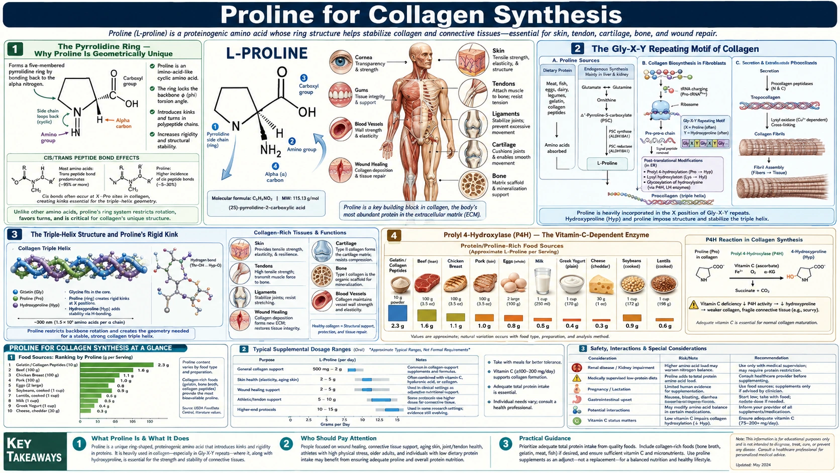
Collagen is the most abundant protein in the human body, accounting for roughly 30% of total body protein. Proline and its post-translationally modified form hydroxyproline together account for approximately 23% of all amino acid residues in collagen — meaning roughly one in every four collagen residues is a proline derivative. The pyrrolidine ring of proline is the only side-chain geometry in nature that can produce the rigid kink required for the collagen triple helix, and the hydroxylation of selected proline residues to hydroxyproline is what locks that helix into a thermally stable conformation at body temperature. This deep-dive walks through the molecular mechanism of proline incorporation, the prolyl-hydroxylase enzymes and their obligate vitamin-C dependence, the cellular machinery of procollagen processing, and what happens when the system fails — from scurvy to the ultra-rare osteogenesis imperfecta caused by mutations in prolyl 3-hydroxylase.
Table of Contents
- The Pyrrolidine Ring — Why Proline Is Geometrically Unique
- The Gly-X-Y Repeating Motif of Collagen
- The Triple-Helix Structure and Proline's Rigid Kink
- Prolyl 4-Hydroxylase (P4H) — The Vitamin-C-Dependent Enzyme
- Prolyl 3-Hydroxylase (P3H) — The Lesser-Known Cousin
- The Four Obligate Cofactors: Oxygen, Iron, Alpha-Ketoglutarate, Ascorbate
- Procollagen Processing in the Endoplasmic Reticulum
- Scurvy — Collagen-Synthesis Failure as a Disease
- Endogenous Proline Biosynthesis Pathways
- 28 Collagen Types and Where Each Lives
- Key Research Papers
- Connections
- Featured Videos
The Pyrrolidine Ring — Why Proline Is Geometrically Unique
Of the 20 standard proteinogenic amino acids, 19 share the same general structure: an alpha-carbon bonded to an amino group (NH2), a carboxyl group (COOH), a hydrogen atom, and a distinct side chain (R group). Proline breaks this pattern. Its side chain — a three-carbon propyl group — folds back and covalently bonds to the nitrogen of the amino group, forming a five-membered ring called a pyrrolidine ring. This ring incorporates the alpha-carbon, the nitrogen, and three of the carbons of the side chain.
This makes proline technically an imino acid rather than an amino acid — the nitrogen has only one hydrogen attached rather than two. In peptide bond formation, the nitrogen of every other amino acid donates a hydrogen as part of the dehydration reaction that links two amino acids together; proline cannot, because its nitrogen is part of a ring. This single difference cascades into a host of structural consequences.
The most important consequence is geometric. In a typical polypeptide chain, the backbone phi angle (the rotation around the N–Calpha bond) can sample a wide range of values. The pyrrolidine ring locks proline's phi angle into a very narrow range around −65° — the ring physically prevents the bond from rotating. This rigidity has two effects: (1) proline cannot participate in alpha-helices or beta-sheets the way other amino acids do, because those secondary structures require specific phi/psi angle ranges that proline cannot achieve; (2) proline introduces a sharp, predictable kink wherever it appears in a polypeptide chain.
Both effects make proline a "structure breaker" in most globular proteins. But in collagen, where the geometry of the triple helix actually requires a sharp kink every third residue, proline is the structure maker. No other amino acid can substitute, which is why every collagen molecule across all 28 known collagen types contains proline at high abundance.
The Gly-X-Y Repeating Motif of Collagen
Every collagen polypeptide chain (called an alpha chain) consists of approximately 1,000 amino acids organized into a strict repeating motif: Gly-X-Y, where X is most frequently proline and Y is most frequently hydroxyproline. About one third of all collagen residues are glycine, and another third are proline or hydroxyproline. The remaining third is distributed among the other 17 amino acids — primarily lysine and hydroxylysine, with smaller amounts of alanine, glutamate, arginine, and others.
The reason for the glycine-every-third-position rule is geometric. Glycine has no side chain (its R group is just a hydrogen atom), making it the smallest possible amino acid. When three collagen alpha chains wind around each other to form the triple helix, the side chains of all the amino acids point outward — except the residues that face the center of the helix. The geometry forces every third residue to be packed against the central axis with essentially no room for any side chain larger than a hydrogen. Glycine is the only amino acid that fits. Any substitution of glycine in the Gly-X-Y motif disrupts the helix and produces severe disease — osteogenesis imperfecta is most commonly caused by glycine-to-larger-residue substitutions in type I collagen.
The X and Y positions face outward and can accommodate larger residues. Proline in the X position and hydroxyproline in the Y position are statistically the most common because their pyrrolidine ring geometry stabilizes the polyproline-II-like local conformation that each alpha chain adopts before winding into the triple helix. Other residues can occupy X and Y, but proline-hydroxyproline pairs are thermally the most stable and represent the evolutionary optimum.
The Triple-Helix Structure and Proline's Rigid Kink
Each individual collagen alpha chain, by itself, adopts a left-handed polyproline-II-like helical conformation with about three residues per turn. Three such chains then wind around each other in a right-handed superhelix — the famous collagen triple helix. The opposing handedness of the individual chains and the superhelix is what gives collagen its extraordinary mechanical strength: pulling on the ends tends to wind the helix tighter rather than unraveling it.
The triple helix is held together by inter-chain hydrogen bonds. Each glycine residue donates a hydrogen from its amide group to an oxygen on a neighboring chain's carbonyl. These hydrogen bonds are direct and require very precise positioning — which is achievable only because the proline residues at the X positions force the chain into the polyproline-II conformation, and the hydroxyproline residues at the Y positions provide additional bridging hydrogen bonds through water molecules that occupy the inter-chain crevices.
The thermodynamic stability of the triple helix is directly proportional to its hydroxyproline content. Collagens with higher hydroxyproline content have higher denaturation temperatures (Tm). Mammalian collagen denatures around 39-42°C — just above body temperature, by design. Cold-water fish collagen, which has lower hydroxyproline content, denatures around 16-20°C, which is why fish gelatin sets at colder temperatures than mammalian gelatin. Sea-cucumber collagen with very low hydroxyproline denatures around 12°C. The relationship between hydroxyproline content and thermal stability is one of the cleanest structure-function correlations in protein biology.
This is the molecular reason why scurvy is a connective tissue disease. Without ascorbic acid, the prolyl-4-hydroxylase enzyme cannot convert proline to hydroxyproline, the inter-chain water-bridged hydrogen bonds cannot form, and the resulting under-hydroxylated procollagen denatures at body temperature. The body cannot build new connective tissue. Bleeding gums, loose teeth, hemorrhage into joints, and poor wound healing follow.
Prolyl 4-Hydroxylase (P4H) — The Vitamin-C-Dependent Enzyme
Prolyl 4-hydroxylase, abbreviated P4H, is the enzyme responsible for converting proline residues in the Y position of the Gly-X-Y motif into 4-hydroxyproline (4-Hyp). It is one of the most important enzymes in the body that most clinicians have never heard of. Without it, collagen cannot form a stable triple helix.
P4H is an alpha-2-beta-2 tetramer. The alpha subunit (encoded in humans by the P4HA1, P4HA2, or P4HA3 genes) contains the catalytic site and the iron-binding domain. The beta subunit is identical to protein disulfide isomerase (PDI) and serves both as a structural component and as a chaperone that ensures correct procollagen folding. The enzyme resides on the luminal side of the endoplasmic reticulum membrane in collagen-producing cells — fibroblasts, chondrocytes, osteoblasts, and others.
The reaction mechanism is an oxygen-dependent hydroxylation. P4H takes a proline residue in the Y position of a procollagen chain, one molecule of oxygen (O2), and one molecule of alpha-ketoglutarate (also called 2-oxoglutarate), and produces hydroxyproline, succinate, and carbon dioxide. The reaction requires ferrous iron (Fe2+) at the active site. Ascorbic acid (vitamin C) is required to keep the iron in the reduced Fe2+ state — the reaction occasionally fails to release the iron back in the reduced form, and accumulated Fe3+ would otherwise inactivate the enzyme. Ascorbate periodically reduces the oxidized iron back to Fe2+, restoring catalytic activity.
This is the molecular role of vitamin C in collagen synthesis. Without it, P4H slowly inactivates as more and more of its active-site iron oxidizes. Hydroxylation slows, procollagen chains accumulate under-hydroxylated proline residues, and the triple helix cannot fold or fold stably. Linus Pauling correctly identified this as the central mechanism in his classic work on ascorbic acid — the connection between vitamin C and collagen runs through prolyl hydroxylase.
Prolyl 3-Hydroxylase (P3H) — The Lesser-Known Cousin
While P4H gets all the textbook attention, there is a second prolyl hydroxylase that hydroxylates a smaller subset of proline residues at the 3-position rather than the 4-position. This enzyme, prolyl 3-hydroxylase (P3H), is encoded by the LEPRE1 gene (now officially P3H1) and uses the same cofactor set as P4H: oxygen, alpha-ketoglutarate, ferrous iron, and ascorbate.
P3H modifies only a handful of proline residues per collagen alpha chain — perhaps one or two in type I collagen, more in type IV collagen. The functional importance of this modest hydroxylation went unappreciated for decades. Then in 2007, researchers discovered that loss-of-function mutations in P3H1 cause a recessively inherited form of severe osteogenesis imperfecta (OI type VIII). Children with biallelic P3H1 mutations have bones so fragile that fractures occur in utero and survival into adulthood is uncommon without aggressive bisphosphonate therapy.
This discovery established that 3-hydroxyproline modification, despite occurring at a small number of sites, is essential for normal type I collagen function. The current model is that P3H1 acts as part of a chaperone complex (together with CRTAP and cyclophilin B) that ensures proper folding of procollagen in the endoplasmic reticulum. Loss of any component of this complex produces clinically indistinguishable severe OI phenotypes.
The clinical implication for proline biology is that even subtle perturbations of the prolyl-hydroxylation system can produce devastating connective tissue disease. The system is engineered for high reliability, with multiple enzymes, chaperones, and cofactor pools all having to work in concert.
The Four Obligate Cofactors: Oxygen, Iron, Alpha-Ketoglutarate, Ascorbate
Both prolyl hydroxylases require the same four cofactors, and any limitation of any single cofactor compromises collagen synthesis. These represent four distinct vulnerabilities in the collagen biosynthetic system.
- Molecular oxygen (O2) — the source of the hydroxyl oxygen added to proline. Hypoxia limits prolyl hydroxylation, which is part of the mechanism by which chronic hypoxic states (chronic obstructive pulmonary disease, advanced heart failure, cyanotic congenital heart disease, high-altitude residency) can produce subtle connective tissue effects over time. The HIF-1-alpha transcription factor is also regulated by a related prolyl hydroxylase (PHD), which is why HIF-1-alpha accumulates under hypoxic conditions and triggers the hypoxic response.
- Ferrous iron (Fe2+) — required at the active site to perform the actual oxygen activation chemistry. Iron deficiency impairs prolyl hydroxylase activity even when serum hemoglobin remains near normal, contributing to the well-documented connection between iron deficiency anemia and impaired wound healing. Iron supplementation in iron-deficient patients with chronic wounds often improves healing more than the change in hemoglobin alone would predict.
- Alpha-ketoglutarate (2-oxoglutarate) — the co-substrate consumed during hydroxylation, becoming succinate and CO2 in the reaction. Alpha-ketoglutarate is produced abundantly by the citric acid cycle and is rarely rate-limiting under normal physiologic conditions, but mitochondrial dysfunction or severe undernutrition can occasionally limit its availability.
- Ascorbic acid (vitamin C) — the iron-reduction cofactor. Required only periodically (to restore oxidized Fe3+ at the active site to Fe2+), but absolutely required at some level. Humans cannot synthesize ascorbate due to a loss-of-function mutation in the gulonolactone oxidase gene roughly 60 million years ago. Without dietary intake of approximately 10 mg/day, prolyl hydroxylase activity falls and scurvy develops over 2-3 months.
Two clinical implications follow. First, addressing collagen-synthesis problems requires assessing all four cofactor pools, not just proline itself. Pure proline supplementation without adequate vitamin C, iron, and oxygenation will not restore impaired collagen synthesis. Second, the redundancy of the cofactor requirements explains why most apparently nutritional connective tissue problems involve multiple simultaneous deficiencies — chronic illness rarely produces an isolated single-nutrient gap.
Procollagen Processing in the Endoplasmic Reticulum
Collagen is not synthesized as a finished molecule. The initial gene product is a procollagen polypeptide that is longer than the final collagen alpha chain at both ends — it has N-terminal and C-terminal "propeptide" extensions that serve essential functions during synthesis and folding, then are cleaved off after the procollagen has been secreted from the cell.
The processing sequence is:
- Translation on rough endoplasmic reticulum ribosomes — the procollagen polypeptide is co-translationally inserted into the ER lumen via a signal peptide that is cleaved upon entry.
- Prolyl and lysyl hydroxylation — P4H, P3H, and lysyl hydroxylase (LH) modify selected proline and lysine residues. This must occur before triple-helix formation, because once the helix forms, the modified residues are buried inside the structure and become inaccessible to the enzymes.
- Glycosylation of hydroxylysines — certain hydroxylysine residues are decorated with galactose or galactose-glucose disaccharides.
- C-terminal propeptide registration — three procollagen chains assemble through their C-terminal propeptide domains, which contain a "registration" sequence that ensures the correct three alpha chains (alpha-1, alpha-1, alpha-2 for type I; three alpha-1 chains for type II; three alpha-1 chains for type III) come together.
- Triple-helix folding zipper — once registered, the three chains zip up from the C-terminus toward the N-terminus, with HSP47 chaperoning the process and preventing premature aggregation.
- Secretion through the Golgi — the assembled procollagen is exported through specialized large COPII vesicles to the cell surface and secreted.
- Propeptide cleavage by extracellular metalloproteases — N-procollagen peptidase (ADAMTS-2) and C-procollagen peptidase (BMP-1) clip off the propeptide extensions, allowing the mature collagen monomers to assemble into fibrils.
- Fibril cross-linking by lysyl oxidase — the copper-dependent enzyme lysyl oxidase oxidizes specific lysine and hydroxylysine residues to aldehydes, which then spontaneously form cross-links between neighboring collagen molecules, giving fibrils their tensile strength.
The entire process can take hours to complete, and a single fibroblast can have thousands of procollagen molecules at various stages of processing at any given moment. The biosynthetic load of maintaining the body's collagen pool — estimated to turn over by approximately 1-3% per day in actively remodeling tissues — is enormous, which is part of why the body invests so much regulatory machinery in protecting this pathway.
Scurvy — Collagen-Synthesis Failure as a Disease
Scurvy is the textbook example of what happens when collagen synthesis fails. The clinical picture — bleeding gums, loose teeth, perifollicular hemorrhage, corkscrew hairs, joint pain from hemarthroses, poor wound healing, fatigue, depression, and eventually death — is entirely explained by failure of new collagen synthesis throughout the body.
The pathophysiology runs through the prolyl-4-hydroxylase enzyme. Vitamin C deficiency depletes the ascorbate that keeps P4H active-site iron in the Fe2+ state. P4H gradually inactivates, prolyl hydroxylation slows, and procollagen chains accumulate with too few hydroxyproline residues. Under-hydroxylated procollagen denatures at body temperature, so the triple helix never forms stably, and the protein is degraded inside the cell rather than secreted. New collagen deposition essentially stops.
The clinical picture is dominated by failure of tissues with high collagen turnover. Periodontal ligament and gingiva have rapid collagen turnover, so bleeding gums and tooth loosening are early signs. Wound healing essentially stops — old surgical scars from years past have been documented to break down in late-stage scurvy because the collagen in those scars was still slowly turning over. Capillary fragility produces the characteristic perifollicular hemorrhages and bruising. The connective tissue of joint capsules weakens, allowing intra-articular bleeding.
Historically, scurvy was the disease that killed more sailors than combat during the age of sail. James Lind's 1747 controlled trial on board HMS Salisbury, demonstrating that citrus fruits cured scurvy where other interventions did not, is often cited as the first modern clinical trial. The British Royal Navy adopted lime juice rations in 1795, which is the origin of the term "limey" for British sailors. The active ingredient (ascorbic acid) was not identified until Albert Szent-Györgyi's work in the 1930s, for which he won the 1937 Nobel Prize in Physiology or Medicine.
Modern scurvy still occurs — in alcoholics with poor diets, in elderly isolated individuals, in patients with restrictive autism-spectrum eating patterns, and in some restrictive-diet experiments. Treatment is straightforward: 100-300 mg vitamin C daily for 1-2 weeks reverses clinical symptoms, with full collagen restoration over a few months as the body has the chance to rebuild its connective tissue stores. The lesson for proline biology is that the proline pool itself is rarely the limiting factor — the hydroxylation machinery and its cofactors are.
Endogenous Proline Biosynthesis Pathways
Proline is classified as a non-essential amino acid because the human body can synthesize it from precursors. There are two major endogenous biosynthesis pathways:
1. The glutamate pathway (P5C route). Glutamate is reduced by the enzyme P5C synthase (encoded by ALDH18A1) to glutamic-gamma-semialdehyde, which spontaneously cyclizes to pyrroline-5-carboxylate (P5C). P5C is then reduced by pyrroline-5-carboxylate reductase (PYCR1, PYCR2, or PYCR3) to proline. This pathway uses NADPH as the reducing equivalent and is the dominant source of endogenous proline in most tissues.
2. The arginine pathway (via ornithine). Arginine is converted to ornithine by arginase, then ornithine is transaminated by ornithine aminotransferase (OAT) to glutamic-gamma-semialdehyde, which spontaneously cyclizes to P5C, which is then reduced to proline as in the first pathway. This pathway is particularly important in wound healing tissue, where activated macrophages produce large amounts of arginase and shunt arginine into proline for collagen synthesis at the repair site.
Loss-of-function mutations in any of the enzymes in these pathways produce rare but informative diseases. ALDH18A1 mutations cause autosomal recessive cutis laxa with neurological involvement — the skin loses elasticity (cutis laxa), and patients also have intellectual disability and cataracts. PYCR1 mutations cause a similar cutis laxa syndrome (ARCL2). Ornithine aminotransferase deficiency causes gyrate atrophy of the choroid and retina (a progressive blindness disorder), because ornithine accumulates and damages retinal cells, and proline synthesis from arginine is impaired.
These rare diseases collectively confirm that endogenous proline synthesis is essential and cannot be fully replaced by dietary proline intake alone — tissue-level synthesis matters because dietary proline must be transported through the bloodstream and into cells, whereas endogenous synthesis can produce proline locally where it is needed.
28 Collagen Types and Where Each Lives
There are at least 28 distinct types of collagen in humans, each encoded by separate genes and expressed in distinct tissues. They share the Gly-X-Y triple-helical motif but differ in chain composition, length, and assembly patterns. The five most clinically important are:
- Type I (encoded by COL1A1 and COL1A2) — the most abundant collagen in the body, accounting for >90% of total collagen. Found in skin, bone, tendon, ligament, dentin, cornea, and the interstitium of most organs. Mutations cause classic osteogenesis imperfecta.
- Type II (COL2A1) — the principal collagen of articular cartilage and the vitreous humor of the eye. Mutations cause spondyloepiphyseal dysplasia, Stickler syndrome, and achondrogenesis type II.
- Type III (COL3A1) — co-distributed with type I in skin, blood vessels, intestine, and uterus. Particularly important in blood vessel walls. Mutations cause vascular Ehlers-Danlos syndrome with spontaneous aortic and intestinal rupture.
- Type IV (COL4A1-A6) — the basement membrane collagen, forming sheet-like networks rather than fibrils. Mutations in COL4A3, COL4A4, or COL4A5 cause Alport syndrome (hereditary nephritis with sensorineural hearing loss).
- Type V (COL5A1, COL5A2, COL5A3) — minor co-fibrillar collagen mixed with type I that helps regulate fibril diameter. Mutations cause classic Ehlers-Danlos syndrome with hyperextensible skin and joint hypermobility.
The remaining 23 collagen types fill specialized roles — type VII anchoring skin to basement membrane (mutations cause epidermolysis bullosa), type IX as a cartilage minor collagen, type X in the hypertrophic zone of growth plate cartilage, type XI in cartilage fibril nucleation, type XII as a fibril-associated collagen with interrupted triple helices (FACIT), and so on. Despite their diversity, all 28 types contain proline at high abundance and require the same prolyl-hydroxylase machinery. A single biosynthetic system serves the entire collagen family.
Key Research Papers
- Shoulders MD, Raines RT (2009). Collagen structure and stability. Annual Review of Biochemistry. — PubMed
- Myllyharju J, Kivirikko KI (2004). Collagens, modifying enzymes and their mutations in humans, flies and worms. Trends in Genetics. — PubMed
- Vranka JA et al. (2004). Prolyl 3-hydroxylase 1, enzyme characterization and identification of a novel family of enzymes. Journal of Biological Chemistry. — PubMed
- Cabral WA et al. (2007). Prolyl 3-hydroxylase 1 deficiency causes a recessive metabolic bone disorder resembling lethal/severe osteogenesis imperfecta. Nature Genetics. — PubMed
- Phang JM et al. (2015). Proline metabolism and microenvironmental stress. Annual Review of Nutrition. — PubMed
- Kivirikko KI, Myllyharju J (1998). Prolyl 4-hydroxylases and their protein disulfide isomerase subunit. Matrix Biology. — PubMed
- Pinnell SR (1985). Regulation of collagen synthesis. Journal of Investigative Dermatology. — PubMed
- Murad S et al. (1981). Regulation of collagen synthesis by ascorbic acid. Proceedings of the National Academy of Sciences. — PubMed
- DePhillips HJ et al. (2015). Modern scurvy: a case report and review. Journal of General Internal Medicine. — PubMed
- Ricard-Blum S (2011). The collagen family. Cold Spring Harbor Perspectives in Biology. — PubMed
- Brodsky B, Persikov AV (2005). Molecular structure of the collagen triple helix. Advances in Protein Chemistry. — PubMed
- Marini JC et al. (2017). Osteogenesis imperfecta. Nature Reviews Disease Primers. — PubMed
PubMed Topic Searches
- PubMed: Prolyl hydroxylase and vitamin C
- PubMed: Collagen triple-helix stability
- PubMed: Osteogenesis imperfecta and type I collagen
- PubMed: Modern scurvy
- PubMed: Hydroxyproline and collagen stability
Connections
- All Amino Acids
- Proline Overview
- Proline Benefits Hub
- Proline for Wound Healing
- Proline for Cardiovascular Health
- Proline for Skin Health
- Vitamin C (Prolyl Hydroxylase Cofactor)
- Iron (Ferrous Cofactor)
- Glycine (Every-Third-Position Residue)
- Lysine (Cross-Link Substrate)
- Arginine (Proline Precursor)
- Glutamine (Glutamate Source)
- Collagen
- Bone Broth
- Osteoporosis