Hypocalcemia (Low Calcium): Heart Rhythm and QT
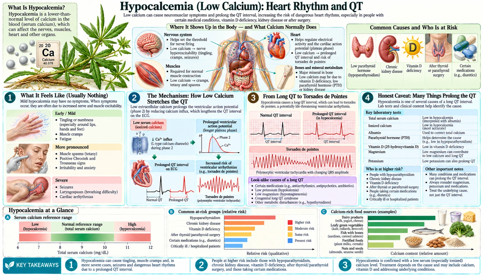
Most people think of calcium as a bone mineral, but a small, tightly guarded pool of calcium circulating in your blood also helps run the electrical timing of your heart. When that blood calcium falls too low — a state doctors call hypocalcemia — one of its quietest yet most dangerous effects is to stretch out the heart's electrical recovery, a change visible on an electrocardiogram (ECG) as a prolonged QT interval. A long QT is usually silent, but it leaves the heart vulnerable to a chaotic rhythm called torsades de pointes that can cause fainting or, rarely, cardiac arrest. This page explains exactly how low calcium lengthens the QT, why it is almost never something you can feel until it tips into a rhythm problem, the many other things that prolong the QT (low calcium is far from the only cause), and how it is diagnosed and corrected safely.
Table of Contents
- What It Feels Like (Usually Nothing)
- The Mechanism: How Low Calcium Stretches the QT
- From Long QT to Torsades de Pointes
- Honest Caveat: Many Things Prolong the QT
- Clues That Point to Calcium
- What Causes the Calcium to Drop
- Getting Diagnosed: Electrocardiogram and Blood Tests
- Correcting It Safely
- When to Seek Care / Red Flags
- Key Research Papers
- Connections
- Featured Videos
What It Feels Like (Usually Nothing)
A calcium-related long QT, by itself, produces no symptom at all. The QT interval is a measurement on an electrocardiogram (ECG) — the time from the start of the heart's electrical squeeze to the end of its electrical recovery — and a stretched-out QT is something a machine sees, not something you feel. Many people walk around with a mildly prolonged QT from low calcium and have no idea, because the heart is still beating in a normal, regular rhythm. The danger is not the long QT itself; it is what the long QT makes possible.
When low calcium does announce itself, it usually does so through the more familiar nerve-and-muscle symptoms that travel with it — tingling around the mouth and in the fingers (numbness and tingling), and muscle twitching, spasm, or cramping (cramps and tetany). Those symptoms are the warning lights; the long QT is the engine trouble happening quietly underneath. So the heart-rhythm story of hypocalcemia is really two separate experiences:
- The silent stretch. A prolonged QT with a normal rhythm — felt as nothing, detected only on an electrocardiogram.
- The dangerous tip. If the QT becomes long enough and the heart is provoked, it can flip into a fast, irregular rhythm. Now there are symptoms: a sudden pounding or racing heartbeat (palpitations), lightheadedness, a feeling of near-fainting, or an actual faint (syncope) that comes on without warning.
“Feeling fine” therefore does not rule out a calcium-related rhythm risk. That is exactly why, when calcium is known to be very low, clinicians put a patient on a heart monitor and check an electrocardiogram rather than waiting for symptoms — the first symptom can be a faint.
The Mechanism: How Low Calcium Stretches the QT
Every heartbeat is an electrical event. Each muscle cell in the ventricles fires an action potential — a precisely shaped voltage spike that drives the cell to contract and then recover. Unlike a nerve's brief blip, the heart cell's action potential has a long, flat middle stretch called the plateau. That plateau is held open largely by calcium flowing into the cell through gates called L-type calcium channels. The plateau is, in effect, the heart deliberately taking its time before resetting — and the QT interval on the electrocardiogram is essentially a measurement of how long that whole fire-and-recover cycle lasts across the ventricles.
Now lower the calcium outside the cell. With less calcium available in the bloodstream, the inward calcium current during the plateau is altered, and the cell takes longer to march through the plateau and reach the point where it can finally repolarize (recover). A 2022 case paper by Tang and Rabkin sets out the likely sequence: less calcium outside means less calcium enters through the L-type channels, so the calcium inside the cell takes longer to reach the level that shuts those channels, and the cell's recovery runs long. Their patient, an 87-year-old man with a QT of 508 ms, saw it shorten when his furosemide dose was reduced and calcium was supplemented. Stretch out that recovery in millions of cells at once and you stretch out the QT interval on the electrocardiogram. The classic electrocardiogram signature of hypocalcemia is therefore a long ST segment (the flat stretch that corresponds to the plateau) pushing out the QT — and notably, the shape of the T wave itself usually stays normal. The relationship is direct and predictable: the lower the blood calcium, the longer the QT tends to run.
An analogy. Picture a relay race where each runner must finish a full lap before the next can start. The calcium plateau is one runner's lap. Normal blood calcium is like a clear, well-lit track — the runner completes the lap on schedule and hands off the baton. Low calcium is like running that same lap in fog and mud: the runner isn't lost and isn't going the wrong way, but each lap takes longer, so the whole relay finishes late. The QT interval is the stopwatch on the whole relay. Restore the calcium — clear the fog — and the lap times snap back to normal, often within hours of correction.
It is worth being precise about which calcium matters. The body guards the ionized (free) calcium in the blood within a narrow band; this is the biologically active fraction that the heart cells actually sense. About half of blood calcium is bound to the protein albumin and is electrically “invisible,” which is why a low albumin level can make total calcium look low when the active ionized calcium is in fact normal — an important point we return to under diagnosis. It is the genuinely low ionized calcium, not a protein-binding artifact, that lengthens the QT.
From Long QT to Torsades de Pointes
A prolonged QT matters because it widens a window of electrical vulnerability. As the ventricles take longer to recover, some heart cells finish recovering before others, leaving the heart briefly in a patchwork state where part of it is ready to fire again and part is not. If a stray early beat lands in that vulnerable window, it can set off a self-sustaining, swirling rhythm called torsades de pointes — French for “twisting of the points,” because on the electrocardiogram the beats appear to twist around the baseline.
Torsades is a form of polymorphic ventricular tachycardia — a very fast, disorganized ventricular rhythm. Because the heart is beating too fast and too chaotically to pump blood effectively, the result is a sudden drop in blood flow to the brain: lightheadedness, then fainting. Most torsades episodes stop on their own within seconds and the person recovers, which is why an unexplained faint can be the only clue. But if an episode is sustained or degenerates into ventricular fibrillation, it can cause cardiac arrest. This is the genuinely dangerous end of the calcium-and-QT story, and it is the reason the seemingly abstract number on an electrocardiogram is taken seriously.
An honest point of proportion: torsades from isolated low calcium is uncommon. The 2010 American Heart Association and American College of Cardiology Foundation statement says low calcium has been linked to torsades "only in rare cases". Hypocalcemia much more often prolongs the QT modestly without ever triggering an arrhythmia. The risk climbs when calcium falls very low, when it falls quickly (as after certain surgeries), and especially when it is layered on top of other QT-prolonging factors — low potassium, low magnesium, or QT-prolonging medications. Documented cases of hypocalcemic torsades exist — including recurrent torsades after thyroid surgery from a rapid post-operative calcium crash — but they are the exception, not the rule. In that thyroid-surgery report, a 49-year-old woman developed torsades even while receiving intravenous calcium, needed repeated electrical cardioversion, temporary pacing and high-dose beta-blockers, and eventually reached a normal calcium level and QT with long-term calcium supplementation. The everyday significance of a calcium-related long QT is as a warning and a risk factor, not a guarantee of catastrophe.
Honest Caveat: Many Things Prolong the QT
It would be a mistake to see a long QT on an electrocardiogram and assume calcium is to blame. A prolonged QT is a final common pathway — many unrelated causes funnel into the same finding, and low calcium is only one of them, and far from the most common. Being honest about this matters, because chasing calcium while missing the real culprit can be dangerous. The major causes of a long QT include:
- Medications — by far the most common acquired cause. A long and varied list of drugs prolongs the QT, including certain antibiotics (some macrolides and fluoroquinolones), antifungals, antipsychotics, the anti-nausea drug ondansetron, methadone, and several antiarrhythmic drugs themselves. Many people develop a long QT purely from a medication, with perfectly normal calcium.
- Other electrolyte disturbances. Low potassium (hypokalemia) and low magnesium (hypomagnesemia) are classic, powerful QT-prolongers — and in practice are more frequent troublemakers than calcium. They often travel together with low calcium, which compounds the risk.
- Congenital long QT syndrome. Inherited mutations in the heart's ion channels lengthen the QT from birth. This is a genetic condition, unrelated to diet or calcium, and is an important cause of fainting and sudden cardiac events in otherwise healthy young people and athletes.
- Heart disease and a slow heart rate. Heart attacks, heart failure, and a very slow pulse (bradycardia) can all stretch the QT.
- Underactive thyroid and other conditions. Hypothyroidism, certain brain injuries, and severe illness can prolong it as well.
So a long QT is best thought of as a flag that prompts a checklist: review every medication, check potassium, magnesium, and calcium, consider thyroid and heart disease, and ask about fainting in the patient or sudden death in the family. Low calcium earns its place on that checklist — but it sits alongside several causes that are individually more common, which is exactly why a doctor tests for several things at once rather than assuming.
Clues That Point to Calcium
Given how many things prolong the QT, what makes a clinician suspect calcium specifically? A few patterns raise the odds:
- The company it keeps. A long QT alongside tingling around the mouth or fingers, muscle twitching or cramps, or frank spasms (tetany) is a strong hint that calcium is low, because those nerve-excitability symptoms and the cardiac QT change come from the same underlying problem — too little free calcium steadying excitable tissues.
- The electrocardiogram shape. In hypocalcemia the lengthening is concentrated in the ST segment, with a normal-looking T wave riding on the end. That “long flat stretch, normal T wave” appearance differs from the broad, abnormal T waves and U waves that low potassium tends to produce, and it can tip a careful reader toward calcium.
- The clinical setting. Recent thyroid or parathyroid surgery, known kidney disease, a history of low vitamin D, or a critical illness all make hypocalcemia more likely as the explanation.
- It tracks with the number. Because the QT lengthens in proportion to how low the calcium is, a QT that shortens as calcium is replaced essentially confirms the link.
Even with these clues, the diagnosis is never made on the electrocardiogram alone. A calcium-related long QT is confirmed by measuring the blood calcium — ideally the ionized fraction — and watching the QT respond as calcium is restored. The clues simply tell the clinician to make sure calcium is on the list of things being checked.
What Causes the Calcium to Drop
Blood calcium is held remarkably steady by a feedback loop involving parathyroid hormone (PTH), vitamin D, and the kidneys. When that system is disrupted, calcium falls. The causes that most often lower it enough to affect the QT include:
- Hypoparathyroidism — too little parathyroid hormone. The most dramatic drops follow damage to or removal of the parathyroid glands, classically after thyroid or parathyroid surgery. Without enough parathyroid hormone, the body cannot defend its calcium, and levels can plunge in the first days after the operation — a setting where rhythm monitoring is routine.
- Vitamin D deficiency. Vitamin D is required to absorb calcium from food. Severe, prolonged vitamin D deficiency is one of the most common reasons calcium runs low worldwide, usually developing gradually.
- Chronic kidney disease. The kidneys perform the final activation step that turns vitamin D into its active form, and they handle phosphate. Failing kidneys disturb both, commonly producing low calcium alongside high phosphate.
- Low magnesium. Magnesium is needed both to release parathyroid hormone and for the body to respond to it. When magnesium is very low, calcium often cannot be corrected until the magnesium is fixed — a tidy parallel to the way magnesium also governs potassium.
- “Hungry bone” and rapid shifts. After surgery for an overactive parathyroid or thyroid, bones starved of mineral can pull calcium out of the blood rapidly (hungry bone syndrome). Severe illness, pancreatitis, large blood transfusions (the citrate preservative binds calcium), and certain drugs can also drop calcium quickly — and a fast fall is more arrhythmogenic than a slow one.
- Denosumab in advanced kidney disease. The osteoporosis drug denosumab can drop calcium severely in people with advanced chronic kidney disease. In US Medicare records of women aged 65 or older on dialysis, severe low calcium developed within 12 weeks in 41.1% on denosumab and 2.0% on oral bisphosphonates. A later analysis of the same program found the risk of hospital or emergency treatment for low calcium rose as kidney disease worsened: on denosumab against oral bisphosphonates, 3.01% against 0.00% on dialysis, and 0.57% against 0.03% in stages 4 and 5 without dialysis. The US label for denosumab products carries a boxed warning for advanced chronic kidney disease (estimated glomerular filtration rate below 30), reporting hospitalization, life-threatening events and deaths.
Identifying which of these is at work changes the treatment entirely — replacing vitamin D is very different from managing post-surgical hypoparathyroidism — which is why diagnosis looks beyond the calcium number to parathyroid hormone, vitamin D, magnesium, phosphate, and kidney function.
Getting Diagnosed: Electrocardiogram and Blood Tests
Two simple, inexpensive tests do most of the work: an electrocardiogram (ECG) to measure the QT, and blood tests to measure calcium and its regulators.
On the electrocardiogram, the QT interval is measured and then corrected for heart rate — reported as the QTc — because the QT naturally shortens when the heart beats faster and lengthens when it slows, so a rate-corrected value is needed to compare fairly. The 2010 American Heart Association and American College of Cardiology Foundation statement treats a QTc above the 99th percentile, about 470 ms in men and 480 ms in women, as abnormally prolonged; other references use lower cut-offs of roughly 450 ms in men and 460–470 ms in women. The same statement calls a QTc above 500 ms highly abnormal, and data it cites from inherited long QT syndrome link that level to a 2- to 3-fold higher risk of torsades, so it prompts urgent attention. In hypocalcemia, the lengthening shows up as a stretched ST segment with a normal T wave.
On the blood side, a routine Comprehensive Metabolic Panel reports total calcium along with albumin, kidney function, and other electrolytes. Two refinements matter here:
- Correct for albumin, or measure ionized calcium. Because about half of blood calcium rides on albumin, a low albumin makes total calcium read low even when the active ionized calcium is normal. Clinicians either apply a correction formula or, better, order an ionized calcium directly — the ionized value is what actually governs the heart, so it is the number that matters for the QT.
- Check the regulators. Because a standard panel does not include magnesium, parathyroid hormone, or vitamin D, those are usually added when low calcium is found — they reveal the cause. Potassium and magnesium are checked specifically because, if they are also low, they compound the QT risk and must be corrected too.
Put together, the electrocardiogram confirms and quantifies the rhythm risk while the blood tests confirm the low calcium and uncover why — and watching the QTc shorten as calcium is restored closes the loop.
Correcting It Safely
How calcium is replaced depends on how low it is, how fast it fell, and whether the QT (or symptoms) signal danger. The guiding principle mirrors other electrolytes: gentle for mild, stable cases; urgent and monitored when the rhythm is at risk.
- Urgent intravenous (IV) calcium for the dangerous picture. A markedly prolonged QT, torsades, seizures, or severe spasm calls for intravenous calcium (calcium gluconate is the usual form), given with continuous heart-rhythm monitoring. Restoring calcium typically shortens the QT and stabilizes the heart, often within hours. This is hospital territory, not something managed at home.
- Correct magnesium alongside. If magnesium is also low — common — calcium often will not stay corrected until magnesium is replaced, because magnesium is needed to release and respond to parathyroid hormone. Intravenous magnesium is also a frontline treatment for torsades in its own right, which is why it is given early when the rhythm is unstable.
- Oral calcium and vitamin D for the stable, chronic case. When calcium is only mildly low and the person is well, treatment is oral calcium supplements plus vitamin D (often the active form, calcitriol, in hypoparathyroidism), titrated by a clinician to bring calcium into the low-normal range. Calcium-rich foods support this, but in true deficiency supplements and vitamin D do the heavy lifting.
- Treat the cause and remove other triggers. Correcting vitamin D deficiency, managing post-surgical hypoparathyroidism, addressing kidney disease — and, crucially, reviewing the medication list to stop or avoid other QT-prolonging drugs — are all part of the fix. Lowering the total QT burden is as important as topping up the calcium.
- Parathyroid hormone replacement. Palopegteriparatide, a once-daily parathyroid hormone replacement injection, is on the US market for adults with hypoparathyroidism; its label says it was not studied for acute post-surgical hypoparathyroidism. In its phase 3 trial, 82 adults were randomized and 73 completed 3.5 years, the last 3 of them open-label with no placebo group. Of those 73, 89% had normal calcium, 96% no longer needed therapeutic doses of calcium and 100% no longer needed active vitamin D.
There is a caution in the other direction too. Too-rapid intravenous correction has its own risks, and people with normal calcium gain nothing from extra. Calcium replacement for a rhythm problem is individualized and monitored.
When to Seek Care / Red Flags
A long QT is often discovered incidentally and managed calmly. But certain features mean get emergency help now — by calling emergency services, not booking a routine visit — because they suggest the rhythm itself may be unstable:
- Fainting or near-fainting — especially if it comes on suddenly, without warning, or during exertion or startle. A faint in someone with low calcium or a known long QT can be a torsades episode and must be taken seriously.
- Palpitations — a sudden racing, pounding, fluttering, or chaotic heartbeat (see heart palpitations and arrhythmia), particularly with lightheadedness.
- Seizures or severe, widespread muscle spasm — signs that calcium may be dangerously low and affecting the brain and muscles as well as the heart.
- A known very low calcium level with any new lightheadedness, chest fluttering, or breathlessness — do not wait to see if it passes.
- Recent thyroid or parathyroid surgery with tingling, twitching, or feeling faint — a classic setup for a rapid post-operative calcium crash that warrants immediate evaluation.
The most dangerous combination is fainting or palpitations in someone with low calcium, because that is the moment a silent long QT may have tipped into a real arrhythmia. An electrocardiogram and a calcium level take minutes and can be lifesaving.
Key Research Papers
- Bushinsky DA, Monk RD (1998). Electrolyte quintet: Calcium. The Lancet;352(9124):306-311. — DOI: 10.1016/s0140-6736(97)12331-5. — doi:10.1016/s0140-6736(97)12331-5
- Cooper MS, Gittoes NJL (2008). Diagnosis and management of hypocalcaemia. BMJ;336(7656):1298-1302. — DOI: 10.1136/bmj.39582.589433.BE. — doi:10.1136/bmj.39582.589433.BE
- Pepe J, Colangelo L, Biamonte F, et al. (2020). Diagnosis and management of hypocalcemia. Endocrine;69(3):485-495. — DOI: 10.1007/s12020-020-02324-2. — doi:10.1007/s12020-020-02324-2
- Gardner JD, Calkins JB, Garrison GE (2014). ECG Diagnosis: The Effect of Ionized Serum Calcium Levels on Electrocardiogram. The Permanente Journal;18(1):e119-e120. — DOI: 10.7812/TPP/13-025. — doi:10.7812/TPP/13-025
- Santana LF, Cheng EP, Lederer WJ (2010). How does the shape of the cardiac action potential control calcium signaling and contraction in the heart? Journal of Molecular and Cellular Cardiology;49(6):901-903. — DOI: 10.1016/j.yjmcc.2010.09.005. — doi:10.1016/j.yjmcc.2010.09.005
- Drew BJ, Ackerman MJ, Funk M, et al. (2010). Prevention of Torsade de Pointes in Hospital Settings: A Scientific Statement From the American Heart Association and the American College of Cardiology Foundation. Circulation;121(8):1047-1060. — DOI: 10.1161/CIRCULATIONAHA.109.192704. — doi:10.1161/CIRCULATIONAHA.109.192704
- Tang JKK, Rabkin SW (2022). Hypocalcemia-Induced QT Interval Prolongation. Cardiology;147(2):191-195. — PubMed PMID: 35078204
- Hagdorn QAJ, Loh P, Velthuis S (2021). Recurrent hypocalcaemic torsades de pointes due to hungry bone syndrome: a rare complication of thyroidectomy. Netherlands Heart Journal;29(7-8):415-416. — DOI: 10.1007/s12471-020-01533-8. — doi:10.1007/s12471-020-01533-8
- Brandi ML, Bilezikian JP, Shoback D, et al. (2016). Management of Hypoparathyroidism: Summary Statement and Guidelines. The Journal of Clinical Endocrinology & Metabolism;101(6):2273-2283. — DOI: 10.1210/jc.2015-3907. — doi:10.1210/jc.2015-3907
- Khan AA, Bilezikian JP, Brandi ML, et al. (2022). Evaluation and Management of Hypoparathyroidism Summary Statement and Guidelines from the Second International Workshop. Journal of Bone and Mineral Research;37(12):2568-2585. — DOI: 10.1002/jbmr.4691. — doi:10.1002/jbmr.4691
- Abate EG, Clarke BL (2017). Review of Hypoparathyroidism. Frontiers in Endocrinology;7:172. — DOI: 10.3389/fendo.2016.00172. — doi:10.3389/fendo.2016.00172
- Bird ST, Smith ER, Gelperin K, et al. (2024). Severe Hypocalcemia With Denosumab Among Older Female Dialysis-Dependent Patients. JAMA;331(6):491-499. — PubMed PMID: 38241060
- Bird ST, Gelperin K, Smith ER, et al. (2025). The Effect of Denosumab on Risk for Emergently Treated Hypocalcemia by Stage of Chronic Kidney Disease: A Target Trial Emulation. Annals of Internal Medicine;178(1):29-38. — PubMed PMID: 39556837
- Khan AA, Clarke BL, Rubin MR, et al. (2026). Palopegteriparatide in Adults With Hypoparathyroidism: 3.5-Year Results From the Phase 3 PaTHway Trial. The Journal of Clinical Endocrinology & Metabolism:dgag440. — PubMed PMID: 42935168
PubMed Topic Searches
Connections
- All Minerals
- Hypocalcemia Symptom Hub
- Hypocalcemia and Muscle Cramps & Tetany
- Hypocalcemia and Numbness & Tingling
- Hypocalcemia and Bone Loss
- Calcium Overview
- Calcium and Cardiovascular Health
- Calcium and Nerve Transmission
- Calcium and Muscle Function
- Magnesium
- Magnesium and Heart Health
- Potassium
- Potassium and Heart Rhythm
- Vitamin D3
- Comprehensive Metabolic Panel
- Arrhythmia
- Heart Palpitations
- Hyperparathyroidism
- Hypoparathyroidism