Calcium for Cardiovascular Health
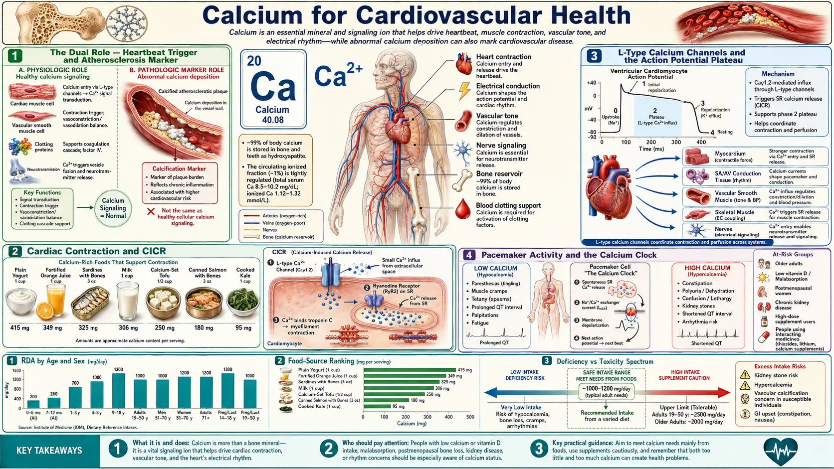
Calcium occupies an uncomfortable dual position in cardiovascular health. On one hand, transient Ca2+ flux is the universal trigger of every cardiac contraction — without calcium-induced calcium release (CICR) through L-type channels and ryanodine receptors, the heart would not beat. On the other hand, ectopic calcium deposition in coronary arteries and aortic valves is the structural signature of advanced atherosclerosis, and a strong predictor of future coronary events. The 2010–2011 Bolland meta-analyses reported that calcium supplements were associated with a modest increase in myocardial infarction risk, and later analyses have pointed in both directions. This page walks through the cellular electrophysiology that makes calcium indispensable to the beating heart, the vitamin-K-dependent proteins (matrix Gla protein in artery walls, osteocalcin in bone) that research links to where calcium is deposited, the calcium supplement controversy in detail, and the calcium channel blocker drug class as a pharmacological counterpoint that demonstrates the consequences of blocking L-type calcium entry into vascular smooth muscle.
Table of Contents
- The Dual Role — Heartbeat Trigger and Atherosclerosis Marker
- Cardiac Contraction and CICR
- L-Type Calcium Channels and the Action Potential Plateau
- Pacemaker Activity and the Calcium Clock
- Vascular Smooth Muscle Tone and Blood Pressure
- Vascular Calcification — The Vitamin K2 Axis
- The Calcium Supplement Controversy (Bolland)
- Calcium Channel Blockers — A Pharmacology Counterpoint
- Coronary Artery Calcium (CAC) Scoring
- Intake, Food and Supplement Findings
- Key Research Papers
- Connections
- Featured Videos
The Dual Role — Heartbeat Trigger and Atherosclerosis Marker
Calcium is unique among nutrients in that it is simultaneously essential for cardiovascular function and a marker of cardiovascular disease. Every cardiac contraction depends on a precisely choreographed rise and fall in intracellular calcium concentration that proceeds 60-100 times per minute for an entire lifetime. The same molecule, when deposited ectopically in arterial walls or heart valves, becomes a structural marker of atherosclerosis that can be quantified on a non-contrast CT scan; in the MESA cohort the score predicted coronary events beyond standard risk factors (Detrano 2008).
This duality creates a complicated clinical picture. Calcium supplements are widely used for bone health, often by older adults who already carry cardiovascular risk. If supplements raise cardiovascular risk, any bone benefit would have to be weighed against it. Research on the question looks at where the calcium ends up, and at the nutrient partners (vitamin D, vitamin K, magnesium) proposed to influence it.
The rest of this page disentangles the cardiovascular calcium story into its constituent parts: the cellular mechanisms that make calcium indispensable to the heartbeat, the molecular biology that determines whether dietary calcium is deposited in bone or in arteries, the supplement-trial evidence that started the calcium-and-cardiovascular-events debate, and the pharmacological calcium channel blocker class that demonstrates what happens when calcium entry into vascular smooth muscle is therapeutically suppressed.
Cardiac Contraction and CICR
Cardiac muscle relies on a mechanism called calcium-induced calcium release (CICR). Unlike skeletal muscle, where the dihydropyridine receptor (DHPR) mechanically couples to and opens the ryanodine receptor (RyR1), in cardiac muscle the L-type DHPR opens during the action potential plateau and admits a small amount of extracellular calcium ("trigger calcium") into the cytoplasm. This trigger calcium then binds to RyR2 channels on the sarcoplasmic reticulum (SR) and amplifies into a much larger release of stored calcium into the cytoplasm.
- Trigger calcium fraction – The L-type calcium influx is a small trigger; much of the calcium that activates contraction comes from release by the SR through RyR2 (Eisner 2017).
- Sarcoplasmic reticulum load – The amplitude of the calcium transient depends on the SR calcium content, which is determined by the balance between SERCA2a reuptake and ryanodine receptor leak. Beta-adrenergic stimulation increases SR load by phosphorylating phospholamban (releasing its inhibition of SERCA2a) and by phosphorylating L-type channels (increasing trigger calcium).
- Frank-Starling mechanism – When end-diastolic volume increases (greater stretch on the cardiac sarcomere), the myofilaments become more sensitive to a given concentration of calcium, increasing force of contraction without requiring a larger calcium transient. This intrinsic length-tension relationship matches cardiac output to venous return on a beat-to-beat basis.
- Calcium removal during diastole – For the heart to relax and fill during diastole, cytoplasmic calcium must be rapidly cleared. Most is pumped back into the SR by SERCA2a; the calcium that entered the cell is extruded across the sarcolemma, mainly by the sodium-calcium exchanger (NCX), with smaller contributions from the plasma membrane calcium ATPase (PMCA) and mitochondrial uptake. In the steady state, calcium influx equals calcium efflux on every beat (Eisner 2017). Reduced SERCA activity has been described in the diseased heart (Eisner 2017) and is one proposed contributor to slower relaxation.
- Arrhythmia from RyR2 leak – Diastolic SR calcium leak through dysfunctional RyR2 channels activates the inward NCX current, generating delayed afterdepolarizations (DADs) that can trigger ectopic beats and ventricular tachycardia. Mutations in the RyR2 gene were identified in 2001 as a cause of catecholaminergic polymorphic ventricular tachycardia (CPVT), a stress-triggered arrhythmia syndrome; in one family described in that report, two sisters of the proband died suddenly at 14 and 16 (Priori 2001).
L-Type Calcium Channels and the Action Potential Plateau
The ventricular myocyte action potential has a distinctive square-wave shape with a prolonged plateau (phase 2). This plateau is the defining electrophysiological feature of cardiac muscle and is created by inward L-type calcium current balanced against delayed-rectifier potassium currents. Three consequences flow from this plateau:
- It powers CICR – Sustained L-type calcium entry during the plateau provides the trigger calcium that opens RyR2 channels on the SR.
- It creates a long refractory period – Because sodium channels remain inactivated throughout the plateau, the cardiac myocyte cannot re-fire until repolarization is complete. This prevents tetanic contraction (sustained, uninterrupted force) of the heart muscle — which would prevent diastolic filling.
- It synchronizes contraction across the ventricle – The long plateau gives the activation wavefront time to traverse the entire ventricle, so that all the working myocardium contracts roughly together rather than as a peristaltic wave.
The L-type calcium channel (CaV1.2 in heart and vascular smooth muscle) is composed of a pore-forming alpha-1c subunit plus regulatory alpha-2/delta, beta, and gamma subunits. It is the target of the dihydropyridine calcium channel blocker class (amlodipine, nifedipine, felodipine) which preferentially blocks vascular smooth muscle channels, and the non-dihydropyridine class (verapamil, diltiazem) which preferentially blocks cardiac channels. These drugs are discussed below under the Calcium Channel Blockers section.
Pacemaker Activity and the Calcium Clock
The sinoatrial (SA) node is the heart's natural pacemaker. Unlike working ventricular myocytes, SA nodal cells have no stable resting membrane potential; they exhibit spontaneous diastolic depolarization that brings the cell to threshold and fires the next action potential. Two complementary mechanisms drive this automaticity:
- The "membrane clock" – The funny current (If) is a slowly activating mixed sodium-potassium inward current carried by HCN4 channels. It begins to flow as the cell repolarizes, slowly depolarizing the membrane. T-type calcium channels (CaV3) activate at intermediate voltages and contribute further to depolarization. Finally, L-type channels open and produce the action potential upstroke.
- The "calcium clock" – Rhythmic spontaneous calcium release events ("local calcium releases" or LCRs) from the SR through RyR2 channels occur during late diastole. These calcium releases activate the inward sodium-calcium exchanger current (3 Na+ in for 1 Ca2+ out is electrogenic, depolarizing the membrane), accelerating the pacemaker depolarization. The membrane clock and the calcium clock are tightly coupled and reinforce each other.
- Beta-adrenergic acceleration – Sympathetic stimulation increases heart rate by accelerating both clocks. PKA phosphorylation enhances If, increases L-type calcium current, and increases SR calcium load (more spontaneous calcium releases per diastolic interval). Parasympathetic stimulation has the opposite effect via M2 muscarinic receptor activation of inward-rectifier potassium channels.
- HCN4 and ivabradine – A mutation in the HCN4 pacemaker channel has been linked to familial sinus bradycardia (Milanesi 2006). Ivabradine selectively inhibits If and slows the heart rate without the negative inotropic effect of beta-blockers; in the SHIFT trial (6,558 patients with heart failure, ejection fraction 35% or lower and heart rate 70 per minute or higher), it reduced the composite of cardiovascular death or heart-failure hospital admission (HR 0.82, 0.75–0.90) (Swedberg 2010).
Vascular Smooth Muscle Tone and Blood Pressure
Vascular smooth muscle cells in the walls of arteries and arterioles maintain partial contraction (tone) at rest. The intracellular calcium concentration in these cells is a primary determinant of vascular resistance and therefore of blood pressure.
- Calcium influx pathways – Vascular smooth muscle cells acquire calcium from extracellular fluid primarily through L-type calcium channels (CaV1.2). Membrane depolarization (e.g., from stretch, alpha-1 adrenergic stimulation, or angiotensin II signaling) opens these channels. Receptor-operated channels and store-operated channels (Orai/STIM) contribute additional calcium entry during sustained agonist stimulation.
- SR calcium release – Vasoconstrictor agonists that signal through Gq-coupled receptors (alpha-1 adrenergic, angiotensin II AT1, endothelin ETA, vasopressin V1a) activate phospholipase C, generating IP3 that opens IP3 receptors on the SR and releases stored calcium.
- Calmodulin-MLCK signaling – Cytoplasmic Ca2+ binds calmodulin, activating myosin light-chain kinase (MLCK), which phosphorylates the regulatory light chain of smooth muscle myosin. Phosphorylated myosin engages cross-bridge cycling with actin, generating force and constricting the vessel.
- Endothelial calcium and nitric oxide – In the endothelial cell layer that lines blood vessels, calcium signaling activates endothelial nitric oxide synthase (eNOS), generating nitric oxide that diffuses into adjacent smooth muscle, activates guanylyl cyclase, raises cGMP, and promotes vasodilation. This is the basis of endothelium-dependent dilation tested clinically by flow-mediated dilation (FMD).
- RhoA / Rho-kinase calcium sensitization – Beyond calcium concentration, vasoconstrictor signaling also activates the RhoA / Rho-kinase pathway, which inhibits myosin light-chain phosphatase (MLCP). This increases the force generated at any given calcium concentration. Fasudil, a Rho-kinase inhibitor, acts on this pathway; a 1992 placebo-controlled trial in Japan tested it against cerebral vasospasm after subarachnoid haemorrhage (Shibuya 1992).
Vascular Calcification — The Vitamin K2 Axis
Vascular calcification is the deposition of calcium phosphate mineral (hydroxyapatite) within the wall of arteries. It comes in two anatomically distinct patterns: intimal calcification within atherosclerotic plaque and medial calcification within the media of the artery wall (Mönckeberg's medial calcific sclerosis), producing stiff but non-occlusive arteries. Both patterns often coincide and occur in people with type 2 diabetes and chronic kidney disease (Lanzer 2014).
Vascular calcification is an actively regulated process, not simple precipitation (Lanzer 2014). Vascular smooth muscle cells make Matrix Gla Protein (MGP); mice bred without it die within two months from arterial calcification that ruptures blood vessels, an animal finding that identified MGP as an inhibitor of arterial calcification (Luo 1997). MGP carries several gamma-carboxyglutamic acid (Gla) residues — five to six in the bovine protein first described in 1983 (Price 1983) — formed by a vitamin-K-dependent carboxylation. Uncarboxylated MGP (ucMGP) is inactive and accumulates at sites of arterial calcification; in a 2008 study, serum ucMGP was lower than in healthy people in patients after angioplasty, with aortic stenosis, on haemodialysis and with calciphylaxis, and the authors proposed it as a biomarker of cardiovascular calcification (Cranenburg 2008; Schurgers 2008).
- The vitamin K1 vs K2 distinction – Vitamin K is the cofactor for carboxylating clotting factors in the liver, osteocalcin in bone and MGP in cartilage and the vessel wall. In healthy volunteers, natto-derived menaquinone-7 (MK-7, a vitamin K2) had a much longer half-life than vitamin K1 (phylloquinone), accumulated to 7- to 8-fold higher levels during prolonged intake and carboxylated osteocalcin more completely; the authors noted that 50 mcg/day or more of MK-7 may interfere with oral anticoagulant treatment (Schurgers 2007). In the Rotterdam Study, higher dietary menaquinone, but not phylloquinone, was associated with lower coronary heart disease mortality (RR 0.43, 0.24–0.77, top versus bottom third), lower all-cause mortality and less severe aortic calcification (Geleijnse 2004). In 564 postmenopausal women, menaquinone intake was associated with less coronary calcification (RR 0.80, 0.65–0.98) and phylloquinone was not (Beulens 2009). Both are observational studies.
- Osteocalcin and the bone-vascular link – Osteocalcin, made by bone-forming cells, is another vitamin-K-dependent Gla protein; Schurgers 2007 used the degree of osteocalcin carboxylation as its measure of vitamin K activity. A shared vitamin K link between bone loss and arterial calcification is a proposed idea rather than an established cause of either.
- Vitamin D context – Vitamin D increases intestinal calcium absorption. The idea that taking calcium and vitamin D without enough vitamin K sends calcium to arteries rather than bone is a hypothesis, not a trial finding.
- Magnesium and calcification – In 2,695 Framingham Heart Study participants free of cardiovascular disease, each 50 mg/day higher self-reported magnesium intake was associated with 22% lower coronary artery calcium, and the highest intake group had 58% lower odds of any coronary calcium than the lowest (Hruby 2014, a cross-sectional study). The authors note that animal and cell studies suggest magnesium may prevent calcification within plaques.
- Phosphate burden in CKD – Vascular calcification is common in chronic kidney disease (Lanzer 2014), where it is studied in relation to high serum phosphate, FGF23 and vitamin K status.
The Calcium Supplement Controversy (Bolland)
In 2010, Mark Bolland and colleagues at the University of Auckland published a meta-analysis in BMJ of 15 placebo-controlled trials of calcium supplements (500 mg/day or more) given without vitamin D. In the five trials with patient-level data (8,151 participants), myocardial infarction was more common with calcium (hazard ratio 1.31, 95% CI 1.02–1.67); the trial-level analysis gave a pooled relative risk of 1.27 (1.01–1.59). In 2011 the same group reanalysed the Women's Health Initiative (WHI) calcium and vitamin D trial and combined it with eight other trials (28,072 participants): calcium with or without vitamin D was associated with a relative risk of 1.24 (1.07–1.45) for myocardial infarction and 1.15 (1.03–1.27) for myocardial infarction or stroke. Both papers called for a reassessment of calcium supplements in osteoporosis management.
- Magnitude – Both papers described the increases as modest; Bolland 2010 noted that, because calcium supplements are widely used, modest increases in risk might translate into a large burden of disease in the population.
- Proposed mechanism – One proposed explanation is the acute rise in serum calcium after a supplement. In a crossover trial of ten women, 500 mg of calcium as citrate raised serum ionised and total calcium over 6 hours; the rise was delayed when the citrate was taken with a meal and smaller after a dairy meal. The authors wrote that whether these differences contribute to cardiovascular risk requires further study (Bristow 2015).
- Dietary versus supplemental calcium – In MESA (5,448 adults, about 10 years of follow-up), the highest fifth of total calcium intake was associated with a lower risk of developing coronary artery calcium (RR 0.73, 0.57–0.93), while calcium supplement use, after accounting for total intake, was associated with a higher risk (RR 1.22, 1.07–1.39) (Anderson 2016). In cohort studies, Chung 2016 found no consistent dose-response relationship between total, dietary or supplemental calcium and cardiovascular mortality.
- Analyses that point the other way, and later ones – Lewis 2015, a meta-analysis of 18 trials (63,563 participants) that counted coronary events only when verified by clinical review, hospital record or death certificate, found a relative risk of 1.02 (0.96–1.09) for coronary heart disease. Chung 2016, reviewing 4 randomized trials and 27 observational studies, concluded that calcium intake within the tolerable upper intake levels (2,000 to 2,500 mg/day) is not associated with cardiovascular risk in generally healthy adults; the accompanying National Osteoporosis Foundation and American Society for Preventive Cardiology guideline (Kopecky 2016) graded the evidence of no relationship as moderate quality. Myung 2021, a meta-analysis of 13 double-blind, placebo-controlled trials, found a 15% higher risk of cardiovascular disease (RR 1.15, 1.06–1.25) in healthy postmenopausal women. After a median 22.3 years, the WHI follow-up found cardiovascular mortality 6% higher with calcium plus vitamin D than with placebo (HR 1.06, 1.01–1.12), cancer mortality 7% lower and no effect on all-cause mortality (Thomson 2024).
- What the trialists concluded – In a 2015 review, Reid, Bristow and Bolland concluded that calcium supplements appear to have a negative risk-benefit effect and argued against their routine use in osteoporosis (Reid 2015); Reid and Bolland wrote in 2020 that current evidence does not support calcium or vitamin D supplements in healthy community-dwelling adults (Reid 2020). The NIH Office of Dietary Supplements states that absorption from supplements is highest at doses of 500 mg or less.
Figures from the abstracts of Bolland 2010, Bolland 2011, Lewis 2015, Myung 2021 and Thomson 2024 (WHI). Outcomes differ between analyses, so the five estimates are not directly comparable.
Related pages: the Atherosclerosis page and the Vitamin K2 page.
Calcium Channel Blockers — A Pharmacology Counterpoint
The calcium channel blocker (CCB) drug class provides a useful counterpoint that illustrates what happens when calcium entry into vascular smooth muscle and cardiac myocytes is therapeutically suppressed.
- Dihydropyridines (DHPs) – Amlodipine, nifedipine, felodipine, nicardipine. These bind preferentially to L-type CaV1.2 channels in vascular smooth muscle, causing peripheral vasodilation and lowering blood pressure. They have minimal direct cardiac effects at therapeutic doses, though reflex sympathetic activation can produce tachycardia.
- Non-dihydropyridines (non-DHPs) – Verapamil and diltiazem. These act on L-type channels in cardiac myocytes and SA/AV nodal cells, producing negative inotropic and negative chronotropic effects in addition to vasodilation. They are used in rate control of atrial fibrillation and for symptomatic supraventricular tachycardia.
- Use in angina – CCBs reduce myocardial oxygen demand by lowering afterload (peripheral vasodilation), reduce heart rate (non-DHPs), and produce coronary vasodilation, all useful in chronic stable angina and vasospastic (Prinzmetal) angina.
- Use in hypertension – Calcium channel blockers are a widely used antihypertensive class. In ALLHAT (33,357 participants aged 55 or older with hypertension), amlodipine did not differ from the diuretic chlorthalidone on the primary outcome of fatal coronary heart disease or nonfatal myocardial infarction (RR 0.98, 0.90–1.07) but had a higher 6-year rate of heart failure (10.2% vs 7.7%).
- Hemiplegic migraine and other channelopathies – Mutations in the neuronal P/Q-type calcium channel (CaV2.1, CACNA1A gene) cause familial hemiplegic migraine type 1 (Ophoff 1996), illustrating that calcium channels have non-cardiovascular roles in neuronal excitability.
- Side effects – Peripheral edema (especially DHPs, due to precapillary arteriolar dilation without postcapillary venular dilation), constipation (especially verapamil), gingival hyperplasia (rare), and bradycardia/AV block (non-DHPs).
- Hypocalcemia is not produced by CCBs – CCBs block voltage-gated channels in specific tissues; they do not lower serum total calcium concentration. Total body calcium balance is governed by the gut/kidney/bone axis and the PTH/vitamin D/calcitonin hormonal system, none of which CCBs affect directly.
Coronary Artery Calcium (CAC) Scoring
Coronary artery calcium scoring (also called the Agatston score) is a non-contrast CT scan that quantifies calcified plaque in the coronary arteries. A 2018 review describes coronary calcium as a highly specific feature of coronary atherosclerosis and CAC scoring as a widely available, consistent and reproducible way of assessing risk, especially useful in people without symptoms (Greenland 2018). The 2018 AHA/ACC cholesterol guideline includes it for selected adults.
- Guideline use of the score – The 2018 AHA/ACC cholesterol guideline suggests considering CAC in adults 40 to 75 years with a 10-year ASCVD risk of 7.5% to 19.9% when a statin decision is uncertain. If CAC is zero, statin therapy may be withheld or delayed, except in cigarette smokers, people with diabetes and those with a strong family history of premature ASCVD; a score of 1 to 99 favors statin therapy, especially at 55 or older; a score of 100 Agatston units or more, or at or above the 75th percentile, is listed as an indication for statin therapy unless deferred after a clinician–patient risk discussion (Grundy 2019).
- What MESA found – In 6,722 people from four racial and ethnic groups followed for a median of 3.8 years, compared with no coronary calcium the adjusted risk of a coronary event was 7.73 times higher with scores of 101 to 300 and 9.67 times higher with scores above 300; a doubling of the score increased the risk of a major coronary event by 15 to 35% across the groups (Detrano 2008).
- Race-ethnic and sex variation – In MESA, compared with White participants, the adjusted relative risk of having any coronary calcium was 0.78 in Black, 0.85 in Hispanic and 0.92 in Chinese participants, and prevalence was lower in women than in men in every group (for example 44.6% of White women against 70.4% of White men) (Bild 2005). Detrano 2008 found no major differences between the groups in the predictive value of the score.
- Limitations – CAC measures calcified plaque only; non-calcified plaque is not counted, so a low score does not rule it out.
- Scan time and radiation – Greenland 2018 states that modern CT scans for CAC take 10 to 15 minutes of total room time at about 1 mSv of radiation, without contrast agents.
Intake, Food and Supplement Findings
- Reference intakes – The NIH Office of Dietary Supplements (ODS) lists an RDA of 1,000 mg/day for adults 19 to 50, 1,000 mg/day for men and 1,200 mg/day for women aged 51 to 70, and 1,200 mg/day over 70; the Tolerable Upper Intake Level is 2,500 mg/day for adults 19 to 50 and 2,000 mg/day from 51. The full table is on the calcium main page.
- Food sources – ODS describes milk, yogurt and cheese as rich natural sources; nondairy sources include canned sardines and salmon eaten with the bones, kale, turnip greens, bok choy, chia seeds and natto (fermented soybeans). The Linus Pauling Institute describes oxalate, found at high concentrations in spinach and rhubarb, as the most potent inhibitor of calcium absorption.
- Supplement forms and dose – Calcium carbonate is 40% calcium by weight and calcium citrate 21%. According to ODS, carbonate dissolves less well in people with low stomach acid unless it is taken with a meal, citrate depends less on stomach acid, and absorption from supplements is highest at doses of 500 mg or less (about 36% of a 300 mg dose and 28% of a 1,000 mg dose).
- Magnesium – The Linus Pauling Institute lists magnesium RDAs of 400 mg/day for men 19 to 30, 420 mg/day for men 31 and older, 310 mg/day for women 19 to 30 and 320 mg/day for women 31 and older, and reports that 57% of men and 52% of women in the 2017–March 2020 NHANES survey had dietary intakes below the Estimated Average Requirement.
- CAC scoring – The 2018 cholesterol guideline's use of CAC applies to adults 40 to 75 years at 7.5% to 19.9% 10-year risk when a statin decision is uncertain (see the CAC section above).
This content is provided for informational purposes only and does not constitute medical advice. Decisions about calcium supplements belong with a clinician.
Key Research Papers
- Bolland MJ, Avenell A, Baron JA, Grey A, MacLennan GS, Gamble GD, Reid IR. Effect of calcium supplements on risk of myocardial infarction and cardiovascular events: meta-analysis. BMJ. 2010;341:c3691. — doi:10.1136/bmj.c3691
- Bolland MJ, Grey A, Avenell A, Gamble GD, Reid IR. Calcium supplements with or without vitamin D and risk of cardiovascular events: reanalysis of the Women's Health Initiative limited access dataset and meta-analysis. BMJ. 2011;342:d2040. — doi:10.1136/bmj.d2040
- Reid IR, Bolland MJ. Calcium and/or vitamin D supplementation for the prevention of fragility fractures: who needs it? Nutrients. 2020;12(4):1011. — doi:10.3390/nu12041011
- Bers DM. Cardiac excitation-contraction coupling. Nature. 2002;415(6868):198–205. — doi:10.1038/415198a
- Eisner DA, Caldwell JL, Kistamás K, Trafford AW. Calcium and excitation-contraction coupling in the heart. Circulation Research. 2017;121(2):181–195. — doi:10.1161/CIRCRESAHA.117.310230
- Geleijnse JM, Vermeer C, Grobbee DE, Schurgers LJ, Knapen MH, van der Meer IM, Hofman A, Witteman JC. Dietary intake of menaquinone is associated with a reduced risk of coronary heart disease: the Rotterdam Study. Journal of Nutrition. 2004;134(11):3100–3105. — doi:10.1093/jn/134.11.3100
- Schurgers LJ, Cranenburg EC, Vermeer C. Matrix Gla-protein: the calcification inhibitor in need of vitamin K. Thrombosis and Haemostasis. 2008;100(4):593–603. — PubMed PMID: 18841280
- Beulens JW, Bots ML, Atsma F, Bartelink ML, Prokop M, Geleijnse JM, Witteman JC, Grobbee DE, van der Schouw YT. High dietary menaquinone intake is associated with reduced coronary calcification. Atherosclerosis. 2009;203(2):489–493. — doi:10.1016/j.atherosclerosis.2008.07.010
- Detrano R, Guerci AD, Carr JJ, Bild DE, Burke G, Folsom AR, et al. Coronary calcium as a predictor of coronary events in four racial or ethnic groups. New England Journal of Medicine. 2008;358(13):1336–1345. — doi:10.1056/NEJMoa072100
- Greenland P, Blaha MJ, Budoff MJ, Erbel R, Watson KE. Coronary calcium score and cardiovascular risk. Journal of the American College of Cardiology. 2018;72(4):434–447. — doi:10.1016/j.jacc.2018.05.027
- Catterall WA. Voltage-gated calcium channels. Cold Spring Harbor Perspectives in Biology. 2011;3(8):a003947. — doi:10.1101/cshperspect.a003947
- Lakatta EG, Maltsev VA, Vinogradova TM. A coupled SYSTEM of intracellular Ca2+ clocks and surface membrane voltage clocks controls the timekeeping mechanism of the heart's pacemaker. Circulation Research. 2010;106(4):659–673. — doi:10.1161/CIRCRESAHA.109.206078
- Reid IR, Bristow SM, Bolland MJ. Calcium supplements: benefits and risks. Journal of Internal Medicine. 2015;278(4):354–368. — doi:10.1111/joim.12394
- Grundy SM, Stone NJ, Bailey AL, Beam C, Birtcher KK, Blumenthal RS, et al. 2018 AHA/ACC/AACVPR/AAPA/ABC/ACPM/ADA/AGS/APhA/ASPC/NLA/PCNA guideline on the management of blood cholesterol: a report of the American College of Cardiology/American Heart Association Task Force on Clinical Practice Guidelines. Circulation. 2019;139(25):e1082–e1143. — doi:10.1161/CIR.0000000000000625
- Lewis JR, Radavelli-Bagatini S, Rejnmark L, Chen JS, Simpson JM, Lappe JM, et al. The effects of calcium supplementation on verified coronary heart disease hospitalization and death in postmenopausal women: a collaborative meta-analysis of randomized controlled trials. Journal of Bone and Mineral Research. 2015;30(1):165–175. — PubMed PMID: 25042841
- Chung M, Tang AM, Fu Z, Wang DD, Newberry SJ. Calcium intake and cardiovascular disease risk: an updated systematic review and meta-analysis. Annals of Internal Medicine. 2016;165(12):856–866. — PubMed PMID: 27776363
- Kopecky SL, Bauer DC, Gulati M, Nieves JW, Singer AJ, Toth PP, et al. Lack of evidence linking calcium with or without vitamin D supplementation to cardiovascular disease in generally healthy adults: a clinical guideline from the National Osteoporosis Foundation and the American Society for Preventive Cardiology. Annals of Internal Medicine. 2016;165(12):867–868. — PubMed PMID: 27776362
- Myung SK, Kim HB, Lee YJ, Choi YJ, Oh SW. Calcium supplements and risk of cardiovascular disease: a meta-analysis of clinical trials. Nutrients. 2021;13(2):368. — PubMed PMID: 33530332
- Thomson CA, Aragaki AK, Prentice RL, Stefanick ML, Manson JE, Wactawski-Wende J, et al. Long-term effect of randomization to calcium and vitamin D supplementation on health in older women: postintervention follow-up of a randomized clinical trial. Annals of Internal Medicine. 2024;177(4):428–438. — PubMed PMID: 38467003
- Anderson JJ, Kruszka B, Delaney JA, He K, Burke GL, Alonso A, Bild DE, Budoff M, Michos ED. Calcium intake from diet and supplements and the risk of coronary artery calcification and its progression among older adults: 10-year follow-up of the Multi-Ethnic Study of Atherosclerosis (MESA). Journal of the American Heart Association. 2016;5(10):e003815. — PubMed PMID: 27729333
- Bristow SM, Gamble GD, Stewart A, Kalluru R, Horne AM, Reid IR. Acute effects of calcium citrate with or without a meal, calcium-fortified juice and a dairy product meal on serum calcium and phosphate: a randomised cross-over trial. British Journal of Nutrition. 2015;113(10):1585–1594. — PubMed PMID: 25851635
- Luo G, Ducy P, McKee MD, Pinero GJ, Loyer E, Behringer RR, Karsenty G. Spontaneous calcification of arteries and cartilage in mice lacking matrix GLA protein. Nature. 1997;386(6620):78–81. — PubMed PMID: 9052783
- Price PA, Urist MR, Otawara Y. Matrix Gla protein, a new gamma-carboxyglutamic acid-containing protein which is associated with the organic matrix of bone. Biochemical and Biophysical Research Communications. 1983;117(3):765–771. — PubMed PMID: 6607731
- Cranenburg EC, Vermeer C, Koos R, Boumans ML, Hackeng TM, Bouwman FG, et al. The circulating inactive form of matrix Gla protein (ucMGP) as a biomarker for cardiovascular calcification. Journal of Vascular Research. 2008;45(5):427–436. — PubMed PMID: 18401181
- Schurgers LJ, Teunissen KJ, Hamulyák K, Knapen MH, Vik H, Vermeer C. Vitamin K-containing dietary supplements: comparison of synthetic vitamin K1 and natto-derived menaquinone-7. Blood. 2007;109(8):3279–3283. — PubMed PMID: 17158229
- Hruby A, O'Donnell CJ, Jacques PF, Meigs JB, Hoffmann U, McKeown NM. Magnesium intake is inversely associated with coronary artery calcification: the Framingham Heart Study. JACC: Cardiovascular Imaging. 2014;7(1):59–69. — PubMed PMID: 24290571
- Lanzer P, Boehm M, Sorribas V, Thiriet M, Janzen J, Zeller T, et al. Medial vascular calcification revisited: review and perspectives. European Heart Journal. 2014;35(23):1515–1525. — PubMed PMID: 24740885
- Bild DE, Detrano R, Peterson D, Guerci A, Liu K, Shahar E, et al. Ethnic differences in coronary calcification: the Multi-Ethnic Study of Atherosclerosis (MESA). Circulation. 2005;111(10):1313–1320. — PubMed PMID: 15769774
- ALLHAT Officers and Coordinators for the ALLHAT Collaborative Research Group. Major outcomes in high-risk hypertensive patients randomized to angiotensin-converting enzyme inhibitor or calcium channel blocker vs diuretic: the Antihypertensive and Lipid-Lowering Treatment to Prevent Heart Attack Trial (ALLHAT). JAMA. 2002;288(23):2981–2997. — PubMed PMID: 12479763
- Priori SG, Napolitano C, Tiso N, Memmi M, Vignati G, Bloise R, Sorrentino V, Danieli GA. Mutations in the cardiac ryanodine receptor gene (hRyR2) underlie catecholaminergic polymorphic ventricular tachycardia. Circulation. 2001;103(2):196–200. — PubMed PMID: 11208676
- Milanesi R, Baruscotti M, Gnecchi-Ruscone T, DiFrancesco D. Familial sinus bradycardia associated with a mutation in the cardiac pacemaker channel. New England Journal of Medicine. 2006;354(2):151–157. — PubMed PMID: 16407510
- Swedberg K, Komajda M, Böhm M, Borer JS, Ford I, Dubost-Brama A, et al. Ivabradine and outcomes in chronic heart failure (SHIFT): a randomised placebo-controlled study. Lancet. 2010;376(9744):875–885. — PubMed PMID: 20801500
- Shibuya M, Suzuki Y, Sugita K, Saito I, Sasaki T, Takakura K, et al. Effect of AT877 on cerebral vasospasm after aneurysmal subarachnoid hemorrhage: results of a prospective placebo-controlled double-blind trial. Journal of Neurosurgery. 1992;76(4):571–577. — PubMed PMID: 1545249
- Ophoff RA, Terwindt GM, Vergouwe MN, van Eijk R, Oefner PJ, Hoffman SM, et al. Familial hemiplegic migraine and episodic ataxia type-2 are caused by mutations in the Ca2+ channel gene CACNL1A4. Cell. 1996;87(3):543–552. — PubMed PMID: 8898206
PubMed Topic Searches
- PubMed: Calcium supplements / CV events
- PubMed: Vascular calcification / MGP / K2
- PubMed: CAC score / Agatston
- PubMed: Cardiac CICR / L-type
- PubMed: Calcium channel blockers / hypertension
- PubMed: Menaquinone / MK-7 / coronary calcium
External Authoritative Resources
Connections
- All Minerals
- Calcium (Main Page)
- Calcium Benefits Hub
- Calcium for Bone Health
- Calcium for Muscle Function
- Calcium for Nerve Transmission
- Vitamin K2 (MGP Cofactor)
- Vitamin D3
- Magnesium
- Magnesium and Heart Health
- Potassium
- Atherosclerosis
- Hypertension
- Arrhythmia
- Stroke
- Osteoporosis
- Kidney Disease (CKD calcification)
- Deficiency
- Toxicity
- History
- Sources