Bladder & Bowel Endometriosis
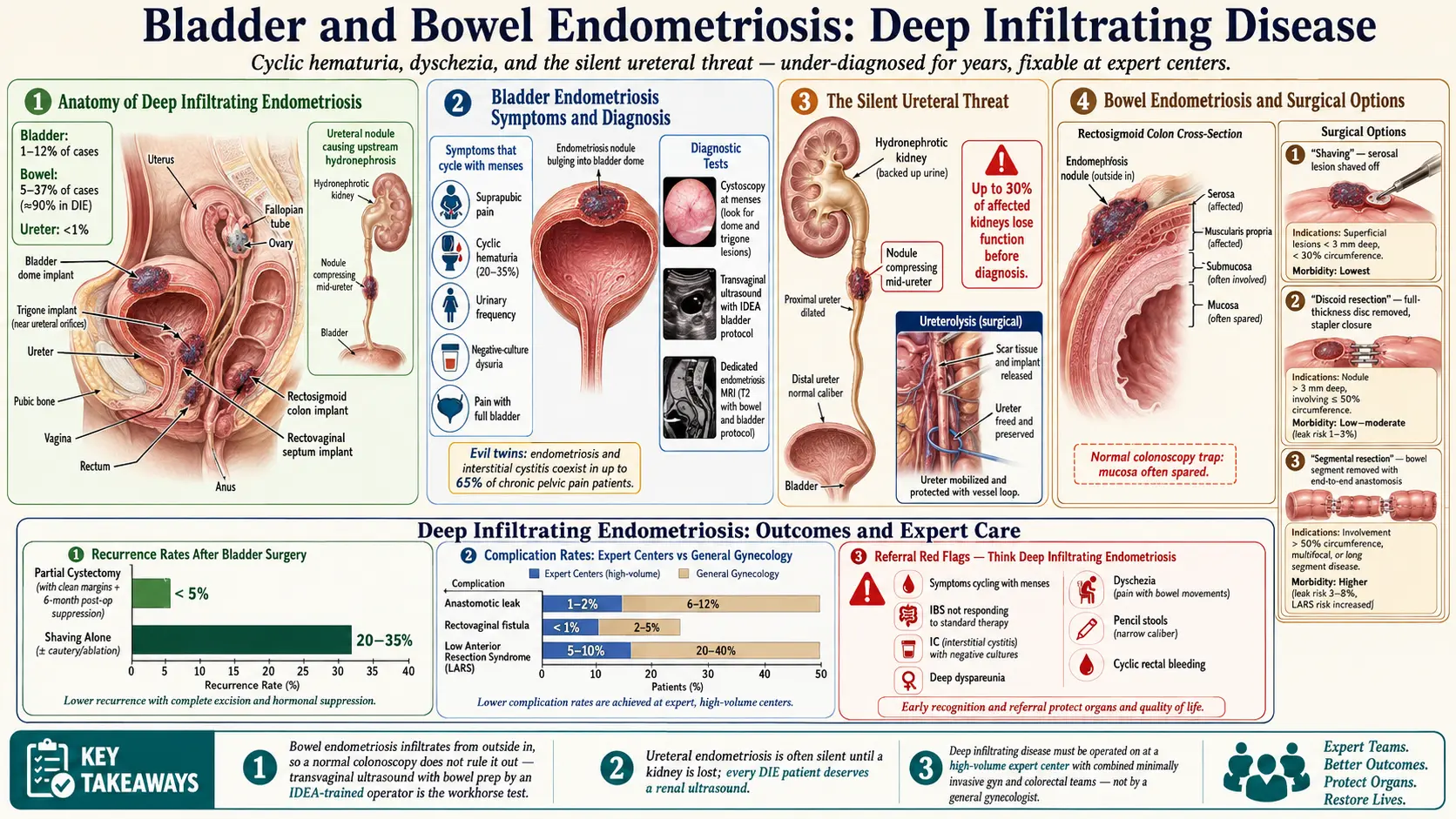
Endometriosis is not confined to the ovaries and peritoneum. In a meaningful minority of patients, the disease invades the wall of the bladder, the ureter, or the bowel — a form called deep infiltrating endometriosis (DIE). These lesions are under-recognized, often misdiagnosed for years as interstitial cystitis, irritable bowel syndrome, or "just bad periods," and they can do real structural damage. The good news: when the right imaging is done and the right surgeon operates, outcomes are generally excellent.
This article is for patients who suspect urinary or bowel involvement, have already been told they "just have irritable bowel syndrome (IBS)" or "just have interstitial cystitis (IC)," or are preparing for surgery and want to know what the options are and what to ask for.
Table of Contents
- How Common Bladder and Bowel Endometriosis Is
- Bladder Endometriosis — Symptoms
- Bladder Endometriosis — Diagnosis
- Bladder Endometriosis — Surgery
- The Silent Danger: Ureteral Endometriosis
- Bowel Endometriosis — Symptoms
- Bowel Endometriosis — Diagnosis
- Bowel Endometriosis — Surgical Options
- Hormonal Suppression, Nutrition, and Expert-Center Referral
- Key Research Papers
- PubMed Topic Searches
- Connections
- Featured Videos
How Common Bladder and Bowel Endometriosis Is
The numbers vary because the disease is chronically under-diagnosed, but published series give a consistent range:
- Bladder endometriosis: roughly 1–12% of endometriosis cases. It is by far the most common urinary-tract site — about 85% of all urinary endometriosis.
- Bowel endometriosis: a 2015 review estimated that deep nodules involve the bowel in 8–12% of women with endometriosis (specialist-referral centers report higher numbers than general gynecology clinics). In a Paris series of 241 women with surgically and microscopically confirmed deep infiltrating endometriosis, who had 344 deep lesions between them, 9.9% of the lesions were in the bowel wall and 6.4% in the bladder; most (69.2%) were in the uterosacral ligaments that hold the uterus in place.
- Ureteral endometriosis: less than 1% of cases, but disproportionately dangerous because it can quietly destroy a kidney.
Two facts worth internalizing: first, most urinary and bowel endometriosis is never diagnosed, because nobody images for it. Second, the average delay from first symptom to correct diagnosis for any form of endometriosis is 7–10 years — and the delay is often longer when the main complaint is urinary or gastrointestinal rather than pelvic pain, because patients get routed to urology or gastroenterology and the cyclic pattern gets missed.
Bladder Endometriosis — Symptoms
The single most important clue is that symptoms cycle with menses. A flare that lines up with your period almost every month is the signature of endometriosis anywhere in the body.
- Suprapubic pain (right above the pubic bone) that worsens in the days leading up to and during your period.
- Cyclic hematuria — visible or microscopic blood in the urine, appearing only around menstruation. This is the most specific sign, but it is present in only about 20–35% of cases. Its absence does not rule out bladder endometriosis.
- Urinary frequency and urgency, often severe enough to wake you at night.
- Dysuria (burning with urination) that is not explained by infection — urine cultures come back negative.
- Pain with a full bladder that eases after emptying.
This symptom cluster is almost identical to interstitial cystitis/bladder pain syndrome (IC/BPS), which is how bladder endometriosis so often gets missed — and why so many women accumulate years of failed IC treatments before anyone images the bladder for endometrial implants. To complicate matters further, the two conditions genuinely coexist: researchers dubbed endometriosis and IC the "evil twins" of chronic pelvic pain. In their first series of 60 women who had both a laparoscopy and a cystoscopy, 96.6% were diagnosed with IC and 93.3% with endometriosis; in a later prospective study of 178 women with chronic pelvic pain, both conditions were diagnosed in 115 women (65%). A 2024 meta-analysis found that the share of women with chronic pelvic pain who had both conditions ranged from 15.5% to 78.3% across studies — higher than IC in the general population, but the studies differ so much that no single figure is reliable. The study authors concluded that both diagnoses need to be considered together in chronic pelvic pain, so bladder pain that does not respond to standard IC treatment, or that flares with the menstrual cycle, is a recognized reason for an endometriosis work-up.
Bladder Endometriosis — Diagnosis
Getting the diagnosis right requires the right test, done by the right person, at the right time in your cycle.
Cystoscopy. A urologist threads a small camera into the bladder. In deep infiltrating bladder endometriosis, they may see a bluish or reddish nodule bulging into the bladder dome. But here is the trap: cystoscopy is often completely normal, because most bladder endometriosis sits in the outer muscle layer (detrusor) and has not yet eroded through the mucosa. A "normal cystoscopy" does not rule out the disease. Ideally cystoscopy is performed in the premenstrual or menstrual window when lesions are most engorged and visible.
Transvaginal ultrasound with bladder protocol. This is the workhorse test, but only in expert hands. The bladder is partially filled (about 50–100 mL), and the scan follows the IDEA (International Deep Endometriosis Analysis) consensus protocol for deep endometriosis, which the sonographer needs specific training to perform. A routine "pelvic ultrasound" is not the same thing. Its accuracy for bladder disease is lower than often assumed: a 2021 meta-analysis (8 studies, 1,052 women) found transvaginal ultrasound picked up 55% of bladder deep endometriosis (sensitivity) and correctly gave a clear result in 99% of women without it (specificity), and a 2026 meta-analysis of 36 studies found a sensitivity of 70% for ultrasound and 77% for magnetic resonance imaging, with specificity of 99–100% for both. In plain words, a normal scan does not rule bladder endometriosis out, but a scan that shows a bladder nodule is almost always right.
Magnetic resonance imaging (MRI) of the pelvis. A dedicated endometriosis magnetic resonance imaging protocol (with bowel and bladder preparation) is the other mainstay, and it maps not just the bladder but the uterus, ovaries, ureters, bowel, and pelvic fascia in one scan. If surgery is on the table, most expert centers want a magnetic resonance imaging for the operative map.
Urinalysis around menses. A simple urine dipstick or microscopy during your period can catch microscopic hematuria that is absent the rest of the month.
Bladder Endometriosis — Surgery
When a bladder nodule is symptomatic and confirmed on imaging, the gold-standard treatment is laparoscopic or robotic partial cystectomy — the surgeon cuts out the full-thickness disc of bladder wall containing the nodule and closes the bladder in two layers. A Foley catheter stays in place for 7–14 days post-op to let the suture line heal. Most patients go home the same day or the next morning and return to normal voiding within two weeks.
Location on the bladder matters a great deal:
- Bladder dome (the top) — the easy zone. Far from the ureters, far from the trigone. Partial cystectomy here is routine for experienced minimally invasive gynecologic surgeons.
- Trigone (the triangle at the base where the ureters enter) — far more technically demanding. Surgery risks ureteral injury, requires ureteral stenting, and is generally done at tertiary expert centers. Reimplantation of a ureter may be needed.
A 2022 systematic review of 28 surgical studies, mostly small case series, found bladder nodules ranged from 0.7 to 5.5 cm and sat most often on the back (posterior) wall of the bladder (63.57%). Most studies used partial cystectomy, and urinary symptoms came back in 7.34% of women after surgery. An earlier review of bladder endometriosis gave an overall recurrence rate of about 30% for surgery combined with hormonal therapy and about 35% for hormonal treatment alone. Surgeons describe a higher recurrence rate when only the visible part of the nodule is shaved off without full-thickness excision.
The Silent Danger: Ureteral Endometriosis
Of all the urinary-tract manifestations, ureteral endometriosis is the one that terrifies specialists, because it is often completely silent until a kidney is already lost. Implants on or around the ureter slowly compress or infiltrate it, urine backs up, the kidney swells (hydronephrosis), and over months to years renal function fades. Some women present with vague flank pain; many have no symptoms at all until imaging for another reason picks up a hydronephrotic kidney. A 2018 systematic review found that ureteral involvement "is often asymptomatic or leads to non-specific symptoms", and that when the diagnosis is delayed it may lead to persistent hydronephrosis and eventually loss of kidney function. A 2012 report described three women in whom ureteral endometriosis had already caused kidney shrinkage (atrophy) and loss of function by the time it was found.
This is why specialists check the ureters and kidneys in women with deep infiltrating endometriosis — especially with endometriomas, bowel involvement, or a palpable nodule. A renal ultrasound is cheap, non-invasive, and can be done at the same visit as the pelvic scan. Dedicated endometriosis magnetic resonance imaging protocols include the ureters. The 2018 review names ultrasound as the first-line test and magnetic resonance imaging as the scan that shows the type of ureteral involvement. If hydronephrosis is found, ureterolysis (freeing the ureter from scar tissue) or, in severe cases, segmental resection with reimplantation is performed — and timing matters, because renal function lost is not recovered.
Bowel Endometriosis — Symptoms
The rectosigmoid colon (the S-shaped last stretch of colon before the rectum) is the most common bowel site by a wide margin, followed by the rectum itself, then the appendix, cecum, and terminal ileum. Because the rectosigmoid sits right behind the uterus and cervix, endometriosis implants from the pouch of Douglas can invade it directly.
The symptom list overlaps heavily with irritable bowel syndrome, which is why women with bowel endometriosis are frequently told for years that they have irritable bowel syndrome:
- Dyschezia — painful bowel movements, especially during menstruation. Often described as a stabbing or "knife-in-the-rectum" sensation.
- Cyclic rectal bleeding — blood in or on the stool, only around your period. Less common than dyschezia but highly specific.
- Alternating constipation and diarrhea that cycles with menses — constipated in the luteal phase, loose during bleeding, for example.
- Small-caliber or "pencil" stools — a narrowed rectosigmoid lumen from an extrinsic nodule.
- Severe bloating — the abdomen distends dramatically, sometimes looking several months pregnant by evening. Commonly called "endo belly." See the Endo Belly article.
- Tenesmus — the constant feeling of needing to defecate even after you just have.
- Deep dyspareunia — pain with deep vaginal penetration, because the nodule sits in the rectovaginal septum.
Two things to note. First, irritable bowel syndrome and endometriosis coexist at far above chance rates — estimates run 30–50% overlap — so having one does not protect you from the other. Second, if "irritable bowel syndrome" symptoms cycle with your period, it is endometriosis until proven otherwise. See the irritable bowel syndrome page for how the two distinguish from each other.
Bowel Endometriosis — Diagnosis
Colonoscopy is usually normal, and that is a trap. Bowel endometriosis typically infiltrates from the outside in — it starts on the serosa and burrows into the muscularis propria, usually stopping before it reaches the mucosa. Because colonoscopy only sees the mucosa, the implants are invisible from inside the bowel. A completely clean colonoscopy does not rule out bowel endometriosis. Colonoscopy is still worth doing to rule out other diseases (inflammatory bowel disease, cancer), but it is not the diagnostic test.
The real tests are:
- Transvaginal ultrasound with bowel prep performed by a sonographer trained in deep endometriosis imaging. A 2018 meta-analysis (6 studies, 424 women) found it detected 85% of rectosigmoid deep endometriosis (sensitivity) with a specificity of 96%, about the same as magnetic resonance imaging (85% and 95%). It is non-invasive and inexpensive, and is commonly used as the first imaging test.
- Magnetic resonance imaging of the pelvis with bowel and rectal preparation, which maps the depth of infiltration (serosa, muscularis, submucosa) and the length of bowel involved — both critical for surgical planning.
- Rectal endoscopic ultrasound in select cases where depth of infiltration is unclear.
The same principle applies as with the bladder: a "routine" pelvic ultrasound at a general imaging center is not the same test as a deep-endometriosis protocol done by a trained operator. The IDEA protocol is the published consensus method for this scan; an ultrasound done without it is not the same test.
Bowel Endometriosis — Surgical Options
Three surgical techniques exist, with different complication rates. A 2021 meta-analysis of 60 studies found a mean complication rate of 2.2% after shaving, 9.7% after disc excision and 9.9% after segmental resection; shaving was less often linked to rectovaginal fistula and to leakage than disc excision, while disc excision and segmental resection did not differ in fistula rates. The choice depends on the depth of infiltration, size of the nodule, and percentage of bowel circumference involved.
- Shaving. The surgeon carefully shaves the nodule off the bowel wall without entering the lumen. Suitable for superficial disease limited to the serosa and outer muscularis. Lowest morbidity; preserves the bowel; recurrence risk is higher if the nodule extends deeper than it appeared.
- Discoid resection. A full-thickness disc of bowel wall containing the nodule is cut out and the defect is closed, usually with a circular stapler. Appropriate for nodules under about 3 cm involving less than one-third of the bowel circumference. Its overall complication rate was about the same as segmental resection in the 2021 meta-analysis, though it was less often followed by narrowing of the joined bowel (anastomotic stenosis).
- Segmental resection. A length of bowel containing the nodule is removed entirely and the two ends are rejoined (anastomosis). Reserved for large nodules (>3 cm), multifocal disease, or involvement of more than a third of the circumference. Complications include anastomotic leak (1–3%), rectovaginal fistula, low anterior resection syndrome (bowel-habit disturbance that can persist), and a small risk of needing a temporary or permanent ostomy.
Functional outcomes after expert bowel endometriosis surgery are generally excellent — most patients report major improvement in pain, dyschezia, and quality of life — but complications are real. Specialist reviews describe a combined team of a minimally invasive gynecologic surgeon and a colorectal surgeon working together at a high-volume center as the standard of care, rather than a general gynecologist or general surgeon operating alone.
Hormonal Suppression, Nutrition, and Expert-Center Referral
Pre-operative hormonal suppression. Some surgeons place patients on 2–3 months of a gonadotropin-releasing hormone (GnRH) antagonist (elagolix, relugolix combination therapy) or continuous progestin (dienogest, norethindrone acetate) before surgery, with the aim of shrinking lesions, reducing inflammation, and making the operation technically easier. A 2017 review of urinary tract endometriosis describes hormonal therapy as a temporary measure that can be useful before surgery. The 2022 European Society of Human Reproduction and Embryology (ESHRE) guideline, however, states that "It is not recommended to prescribe preoperative hormone treatment to improve the immediate outcome of surgery for pain in women with endometriosis" (a strong recommendation). See the hormonal therapy page for how each class works.
Post-operative suppression. After excision of deep infiltrating disease, continuous progestin or a GnRH antagonist for at least 6–12 months is widely used to reduce recurrence. The ESHRE 2022 guideline states that women not seeking immediate pregnancy "may be offered" postoperative hormone treatment, and that for preventing recurrence of deep endometriosis "long-term administration of postoperative hormone treatment can be considered" (both weak recommendations). Combined oral contraceptives used cyclically are generally not adequate for DIE — continuous or progestin-only regimens suppress more completely.
Relugolix combination therapy in trials. Two placebo-controlled trials (SPIRIT 1 and 2, with 638 and 623 women with endometriosis pain) tested a daily tablet of relugolix 40 mg with estradiol 1 mg and norethisterone acetate 0.5 mg for 24 weeks. Period pain met the trials’ response criteria in 75% of women on the tablet in both trials, against 27% and 30% on placebo; pelvic pain between periods responded in 58% and 66%, against 40% and 43%. Bone density at the lower spine fell by 0.70% and 0.78%, against small rises of 0.21% and 0.02% on placebo. The most common side effects were headache, common colds (nasopharyngitis) and hot flushes.
Nutrition around surgery. Bowel surgery in particular benefits from prehabilitation: adequate protein (1.2–1.5 g/kg/day), iron repletion if deficient, vitamin D optimization, and reducing inflammatory dietary triggers for the 4–8 weeks before the operation. Post-operatively, a low-residue diet for the first 2–4 weeks gives the anastomosis time to heal. See the diet and lifestyle page for longer-term nutritional support.
Expert-center referral. The ESHRE 2022 guideline recommends that women with deep endometriosis are referred to a centre of expertise. Outcomes at high-volume endometriosis centers — places doing hundreds of these operations a year, with dedicated radiology, colorectal, urology, and pelvic floor physical therapy teams — are described as dramatically better than community outcomes. Questions patients put to a surgeon include how many of these operations they do a year, what their complication rate is, and whether they can share their data.
In the U.S., one list patients use is the AAGL-accredited Centers of Excellence in Minimally Invasive Gynecology (COEMIG) list (AAGL was founded as the American Association of Gynecologic Laparoscopists); patient organizations such as Endometriosis.org and patient-run online groups also share information about surgeons. Insurance coverage of out-of-network expert centers is sometimes negotiable, particularly when local treatment failures are documented, and an appeal may be needed.
Key Research Papers
- Chapron C, et al. Anatomical distribution of deeply infiltrating endometriosis: surgical implications and proposition for a classification. Hum Reprod. 2003. — doi:10.1093/humrep/deg009
- Abrao MS, et al. Deep endometriosis infiltrating the recto-sigmoid: critical factors to consider before management. Hum Reprod Update. 2015. — doi:10.1093/humupd/dmv003
- Maccagnano C, et al. Diagnosis and treatment of bladder endometriosis: state of the art. Urol Int. 2012;89(3):249-58. — doi:10.1159/000339519
- Guerriero S, et al. Systematic approach to sonographic evaluation of the pelvis in women with suspected endometriosis, including terms, definitions and measurements: a consensus opinion from the IDEA group. Ultrasound Obstet Gynecol. 2016. — doi:10.1002/uog.15955
- Nezhat C, Falik R, McKinney S, King LP. Pathophysiology and management of urinary tract endometriosis. Nat Rev Urol. 2017;14(6):359-372. — doi:10.1038/nrurol.2017.58
- Chung MK, Chung RR, Gordon D, Jennings C. The evil twins of chronic pelvic pain syndrome: endometriosis and interstitial cystitis. JSLS. 2002;6(4):311-4. — PubMed PMID: 12500828
- Chung MK, Chung RP, Gordon D. Interstitial cystitis and endometriosis in patients with chronic pelvic pain: the "Evil Twins" syndrome. JSLS. 2005;9(1):25-9. — PubMed PMID: 15791965
- Inzoli A, et al. The Evil Twins of Chronic Pelvic Pain Syndrome: A Systematic Review and Meta-Analysis on Interstitial Cystitis/Painful Bladder Syndrome and Endometriosis. Healthcare (Basel). 2024;12(23):2403. — doi:10.3390/healthcare12232403
- Gerges B, et al. Meta-analysis and systematic review to determine the optimal imaging modality for the detection of bladder deep endometriosis. Eur J Obstet Gynecol Reprod Biol. 2021;261:124-133. — doi:10.1016/j.ejogrb.2021.04.030
- Pitsillidi A, et al. Diagnostic performance of MRI and transvaginal ultrasound for urinary tract endometriosis: a systematic review and meta-analysis. Arch Gynecol Obstet. 2026;313(1):307. — doi:10.1007/s00404-026-08553-6
- Guerriero S, et al. Transvaginal ultrasound vs magnetic resonance imaging for diagnosing deep infiltrating endometriosis: systematic review and meta-analysis. Ultrasound Obstet Gynecol. 2018;51(5):586-595. — doi:10.1002/uog.18961
- Tomasi MC, et al. Symptoms and Surgical Technique of Bladder Endometriosis: A Systematic Review. J Minim Invasive Gynecol. 2022;29(12):1294-1302. — doi:10.1016/j.jmig.2022.10.003
- Barra F, et al. Ureteral endometriosis: a systematic review of epidemiology, pathogenesis, diagnosis, treatment, risk of malignant transformation and fertility. Hum Reprod Update. 2018;24(6):710-730. — doi:10.1093/humupd/dmy027
- Nezhat C, Paka C, Gomaa M, Schipper E. Silent loss of kidney seconary to ureteral endometriosis. JSLS. 2012;16(3):451-5. — doi:10.4293/108680812X13462882736213
- Bendifallah S, et al. Surgical Outcomes after Colorectal Surgery for Endometriosis: A Systematic Review and Meta-analysis. J Minim Invasive Gynecol. 2021;28(3):453-466. — doi:10.1016/j.jmig.2020.08.015
- Becker CM, et al. ESHRE guideline: endometriosis. Hum Reprod Open. 2022;2022(2):hoac009. — doi:10.1093/hropen/hoac009
- Giudice LC, et al. Once daily oral relugolix combination therapy versus placebo in patients with endometriosis-associated pain: two replicate phase 3, randomised, double-blind, studies (SPIRIT 1 and 2). Lancet. 2022;399(10343):2267-2279. — doi:10.1016/S0140-6736(22)00622-5
PubMed Topic Searches
For deeper reading, the following PubMed searches return current peer-reviewed literature on urinary- and bowel-tract endometriosis:
- Bladder endometriosis
- Ureteral endometriosis and hydronephrosis
- Bowel endometriosis and rectosigmoid disease
- Deep infiltrating endometriosis and MRI imaging
- Transvaginal ultrasound and the IDEA protocol
- Segmental resection outcomes for bowel endometriosis
- Shaving versus discoid resection for bowel endometriosis
- Interstitial cystitis and endometriosis overlap
- Irritable bowel syndrome and endometriosis
- Partial cystectomy for bladder endometriosis
Connections
- Reproductive Medicine
- Diet and Lifestyle
- Irritable Bowel Syndrome
- Hormonal Therapy Options
- Endo Belly and Gut Connection
- Endometriosis
- Laparoscopy: Excision vs Ablation
- Diagnosis Delay and Imaging
- Pelvic Floor Therapy and Central Sensitization
- Fertility and Endometriosis
- Adenomyosis Overlap
- Interstitial Cystitis
- Full Body Magnetic Resonance Imaging
- Urinalysis
- Constipation
- Bloating
- Inflammatory Bowel Disease
- Iron