Restless Legs Syndrome (Willis-Ekbom Disease)
Overview
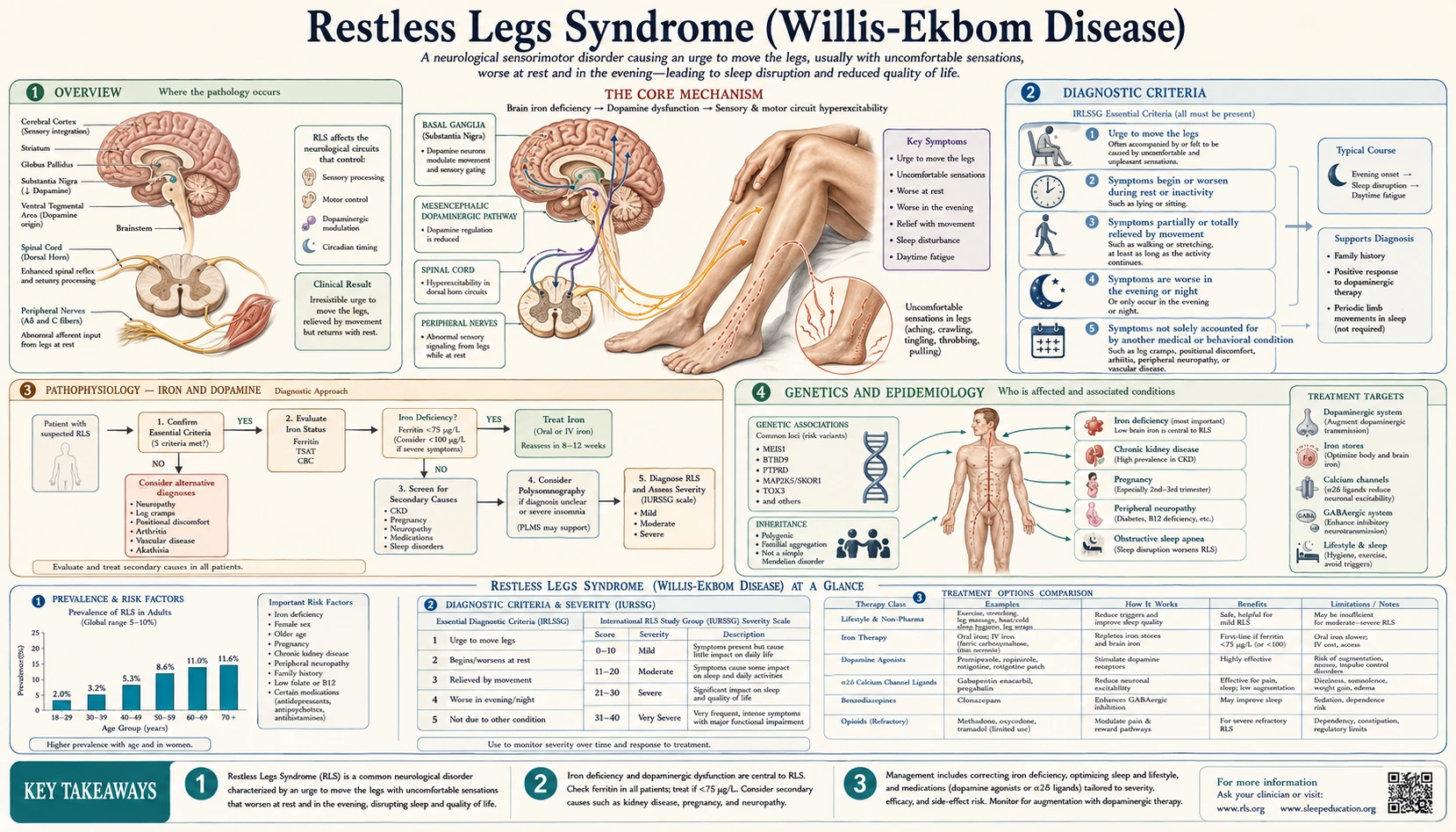
Restless Legs Syndrome (RLS), now formally called Willis-Ekbom Disease, is a neurological sensorimotor disorder characterized by an uncomfortable urge to move the legs that worsens at rest and in the evening. It affects 5–15% of adults in Western populations, making it one of the most common sleep and neurological disorders — yet it is frequently misdiagnosed or dismissed by clinicians who are unfamiliar with its distinct clinical signature. RLS can range from mild inconvenience to a severely disabling condition causing profound insomnia and reduced quality of life.
The condition was formally described by neurologist Karl-Axel Ekbom in 1945, building on earlier observations by Thomas Willis in 1685 — hence the dual eponym. Despite this centuries-long recognition, widespread clinical awareness remains inconsistent, and many patients suffer for years before receiving a correct diagnosis and appropriate treatment.
Diagnostic Criteria
The International Restless Legs Syndrome Study Group (IRLSSG) 2012 revised criteria require ALL FIVE of the following:
- Urge to move the legs — usually accompanied by uncomfortable sensations in the legs (sometimes described as crawling, creeping, pulling, throbbing, aching, itching, or electric). The discomfort is deep inside the limb, not on the skin surface.
- Worsening at rest — symptoms begin or worsen during periods of inactivity (sitting, lying down). This is what makes sleep impossible and sitting through movies or long flights torturous.
- Relief by movement — partial or complete relief when moving the legs (walking, stretching, rubbing). The relief lasts only as long as movement continues.
- Circadian pattern — symptoms are worse in the evening and night compared to daytime. This is the hallmark that distinguishes RLS from peripheral neuropathy, arthritis, and other mimics.
- Not solely accounted for by another medical or behavioral condition — excludes leg cramps, positional discomfort, myalgia, venous stasis, leg edema, arthritis, and habitual foot tapping.
Supportive clinical features (not required but strengthen diagnosis): family history, dopaminergic response (improvement with a low-dose dopamine agonist confirms diagnosis therapeutically), and periodic limb movements of sleep (PLMS) on polysomnogram.
Pathophysiology — Iron and Dopamine
The central mechanism linking the two main pathophysiological threads of RLS is iron deficiency in the brain, particularly in the substantia nigra.
Iron's Role in Dopamine Synthesis
Iron is an essential cofactor for tyrosine hydroxylase, the rate-limiting enzyme that converts tyrosine to L-DOPA and ultimately dopamine. When brain iron stores are insufficient, dopaminergic function in the striatal pathways governing sensorimotor integration is impaired. This produces the classic RLS symptoms: the sensory discomfort, the urge to move, and the failure of normal inhibitory gating during rest.
The Brain-Serum Iron Disconnect
Critically, brain iron deficiency can exist even when serum ferritin appears low-normal. The blood-brain barrier has a saturable iron transport system. Studies of post-mortem RLS brains show decreased iron in the substantia nigra despite normal peripheral iron stores in some patients. This is why serum ferritin cutoffs matter even in the “normal” laboratory range — a ferritin of 40 μg/L may be clinically normal for a hematologist but inadequate for optimal dopamine function in a susceptible individual.
Circadian Dopamine Variation
Both dopamine and iron metabolism have circadian rhythmicity that peaks during daytime and troughs at night. This biological rhythm directly explains the characteristic evening and nighttime worsening of RLS symptoms. The dopamine system's natural nighttime ebb unmasks the underlying sensorimotor dysregulation that is held in check during the day.
Opioid System Involvement
The endogenous opioid system modulates sensorimotor gating and appears dysregulated in RLS. This explains why low-dose opioids are effective in refractory cases and why some patients report worsening with opioid antagonists. PET imaging studies have demonstrated altered opioid receptor availability in RLS patients, suggesting this is a distinct neurobiological feature rather than a side effect of treatment.
Genetics and Epidemiology
RLS has a strong genetic component. Six susceptibility loci have been replicated in genome-wide association studies:
- BTBD9 (chromosome 6p21): The strongest common-variant signal; also independently associated with PLMS. The protein product is involved in ubiquitination and protein quality control.
- MEIS1 (chromosome 2p14): A homeobox transcription factor that regulates iron metabolism and limb development. Functional studies implicate it in dopaminergic neuron patterning.
- MAP2K5/SKOR1 (chromosome 15q23): Involved in MAPK signaling and neuronal development.
- PTPRD (chromosome 9p24): A receptor tyrosine phosphatase expressed in the central nervous system; also associated with restless sleep in children.
- TOX3 (chromosome 16q12): A transcription factor previously associated with breast cancer risk; its role in RLS neurobiology is incompletely understood.
- MEIS2 (chromosome 15q14): A paralog of MEIS1 with overlapping developmental roles.
Epidemiology
- Prevalence 5–15% in North American and European adults; consistently lower in Asian populations (approximately 1–3%), suggesting genuine genetic-ethnic differences beyond diagnostic variation.
- Women are approximately twice as likely as men to be affected — partly explained by iron loss through menstruation and the RLS risk conferred by pregnancy.
- Prevalence increases with age, though RLS can present at any age, including childhood, where it is often misdiagnosed as growing pains, attention deficit disorder, or anxiety.
- Strong familial aggregation: first-degree relatives have a 3–5 times higher risk. Earlier age of onset is associated with stronger family history and a more genetic (rather than secondary/acquired) form of the disease.
Secondary Causes
Several medical conditions cause or significantly worsen RLS. Identifying and treating secondary causes can dramatically reduce — or fully resolve — symptoms.
Iron Deficiency (Most Treatable Cause)
Any cause of iron deficiency — including GI bleeding, inadequate dietary intake, malabsorption, menorrhagia, or frequent blood donation — can trigger or worsen RLS. Serum ferritin below 75 μg/L correlates with increased RLS severity, and iron supplementation often substantially reduces symptoms. This is the first thing to assess and treat in every RLS patient.
Chronic Kidney Disease
The most important secondary cause after iron deficiency. Uremia impairs dopamine metabolism and iron utilization simultaneously. Hemodialysis patients have RLS prevalence of 25–50%. RLS in CKD responds to iron repletion, erythropoietin therapy, and dopaminergic medications, though management is complicated by renal constraints on drug dosing and the dialysis schedule.
Pregnancy
RLS emerges or dramatically worsens in the third trimester in 20–25% of pregnant women. Contributing factors include iron and folate deficiency, hormonal changes (particularly elevated estrogen and progesterone), and mechanical factors. Symptoms typically resolve within weeks of delivery. Management in pregnancy is limited because most RLS medications lack pregnancy safety data: iron supplementation, folate repletion, and non-pharmacologic measures are the foundation.
Peripheral Neuropathy
Particularly small-fiber neuropathy. The mechanism is independent of central dopamine pathways — peripheral sensitization of nociceptors drives the sensory discomfort. Workup for underlying cause is essential: diabetes, vitamin B12 deficiency, monoclonal gammopathy of undetermined significance (MGUS), hereditary neuropathy, and autoimmune neuropathy.
Spinal Cord Disorders
Multiple sclerosis, spinal cord injury, lumbar spinal stenosis, and myelopathy can all produce or mimic RLS through disruption of the descending inhibitory pathways that normally suppress sensorimotor signals during rest.
Medications That Worsen RLS
This is clinically critical — many commonly prescribed medications dramatically exacerbate RLS:
- Dopamine antagonists: antipsychotics (haloperidol, risperidone, quetiapine, olanzapine), antiemetics (metoclopramide, prochlorperazine, promethazine). These are potent RLS triggers — any patient whose RLS began or worsened after starting one of these should have the medication reconsidered.
- Antihistamines: diphenhydramine (Benadryl) — an extremely common and underrecognized trigger. Present in most over-the-counter sleep aids (ZzzQuil, Unisom, Tylenol PM, Benadryl). Patients with RLS must avoid all antihistamine sleep aids.
- Antidepressants: mirtazapine is the strongest aggravator (5-HT2C and histamine antagonism). SSRIs cause moderate worsening. TCAs cause mild worsening. Notable exceptions that do NOT worsen RLS: bupropion (may actually improve RLS through dopamine reuptake inhibition); venlafaxine (variable — mild worsening in some patients).
- Lithium: can worsen or trigger RLS through unclear mechanisms.
Periodic Limb Movements of Sleep (PLMS)
PLMS are repetitive, stereotyped limb movements during sleep, documented on polysomnography. They occur in clusters every 20–40 seconds and typically involve extension of the big toe, dorsiflexion of the ankle, and sometimes flexion at the knee and hip — a pattern reminiscent of the Babinski reflex. Each movement lasts 0.5–10 seconds.
Key Distinctions
- PLMS are present in 80–90% of RLS patients but are also common in other sleep disorders (narcolepsy, REM sleep behavior disorder, obstructive sleep apnea) and in healthy older adults without RLS.
- PLMS are not required for RLS diagnosis — clinical history alone is sufficient when the 5 IRLSSG criteria are met.
- PLMS can disturb sleep architecture independently by causing cortical arousals (detectable on EEG as brief wakings, even without the patient's awareness), reducing deep sleep and overall sleep quality — even in individuals who do not have classic daytime RLS symptoms.
- Isolated PLMS without RLS symptoms is called “Periodic Limb Movement Disorder” (PLMD) — a separate diagnosis of exclusion requiring ruling out RLS, obstructive sleep apnea, and other causes of PLMS.
- A PLMS index greater than 15 events per hour of sleep is considered clinically significant. PLMS associated with arousals (PLMS-arousal index) is the more clinically relevant measure for sleep quality impact.
Diagnosis
Clinical Diagnosis (Primary)
RLS is fundamentally a clinical diagnosis based on the five IRLSSG criteria. A careful history asking specifically about the four cardinal features — urge to move, rest worsening, relief by movement, and circadian (evening/night) pattern — makes the diagnosis in most cases. The key is knowing to ask: patients rarely volunteer these symptoms in standard medical history-taking, and providers rarely think to ask.
IRLSSG Severity Scale
The validated 10-item International RLS Study Group Rating Scale (IRLS) quantifies symptom severity on a 0–40 scale: mild (1–10), moderate (11–20), severe (21–30), very severe (31–40). It is used in clinical trials and as a practical tool to track treatment response over time. Ask patients to complete it before and after therapeutic interventions.
Laboratory Testing
- Serum ferritin (not just serum iron or hemoglobin): Target above 75 μg/L for RLS. Below 50 μg/L is clearly inadequate. Below 75 μg/L warrants supplementation even if hemoglobin is normal — iron deficiency can be present without anemia.
- Transferrin saturation (TSAT): Assesses iron utilization; below 20% suggests functional iron deficiency.
- Complete blood count: Identify frank anemia and macrocytosis (B12/folate deficiency).
- Serum iron and TIBC: Complement ferritin for a complete picture of iron status.
- Comprehensive metabolic panel: Evaluate kidney function (creatinine, BUN, estimated GFR) to identify CKD as a secondary cause.
- HbA1c: Screen for diabetes-associated peripheral neuropathy.
- B12 and folate: Deficiency causes neuropathy that may mimic or exacerbate RLS; folate deficiency is particularly important in pregnancy.
- Peripheral neuropathy workup if clinically indicated: nerve conduction studies and electromyography to identify large-fiber neuropathy; skin punch biopsy for small-fiber neuropathy (intraepidermal nerve fiber density).
Polysomnography
Not required for diagnosis but indicated when: the diagnosis is uncertain and objective sleep data is needed; a coexisting sleep disorder (particularly obstructive sleep apnea) is suspected; PLMS documentation and severity quantification is needed; or treatment evaluation requires objective data. PSG demonstrates PLMS and can identify sleep-stage distribution abnormalities.
Actigraphy
Wrist or ankle actigraphy may help document limb movements in the home setting over multiple nights, providing a more representative sample than a single-night lab PSG. Less definitive than PSG but useful for monitoring treatment response over time.
Treatment — Non-Pharmacologic and Iron
Non-Pharmacologic Approaches (First-Line for Mild RLS)
For patients with mild RLS (IRLS score 1–10) or who prefer to avoid medications, behavioral and lifestyle interventions can provide meaningful benefit:
- Sleep hygiene: Consistent sleep schedule, cool bedroom temperature, avoiding screens in the evening (blue light suppresses melatonin and may amplify circadian symptom peaks).
- Moderate regular exercise: Particularly walking and stretching in the morning or early afternoon. Vigorous exercise close to bedtime can paradoxically worsen symptoms — timing matters.
- Avoid triggers: Alcohol (worsens RLS despite initial sedation), caffeine in the afternoon and evening, antihistamines (Benadryl and OTC sleep aids), and all medications listed in the secondary causes section above.
- Mental engagement: Mentally stimulating activities — reading, video games, puzzles, engaging conversation — can suppress mild symptoms during sedentary periods by competing for attentional resources. This is a practical strategy for managing symptoms during travel or long events.
- Hot bath or leg massage at bedtime: Temporary sensory stimulation provides relief in some patients; effects are short-lived but can help initiate sleep.
- Pneumatic compression devices: FDA-cleared devices providing repetitive sequential pressure to the legs have randomized trial support and are an evidence-based non-pharmacologic option for patients who prefer to avoid medications or when medications are contraindicated (pregnancy).
Iron Supplementation
Oral iron: Ferrous sulfate 325 mg (65 mg elemental iron) taken with vitamin C (250–500 mg) on an empty stomach to maximize absorption. Alternate-day dosing — taking iron every other day rather than daily — has been shown in randomized studies to produce higher net absorption by allowing the intestinal mucosal iron transporter (ferroportin) to recover between doses. Target serum ferritin above 75 μg/L. Takes 3–6 months of sustained supplementation to see full RLS benefit. GI side effects (constipation, nausea, dark stools) limit adherence — bisglycinate forms may be better tolerated.
Intravenous iron: Indicated when serum ferritin is below 100 μg/L with oral iron intolerance or inadequate response; in CKD (where oral absorption is impaired and GI tolerance is worse); in malabsorption syndromes (celiac disease, gastric bypass); or when a faster therapeutic response is needed. Ferric carboxymaltose (1000 mg in a single infusion) and ferumoxytol have both demonstrated efficacy for RLS in randomized controlled trials. Target ferritin with IV iron replacement is above 100–150 μg/L. Response can be dramatic — significant symptom reduction within 2–4 weeks in iron-responsive cases.
Treatment — Pharmacologic
Alpha-2-Delta Calcium Channel Ligands (First-Line Pharmacotherapy)
Current guidelines from the American Academy of Sleep Medicine and the Mayo Clinic updated algorithm recommend alpha-2-delta ligands as first-line pharmacotherapy for moderate-to-severe RLS, superseding dopamine agonists due to the augmentation problem.
Gabapentin enacarbil (Horizant): The only medication with an FDA indication specifically for moderate-to-severe primary RLS. It is a prodrug of gabapentin designed for improved and more predictable GI absorption via active transporter-mediated uptake. Standard dose: 600 mg once daily at approximately 5 pm. Benefits: treats both RLS symptoms and comorbid restless sleep, pain, and anxiety; no risk of augmentation; once-daily dosing improves adherence. Principal side effects: dizziness and somnolence, particularly when starting.
Pregabalin: Not FDA-approved specifically for RLS but extensively studied in high-quality randomized trials and widely used off-label. Dose range 75–300 mg per night, titrated from a low starting dose. A landmark 52-week head-to-head trial (Trenkwalder et al., Lancet 2013) demonstrated that pregabalin provided sustained RLS symptom control with significantly lower augmentation risk compared to pramipexole — establishing alpha-2-delta ligands as the preferred first-line class for long-term management.
Gabapentin: The parent compound; lowest bioavailability and most variable absorption of the three (requires higher doses, typically 900–2400 mg per night split into two or three doses). Effective but less convenient. Least expensive option, which matters for some patients.
Dopamine Agonists (Effective but Second-Line Due to Augmentation)
Dopamine agonists were the dominant RLS treatment for two decades and remain highly effective short-term. However, augmentation — a paradoxical treatment-induced worsening of RLS — has moved them to second-line status for most patients.
Pramipexole (Mirapex): Dose 0.125–0.75 mg taken 1–3 hours before bedtime. Rapid and substantial relief in most patients.
Ropinirole (Requip): Dose 0.25–4 mg taken 1–3 hours before bedtime. Similar efficacy to pramipexole.
Augmentation: This is the critical long-term problem with dopamine agonists. Augmentation affects up to 50% of patients on chronic therapy and manifests as: symptoms appearing earlier in the day (afternoon or even morning); spread of symptoms to the arms, trunk, or face; shorter latency before symptoms appear with rest; more severe symptoms at onset; reduced duration of relief from each dose. Augmentation is not tolerance — it is an actual worsening of the underlying condition driven by dopaminergic overstimulation. Managing established augmentation typically requires opioid therapy, making it a serious long-term complication.
Rotigotine transdermal patch (Neupro): Provides continuous (non-pulsatile) dopaminergic stimulation via a 24-hour patch. Lower augmentation risk than pulsatile oral dopamine agonists, though augmentation still occurs. FDA-approved for RLS at doses of 1–3 mg/24 hours. Particularly useful for patients with early-morning symptom breakthrough.
If dopamine agonists are used: employ the lowest effective dose, monitor closely for augmentation at every visit, have a clear exit plan, and inform patients that switching to alpha-2-delta ligands may be necessary.
Low-Dose Opioids (Refractory RLS)
For patients who fail alpha-2-delta ligands and dopamine agonists, or who have developed augmentation on chronic dopamine agonist therapy, low-dose opioids are an evidence-based and often dramatically effective option. The endogenous opioid system's role in RLS pathophysiology makes this mechanistically sound, not merely empirical.
- Oxycodone/naloxone extended-release: The only opioid formulation studied in a rigorous randomized controlled trial for RLS (Trenkwalder et al. 2016). The naloxone component reduces constipation and abuse potential while not crossing the blood-brain barrier at standard doses. Doses of 5–20 mg oxycodone equivalent nightly.
- Tramadol: Weak mu-opioid agonist plus serotonin-norepinephrine reuptake inhibitor; 50–100 mg at bedtime. Useful for milder refractory cases.
- Oxycodone or hydrocodone: 5–10 mg nightly for more severe refractory cases.
- Methadone: Extremely effective for very severe, refractory RLS — particularly after augmentation on dopamine agonists — due to its long half-life providing continuous opioid coverage. Requires specialist management; doses of 5–15 mg daily. Reserved for cases where other treatments have failed.
Benzodiazepines
Clonazepam is sometimes used as an adjunct for PLMS-associated sleep disruption in patients who cannot tolerate other treatments, but it has no specific RLS mechanism (does not reduce the urge to move), carries dependency risk, worsens sleep apnea, and causes residual morning sedation. It is not preferred as a primary RLS treatment by any major guideline.
Key Research Papers
- Allen RP, et al. (2003). Restless legs syndrome: Diagnostic criteria, special considerations, and epidemiology. Sleep Medicine, 4(2), 101–119. PMID 14592341
- Earley CJ, et al. (2000). Abnormalities in CSF concentrations of ferritin and transferrin in restless legs syndrome. Neurology, 54(8), 1698–1700. — Search PubMed
- Winkelmann J, et al. (2007). Genome-wide association study of restless legs syndrome identifies common variants in three genomic regions. Nature Genetics, 39(8), 1000–1006. PMID 17637780
- Trenkwalder C, et al. (2013). Pregabalin versus pramipexole: Results from a double-blind, randomised trial. Lancet, 383(9925), 1302–1310. — Search PubMed
- Allen RP, et al. (2014). Restless legs syndrome/Willis-Ekbom disease diagnostic criteria: Updated International Restless Legs Syndrome Study Group (IRLSSG) consensus criteria. Sleep Medicine, 15(8), 860–873. PMID 25023924
- Oertel W, et al. (2011). Long-term safety and efficacy of rotigotine transdermal patch for moderate-to-severe idiopathic restless legs syndrome. Sleep Medicine, 12(10), 990–994. — Search PubMed
- Silber MH, et al. (2021). The management of restless legs syndrome: An updated algorithm. Mayo Clinic Proceedings, 96(7), 1921–1937. PMID 34218864
- Ferini-Strambi L, et al. (2014). Pharmacological treatment for restless legs syndrome. Expert Opinion on Pharmacotherapy, 15(8), 1127–1138. — Search PubMed
- Winkelman JW, et al. (2016). Restless legs syndrome: Therapeutics. Annals of Neurology, 80(4), 518–530. — Search PubMed
- Trenkwalder C, et al. (2016). Oxycodone-naloxone is effective in the long-term treatment of severe restless legs syndrome. Sleep Medicine, 21, 33–40. — Search PubMed
- Earley CJ, et al. (2015). Intravenous ferric carboxymaltose in restless legs syndrome. Sleep Medicine, 16(11), 1340–1345. — Search PubMed
- Picchietti DL, et al. (2013). Pediatric restless legs syndrome diagnostic criteria: An update by the International Restless Legs Syndrome Study Group. Sleep Medicine, 14(12), 1253–1259. PMID 24184054
Connections
- Neurology
- Dopamine, Reward & How Habits Hijack You — interactive animation
- Narcolepsy
- Circadian Rhythm Sleep-Wake Disorders
- Insomnia
- Obstructive Sleep Apnea
- Iron
- Magnesium
- Vitamin B12
- Chronic Kidney Disease