Fabry Disease
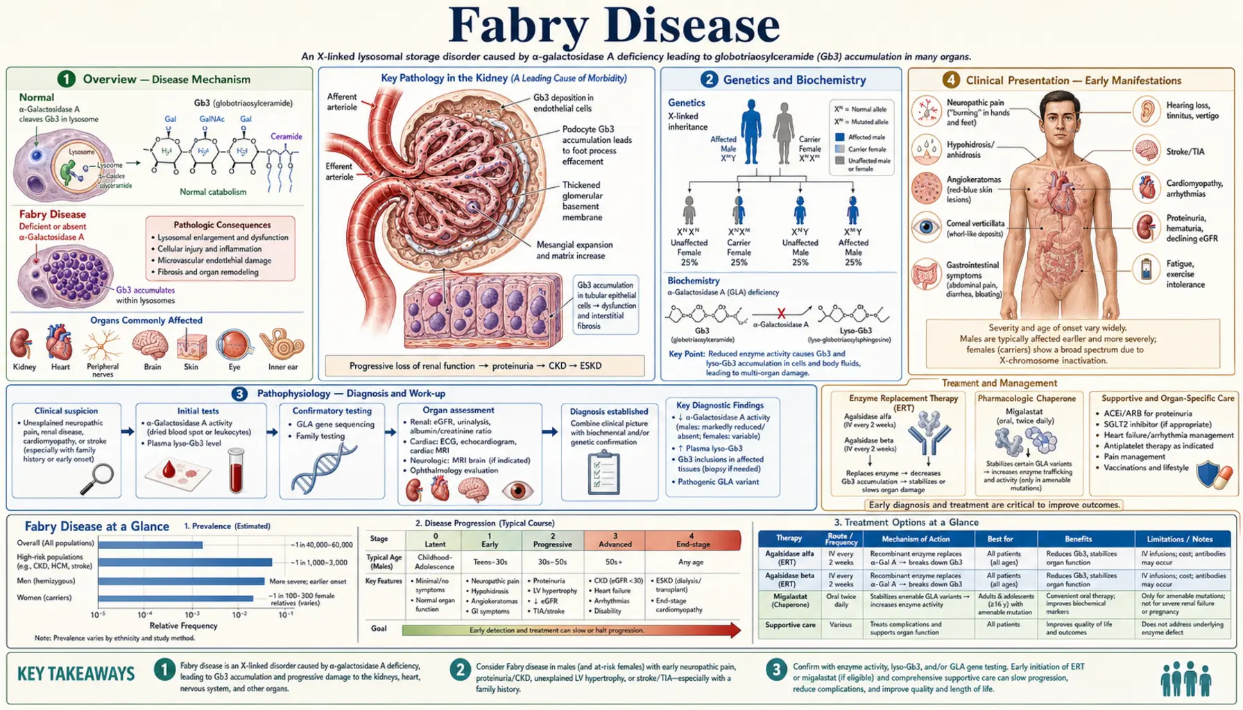
Fabry disease is an X-linked lysosomal storage disorder caused by deficiency of alpha-galactosidase A (alpha-Gal A, encoded by the GLA gene), leading to progressive intracellular accumulation of globotriaosylceramide (Gb3/GL-3) and its deacylated form lyso-Gb3 in endothelial, smooth muscle, cardiac, and renal cells. The disorder causes a multisystem vasculopathy affecting the kidneys, heart, nervous system, and skin, with untreated males typically developing end-stage renal disease and premature cardiovascular death.
Table of Contents
1. Overview
Fabry disease (Anderson-Fabry disease, OMIM #301500) is a rare X-linked lysosomal storage disorder with an estimated incidence of 1 in 40,000–120,000 male births in classic form; newborn screening studies suggest a broader prevalence of 1 in 1,500–3,100 when atypical/late-onset variants are included. Described independently by Johannes Fabry (Germany) and William Anderson (UK) in 1898. The disorder is caused by mutations in the GLA gene (chromosome Xq22.1), producing deficient or absent alpha-galactosidase A (alpha-Gal A) enzyme activity. This results in lysosomal accumulation of globotriaosylceramide (Gb3, also called GL-3) throughout the body, particularly in vascular endothelial and smooth muscle cells, podocytes, cardiomyocytes, and neurons. Classic Fabry disease in hemizygous males causes neuropathic pain, angiokeratomas, cornea verticillata, progressive kidney disease (end-stage renal disease by the 40s in untreated males), hypertrophic cardiomyopathy, and early stroke. Enzyme replacement therapy and the oral chaperone migalastat have transformed the prognosis for treatable patients.
A 2025 review by Lenders and colleagues states that, left untreated, Fabry disease shortens life expectancy by around 20 years in men and around 10 years in women, through kidney failure, thickened heart muscle (hypertrophic cardiomyopathy), heart rhythm problems and early strokes.
2. Genetics and Biochemistry
X-linked inheritance: GLA gene on chromosome Xq22.1; >1,000 pathogenic variants identified (missense, nonsense, splicing, insertions/deletions). Hemizygous males (one X chromosome): classic or atypical disease depending on residual enzyme activity. Heterozygous females: phenotype ranges from asymptomatic carrier to severe classic disease (X-inactivation determines severity). Neither parent-of-origin effect applies uniformly.
Classic vs. atypical variants:
- Classic Fabry (males): alpha-Gal A activity <1% of normal. Full multisystem involvement. Childhood/adolescent onset of acroparesthesias, angiokeratomas.
- Atypical/late-onset variants (commonly p.A143T, p.R112H, IVS4+919 G→A): residual alpha-Gal A activity 1–15%. Predominantly cardiac or renal involvement (late-onset cardiomyopathy or nephropathy); absent or minimal early neuropathic symptoms. Particularly prevalent in certain populations (p.A143T in Black males — uncertain pathogenicity; IVS4+919 common in Taiwan).
Biochemistry: Alpha-Gal A is a lysosomal hydrolase that cleaves terminal galactose residues from glycosphingolipids, primarily converting Gb3 (globotriaosylceramide, ceramide-glucose-galactose-galactose) to lactosylceramide. Deficiency causes Gb3 to accumulate within lysosomes. Gb3 accumulation in endothelial and smooth muscle cells causes vasculopathy; in podocytes causes progressive nephropathy; in cardiomyocytes causes hypertrophic cardiomyopathy; in neurons causes neuropathic pain. Lyso-Gb3 (deacylated form) is a more sensitive biomarker (elevated in males and female carriers) and promotes additional cellular injury pathways.
3. Pathophysiology
Gb3 accumulates within secondary lysosomes in virtually all cell types. The predominant pathological consequences:
Vascular endothelium and smooth muscle: Gb3-laden endothelial cells swell and occlude small vessel lumens (microangiopathy). This drives ischemia in the kidney (tubular atrophy, glomerulosclerosis), heart (myocardial ischemia despite normal coronaries), and brain (lacunar infarcts, white matter lesions, posterior circulation strokes). Vasculopathy is progressive and underlies the multiorgan failure.
Podocyte injury: Podocytes are among the most heavily Gb3-loaded cells in the kidney. Gb3 disrupts lysosomal function, induces autophagy failure, and triggers podocyte apoptosis → podocyte depletion → focal segmental glomerulosclerosis (FSGS) pattern → proteinuria → progressive chronic kidney disease (CKD).
Cardiomyocytes: Gb3 accumulation causes myocyte hypertrophy and fibrosis → concentric left ventricular hypertrophy (LVH). Gb3 in the conduction system → arrhythmias (AV block, atrial fibrillation, Wolff-Parkinson-White-like patterns).
Dorsal root ganglia neurons: Gb3 accumulation → small fiber neuropathy → burning neuropathic pain (acroparesthesias), autonomic dysfunction (hypohidrosis, gastroparesis, orthostatic hypotension).
Lyso-Gb3: More water-soluble than Gb3; more readily detectable in plasma and urine. Acts as a signaling molecule that activates smooth muscle cell proliferation, inhibits K+ channels in sensory neurons (contributing to pain), and promotes Gb3 synthesis — amplifying disease.
4. Clinical Presentation — Early Manifestations
Classic Fabry disease in males presents in childhood and adolescence with:
Neuropathic pain (acroparesthesias): Burning, lancinating pain in hands and feet, characteristically worse with heat, fever, exercise, or emotional stress. Episodic “Fabry crises” (severe pain attacks lasting hours to days, precipitated by fever or exercise). Cause: small fiber neuropathy from Gb3 in dorsal root ganglia. Often the first and most disabling symptom; may begin as early as age 3–7. Frequently misdiagnosed as arthritis, rheumatic fever, or growing pains.
Hypohidrosis (reduced sweating): Due to Gb3 in eccrine sweat glands and autonomic ganglia. Causes heat intolerance — patients cannot regulate temperature during exercise or hot weather. Heat intolerance combined with acroparesthesias in a young male is a classic Fabry presentation.
Angiokeratomas: Small dark red to blue-black telangiectatic papules that do not blanch on pressure. Distributed classically in the “bathing trunk” area — lower abdomen, buttocks, genitalia, and upper thighs — and on lips/oral mucosa. Present in 70% of classic males, developing in teens. Pathognomonic when combined with acroparesthesias.
Cornea verticillata (corneal whorl): Whorled pattern of cream-colored corneal epithelial deposits (best seen on slit-lamp). Found in >90% of hemizygous males and >70% of carrier females. Asymptomatic. Also caused by amiodarone and chloroquine, which are told apart from Fabry disease by the medicine history.
Gastrointestinal: Nausea, vomiting, diarrhea, early satiety (Gb3 in autonomic gastrointestinal (GI) ganglia). Often dismissed as irritable bowel. Common in childhood and adolescence.
5. Organ Involvement — Kidney
Renal disease is the second leading cause of death in untreated Fabry males (after cardiac disease).
Progression: Microalbuminuria typically appears in the late teens/20s in males. Proteinuria increases through 20s–30s. Chronic kidney disease progresses: estimated glomerular filtration rate (eGFR) decline rate ~3–5 mL/min/1.73m²/year in untreated males. End-stage renal disease in untreated males: median age 38–42 years. In treated males: enzyme replacement therapy (ERT) slows decline but does not stop it; stabilization best in those with estimated glomerular filtration rate >60 at treatment start and minimal proteinuria.
Histopathology: Kidney biopsy pathognomonic — electron microscopy shows zebra bodies (lamellar, concentric myelin-like inclusions within lysosomes of podocytes, mesangial cells, endothelial cells, tubular cells). Light microscopy: podocyte cytoplasm appears foamy/vacuolated on hematoxylin and eosin (H&E) stain. Periodic acid–Schiff (PAS) stain shows glycolipid-laden cells. Immunofluorescence: Gb3 staining positive. Pattern: focal segmental glomerulosclerosis (FSGS) appearance + tubular atrophy as disease advances.
Proteinuria as key prognostic marker: Proteinuria >1 g/day strongly predicts faster progression to end-stage renal disease. Blocking the renin–angiotensin–aldosterone system (RAAS) reduces proteinuria and slows chronic kidney disease in Fabry as in other proteinuric nephropathies.
Dialysis and transplant: Renal replacement therapy outcomes are good in Fabry patients. Kidney transplant — allograft is free of Fabry disease (no GLA mutation). However, extrarenal manifestations (cardiac, neurological) continue to progress post-transplant, because the new kidney does not supply the missing enzyme to the rest of the body — ERT is therefore typically continued after transplant. Recurrence in allograft does not occur, unlike most hereditary glomerulonephritis.
6. Organ Involvement — Heart
Cardiac disease is the leading cause of death in Fabry patients (responsible for ~40–50% of deaths).
Hypertrophic cardiomyopathy (HCM): Concentric LVH develops in the 3rd–4th decade in males (later in females). LV wall thickness increases progressively. Clinically: exertional dyspnea, chest pain, near-syncope. Echo: increased interventricular septal thickness and posterior wall thickness; preserved or reduced systolic function with impaired diastolic function in advanced disease. Cardiac magnetic resonance imaging (CMR): late gadolinium enhancement (LGE) in infero-lateral mid-myocardium (“Fabry pattern”) distinguishes from sarcomeric HCM. LGE correlates with fibrosis and predicts arrhythmia risk.
Arrhythmias: Bradyarrhythmias (sinus bradycardia, AV block from Gb3 in conduction system); tachyarrhythmias (atrial fibrillation in 10–20% of Fabry adults; ventricular tachycardia/fibrillation — rare but risk of sudden death). Electrocardiogram (EKG): shortened PR interval (pseudo-WPW pattern), LVH voltage criteria, ST-T changes.
Heart failure: Late complication. Mixed systolic + diastolic dysfunction. Patients may require implantable cardiac defibrillator (ICD) for sudden death prevention.
Important clinical pearl: Fabry disease may cause HCM phenotype in 0.5–1% of patients presenting with “unexplained HCM”. For this reason, testing for Fabry disease is part of the work-up of unexplained HCM (in males, by measuring alpha-Gal A activity on a dried blood spot).
7. Organ Involvement — Nervous System and Stroke
Cerebrovascular disease: Stroke and transient ischemic attack (TIA) occur in Fabry patients at a mean age of 33–46 years in males — far younger than the general population. Mechanism: small vessel vasculopathy (cerebral microangiopathy) + cardiac embolism (AF, LV thrombus). Pattern: posterior circulation strokes (basilar artery territory, posterior cerebral artery) are disproportionately common in Fabry. White matter lesions (leukoencephalopathy) on magnetic resonance imaging (MRI) T2/FLAIR — dolichoectasia of basilar artery is a characteristic finding. Fabry disease is among the conditions tested for when an adult under 50 has a stroke with no clear cause (cryptogenic stroke). In the Fabry Registry (2,446 patients, Sims 2009), 50% of males and 38.3% of females had their first stroke before Fabry disease was diagnosed.
Small fiber neuropathy: Reduced intraepidermal nerve fiber density on skin biopsy. Autonomic neuropathy: reduced heart rate variability, orthostatic hypotension, hypohidrosis, gastroparesis, neurogenic bladder.
Central nervous system (CNS): Cognitive impairment, depression, anxiety are common but often under-recognized. May relate to chronic pain, white matter disease, and/or direct Gb3 effects on central nervous system.
Tinnitus and hearing loss: Sensorineural hearing loss and tinnitus occur in ~50% of Fabry patients; usually mild-to-moderate.
8. Diagnosis
Diagnosis requires a high index of suspicion given the rarity and multisystem nature.
Alpha-galactosidase A enzyme activity:
- Hemizygous males: alpha-Gal A activity in white blood cells (leukocytes) or a dried blood spot (DBS) — markedly low or absent in classic disease; may be normal in females and some atypical males.
- Heterozygous females: enzyme activity overlaps with normal in up to 30–40% — enzyme assay alone CANNOT exclude Fabry in females. Genetic testing is needed to confirm or exclude Fabry in females.
- A 2025 review (Lenders and colleagues) names low alpha-Gal A activity in white blood cells as the method of choice for confirming the diagnosis in men, and finding the disease-causing GLA gene change as the method in women.
GLA gene sequencing: Definitive diagnosis. Detects pathogenic variants; classifies as classic vs. atypical. Important for variants of uncertain significance (VUS) — functional studies or lyso-Gb3 measurement needed.
Biomarkers:
- Urine and plasma Gb3 (GL-3): Elevated in classic males. May be near-normal in atypical/late-onset and carrier females.
- Plasma lyso-Gb3: More sensitive biomarker — elevated in virtually all affected males AND in heterozygous females regardless of enzyme activity. Best biomarker for female carriers and atypical variants. Also used to monitor treatment response.
Kidney biopsy: If diagnosis uncertain or to stage nephropathy. Electron microscopy shows pathognomonic zebra bodies.
Ophthalmology: Slit-lamp for cornea verticillata (confirms Gb3 storage but not specific to Fabry — also with amiodarone).
Organ-specific evaluation at diagnosis:
- Estimated glomerular filtration rate, urine albumin/creatinine ratio (renal)
- Electrocardiogram (ECG), echocardiogram, cardiac magnetic resonance imaging if LVH present (cardiac)
- Brain magnetic resonance imaging with DWI and FLAIR (cerebrovascular)
- Audiogram (hearing)
- Pain score / quality of life questionnaire
Newborn screening: Several countries now screen alpha-Gal A activity on DBS at birth (Taiwan, Italy, Austria). Cascade family screening after index case diagnosis.
9. Treatment
Enzyme Replacement Therapy (ERT): The current standard of care for Fabry patients with classic disease.
Agalsidase alfa: 0.2 mg/kg intravenous (IV) every 2 weeks. Approved in Europe, Canada, Asia. Not approved by the U.S. Food and Drug Administration (FDA).
Agalsidase beta: 1 mg/kg intravenous every 2 weeks. FDA-approved (initial U.S. approval 2003; the U.S. label covers adults and children aged 2 years and older) and approved worldwide. Higher dose delivers more Gb3 clearance per infusion.
Pegunigalsidase alfa: a PEGylated alpha-galactosidase A (the enzyme with polyethylene glycol chains attached), 1 mg/kg intravenous every 2 weeks. FDA-approved in 2023 (U.S. label: adults with confirmed Fabry disease). In the 2-year BALANCE trial (Wallace 2024), 77 adults whose kidney function was falling by more than 2 mL/min/1.73 m² a year after at least a year on agalsidase beta were assigned to pegunigalsidase alfa (52) or to stay on agalsidase beta (25). Kidney function declined at a similar rate in both groups (difference in yearly eGFR slope −0.36 mL/min/1.73 m², within the trial’s non-inferiority margin). Mild or moderate infusion reactions, adjusted for exposure, were 7.8-fold more frequent with agalsidase beta. At the end of the trial, neutralising antibodies were found in 7 of 47 (15%) patients on pegunigalsidase alfa and 6 of 23 (26%) on agalsidase beta. There were no deaths.
ERT efficacy: Reduces plasma and urine Gb3/lyso-Gb3. Stabilizes or slows renal function decline. In the placebo-controlled trial by Banikazemi and colleagues (2007; 82 adults with advanced Fabry disease and mild-to-moderate kidney disease, median 18.5 months), agalsidase beta delayed a combined outcome of kidney, heart or brain events or death: 14 of 51 treated patients (27%) and 13 of 31 placebo patients (42%) had such an event. In the main (intention-to-treat) analysis the hazard ratio was 0.47 (P = 0.06, short of statistical significance); the often-quoted 61% lower risk comes from the secondary analysis of protocol-adherent patients (hazard ratio 0.39, P = 0.034). Reduces LV mass in cardiomyopathy (with early treatment). Reduces neuropathic pain. Best outcomes with early initiation (before significant proteinuria, before estimated glomerular filtration rate <60, before significant LGE on CMR). ERT does NOT fully reverse established fibrosis/sclerosis.
Contraindications/challenges: Infusion-related reactions are common: in the 2007 trial, mostly mild or moderate infusion reactions were reported by 55% of patients on agalsidase beta and 23% on placebo. The agalsidase beta label lists considering pretreatment with antihistamines, fever reducers (antipyretics) and/or corticosteroids before infusions. True allergic antibodies (immunoglobulin E, IgE) are uncommon: in the label’s clinical trials, 10 of 238 patients developed IgE antibodies or skin-test reactivity to agalsidase beta. Long-term ERT antibody formation may reduce efficacy in some patients.
Antidrug antibodies explained: Because many men with classic Fabry disease make little or no enzyme of their own, the immune system can treat the infused enzyme as foreign and make antibodies against it (antidrug antibodies, mostly of the IgG type). A 2025 review (Gómez-Cerezo and colleagues) reports that neutralising antibodies — ones that block the enzyme’s action — are present in about 40% of men treated with agalsidase alfa or agalsidase beta. In laboratory tests they react with both enzymes. They seem to be permanent: most men who test positive remain positive for up to 10 years. They are often linked to a worse course and faster disease progression, which is why international recommendations include checking for them. In some patients, these pre-existing antibodies bind pegunigalsidase alfa less strongly.
Migalastat: Oral pharmacological chaperone. FDA-approved 2018 under accelerated approval; the U.S. label covers adults with confirmed Fabry disease and an amenable GLA variant (amenable variants are defined by an in vitro human embryonic kidney (HEK) cell assay — ~35–50% of all variants are amenable). 123 mg orally every other day. Stabilizes misfolded alpha-Gal A and improves lysosomal trafficking. ATTRACT trial (Hughes et al., J Med Genet 2017; 57 adults switched from ERT or kept on it for 18 months): similar effects to ERT on kidney function; left ventricular mass index fell with migalastat (−6.6 g/m²) with no significant change on ERT; plasma lyso-Gb3 stayed low and stable after the switch. Advantages over ERT: oral, no infusion reactions, home administration. Does NOT work for null mutations (no protein to stabilize).
Symptomatic management:
- Neuropathic pain: carbamazepine (first-line for Fabry neuropathic pain — reduces frequency/severity of crises), gabapentin/pregabalin, mexiletine (severe cases); opioids are generally kept as a last option.
- Renal protection: angiotensin-converting enzyme inhibitor (ACE inhibitor) or angiotensin receptor blocker (ARB) — reduces proteinuria and slows chronic kidney disease progression (standard of care alongside ERT).
- Cardiac: antihypertensives; amiodarone (or ICD) for arrhythmia; anticoagulation for atrial fibrillation (AF); heart failure management.
- Stroke prevention: antiplatelet therapy (aspirin/clopidogrel); anticoagulation for atrial fibrillation; blood pressure control.
- Skin: angiokeratomas — laser therapy for cosmetic improvement.
Pipeline: Venglustat (glucosylceramide synthase inhibitor, substrate reduction therapy — Phase 2/3 in Fabry); moss-derived agalsidase alfa (improved mannose-6-phosphate receptor targeting — Phase 3); gene therapy approaches (AAV-GLA) in preclinical/Phase 1.
Lucerastat (phase 3 failure): Lucerastat, a pill meant to lower Gb3 production (substrate reduction therapy), did not meet its main goal in the phase 3 MODIFY trial (Nordbeck 2026). In 117 adults with moderate-to-severe nerve pain, 6 months of lucerastat 1000 mg twice daily (80 people) did not reduce pain more than placebo (37 people), even though plasma Gb3 fell in the lucerastat group. Its possible effect on kidney function, seen in an unplanned analysis of the open-label extension, is described by the authors as needing further study.
10. Monitoring and Follow-Up
European Fabry Working Group and international guidelines recommend:
Renal: estimated glomerular filtration rate and urine albumin/creatinine ratio every 6–12 months. 24-hour urine protein if significant proteinuria. Kidney biopsy at baseline if diagnosis uncertain or to stage disease; repeat to assess ERT response debated.
Cardiac: electrocardiogram annually. Echocardiogram every 1–2 years. Cardiac magnetic resonance imaging with gadolinium every 3–5 years (assess LGE fibrosis — strongest predictor of arrhythmia and sudden death). Holter if palpitations or syncope.
Neurological: Brain magnetic resonance imaging at baseline and every 3–5 years (or after neurological events). Neurological exam; cognitive screening.
Pain: Validated questionnaire (Brief Pain Inventory — Fabry version). Assess pain triggers, analgesic use.
Biomarkers: Plasma lyso-Gb3 every 6–12 months (treatment response monitoring). Alpha-Gal A activity in males on ERT (to detect antibody interference).
Pregnancy: Female Fabry patients — ERT can be continued during pregnancy (risk-benefit discussion; limited data). Genetic counseling pre-conception for X-linked inheritance.
Key Research Papers
- Schiffmann R, et al. Enzyme replacement therapy in Fabry disease: a randomized controlled trial. JAMA. 2001;285(21):2743–2749. — PubMed PMID: 11386930 · doi:10.1001/jama.285.21.2743
- Banikazemi M, Bultas J, Waldek S, et al. Agalsidase-beta therapy for advanced Fabry disease: a randomized trial. Ann Intern Med. 2007;146(2):77–86. — PubMed PMID: 17179052 · doi:10.7326/0003-4819-146-2-200701160-00148
- Hughes DA, et al. Oral pharmacological chaperone migalastat compared with enzyme replacement therapy in Fabry disease: 18-month results from the randomised phase III ATTRACT study. J Med Genet. 2017;54(4):288–296. — PubMed PMID: 27834756 · doi:10.1136/jmedgenet-2016-104178
- Eng CM, et al. Fabry disease: baseline medical characteristics of a cohort of 1765 males and females in the Fabry Registry. J Inherit Metab Dis. 2007;30(2):184–192. — PubMed PMID: 17347915 · doi:10.1007/s10545-007-0521-2
- Germain DP, et al. Treatment of Fabry’s disease with the pharmacologic chaperone migalastat. N Engl J Med. 2016;375(6):545–555. — PubMed PMID: 27509102 · doi:10.1056/NEJMoa1510198
- Weidemann F, et al. Improvement of cardiac function during enzyme replacement therapy in patients with Fabry disease: a prospective strain rate imaging study. Circulation. 2003;108(11):1299–1301. — PubMed PMID: 12952834 · doi:10.1161/01.cir.0000091253.71282.04
- Sims K, et al. Stroke in Fabry disease frequently occurs before diagnosis and in the absence of other clinical events: natural history data from the Fabry Registry. Stroke. 2009;40(3):788–794. — PubMed PMID: 19150871 · doi:10.1161/STROKEAHA.108.526293
- Desnick RJ, et al. α-Galactosidase A deficiency: Fabry disease. In: Valle DL, et al., eds. The Online Metabolic and Molecular Bases of Inherited Disease. McGraw-Hill; 2001. Accessed at OMIM #301500.
- Nowak A, Mechtler TP, Desnick RJ, Kasper DC. Plasma LysoGb3: A useful biomarker for the diagnosis and treatment of Fabry disease heterozygotes. Mol Genet Metab. 2017;120(1–2):57–61. — PubMed PMID: 27773586 · doi:10.1016/j.ymgme.2016.10.006
- Ortiz A, et al. Fabry disease revisited: Management and treatment recommendations for adult patients. Mol Genet Metab. 2018;123(4):416–427. — PubMed PMID: 29530533 · doi:10.1016/j.ymgme.2018.02.014
- Wallace EL, Goker-Alpan O, Wilcox WR, et al. Head-to-head trial of pegunigalsidase alfa versus agalsidase beta in patients with Fabry disease and deteriorating renal function: results from the 2-year randomised phase III BALANCE study. J Med Genet. 2024;61(6):520–530. — PubMed PMID: 37940383 · doi:10.1136/jmg-2023-109445
- Gómez-Cerezo JF, Fernández-Martín J, Barba-Romero MÁ, et al. Current status of the immunogenicity of enzyme replacement therapy in fabry disease. Orphanet J Rare Dis. 2025;20(1):253. — PubMed PMID: 40420145 · doi:10.1186/s13023-025-03705-4
- Lenders M, Menke ER, Brand E. Progress and Challenges in the Treatment of Fabry Disease. BioDrugs. 2025;39(4):517–535. — PubMed PMID: 40310476 · doi:10.1007/s40259-025-00723-3
- Nordbeck P, Goker-Alpan O, Bernat JA, et al. Lucerastat, an oral therapy for Fabry disease: results from a pivotal randomized phase 3 study and its open-label extension. Nat Commun. 2026;17(1):1534. — PubMed PMID: 41519901 · doi:10.1038/s41467-025-68256-5
PubMed Topic Searches
- PubMed: Fabry disease alpha-galactosidase A enzyme replacement therapy
- PubMed: Fabry disease migalastat ATTRACT trial GLA variants
- PubMed: Fabry disease cardiomyopathy cardiac MRI late gadolinium enhancement
- PubMed: Fabry disease stroke cryptogenic young adults cerebrovascular
- PubMed: Fabry disease renal nephropathy proteinuria progression
- PubMed: Lyso-Gb3 biomarker Fabry disease females heterozygous