Immunoglobulin A (IgA) Nephropathy

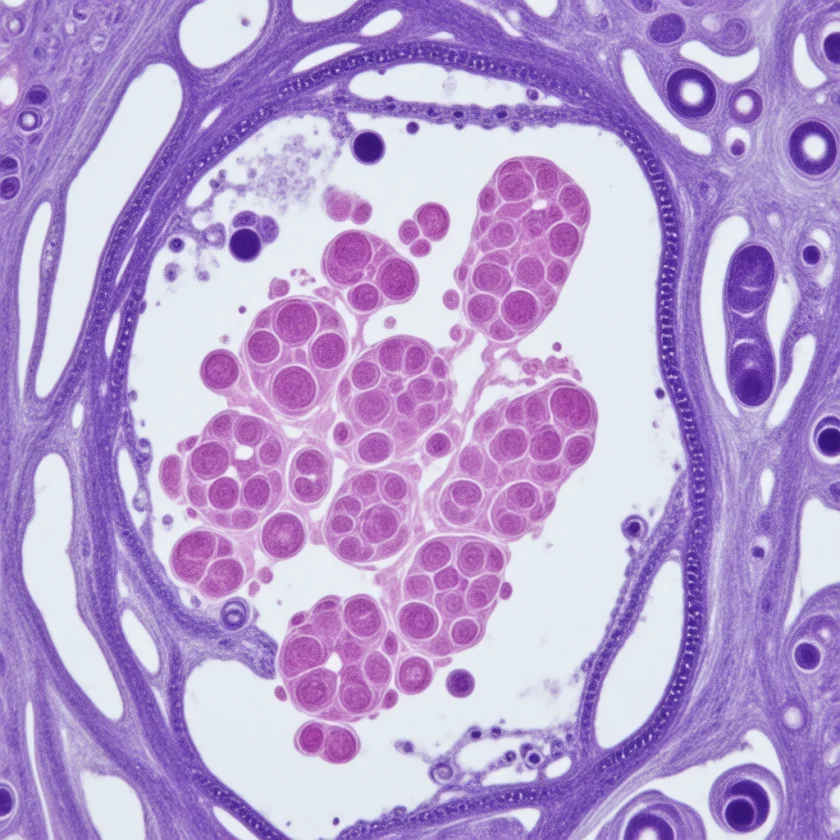
Immunoglobulin A (IgA) Nephropathy (IgAN), also known as Berger's disease, is the most common primary glomerulonephritis worldwide. It is defined by a defect in IgA1 O-glycosylation that produces galactose-deficient IgA1 (Gd-IgA1), which triggers autoantibody formation, circulating immune complex deposition in the glomerular mesangium, complement activation, and progressive kidney injury. The disease affects people of all ages but peaks in the second and third decades of life, and up to 50% of high-risk patients develop end-stage renal disease within 20–25 years.
Table of Contents
1. Overview
Immunoglobulin A Nephropathy (IgAN), also known as Berger's disease (after Jean Berger who first described it in 1968), is the most common primary glomerulonephritis worldwide, accounting for 30–40% of primary GN in developed countries. The disease is characterized by predominant IgA1 immune deposits in the glomerular mesangium, leading to mesangial cell proliferation, complement activation, and progressive kidney injury. IgAN affects people of all ages but peaks in the second and third decades of life with a 2:1 male predominance. Though often considered a mild condition, up to 50% of high-risk patients develop end-stage renal disease (ESRD) within 20–25 years.
2. Epidemiology
IgAN exhibits marked geographic variation, with the highest prevalence in Asia (particularly Japan and China, where it accounts for up to 40–50% of primary GN biopsies) and lower prevalence in Black populations. Annual incidence is approximately 2.5 cases per 100,000 in Western countries. The disease accounts for approximately 10% of patients on dialysis in developed nations. Family clustering occurs in 5–10% of cases, suggesting genetic susceptibility involving human leukocyte antigen (HLA) loci (HLA-DR4, HLA-B35) and genes regulating galactose-deficient IgA1 production.
3. Pathophysiology
The multi-hit hypothesis for IgAN pathogenesis involves four sequential steps:
Hit 1 — Elevated circulating galactose-deficient IgA1 (Gd-IgA1): O-glycan chains on the hinge region of IgA1 are underglycosylated due to reduced activity of the enzyme C1GALT1 and its chaperone COSMC, leading to exposure of N-acetylgalactosamine (GalNAc) residues. Elevated Gd-IgA1 is found in patients and their first-degree relatives.
Hit 2 — Autoantibody formation: Gd-IgA1 is recognized as a neoantigen, triggering production of immunoglobulin G (IgG) and immunoglobulin A autoantibodies directed against the abnormal hinge-region glycans.
Hit 3 — Immune complex formation: Autoantibodies bind Gd-IgA1, forming large immune complexes with high mesangial affinity.
Hit 4 — Mesangial injury: Immune complex deposition in the mesangium activates the lectin pathway of complement (C4d deposition) and the alternative pathway, triggering mesangial cell proliferation, cytokine release (TGF-β, PDGF, IL-6), and eventual glomerulosclerosis and tubular atrophy.
4. Clinical Presentation
Three classic presentations:
Synpharyngitic gross hematuria: Episodic gross hematuria occurring within 24–72 hours of a mucosal infection (typically upper respiratory tract infection), distinguishing IgAN from post-streptococcal GN (which has a 2–3 week latency). Tea-colored or brown urine with flank pain. Common in younger patients (30–40% of presentations in Asian populations).
Asymptomatic microscopic hematuria ± proteinuria: Detected incidentally on routine urinalysis. Most common presentation in Western populations (50–60%). May have low-grade proteinuria.
Chronic GN with nephrotic-range proteinuria: Hypertension, progressive chronic kidney disease (CKD), proteinuria >1 g/day. Associated with the worst prognosis. Rare presentation as nephrotic syndrome proper.
Rare presentations include rapidly progressive GN (crescentic IgAN) with rapid glomerular filtration rate (GFR) decline, and macroscopic hematuria causing acute kidney injury (AKI) from tubular obstruction by RBC casts.
5. MEST-C Oxford Classification
The Oxford Classification (2009, revised 2017) provides a standardized histological scoring system predicting renal outcomes independently of clinical parameters:
M — Mesangial hypercellularity: M0 = ≤50% of glomeruli with mesangial hypercellularity; M1 = >50%. M1 predicts faster glomerular filtration rate decline.
E — Endocapillary hypercellularity: E0 = absent; E1 = present in any glomerulus. E1 predicts increased risk of progression especially without immunosuppression.
S — Segmental glomerulosclerosis: S0 = absent; S1 = present (any segmental sclerosis, adhesion, or tip lesion). S1 independently predicts glomerular filtration rate decline and ESRD.
T — Tubular atrophy/interstitial fibrosis: T0 = 0–25%; T1 = 26–50%; T2 = >50% of cortical area. T-score is the strongest predictor of ESRD. T2 carries very high risk of progression to ESRD.
C — Crescents (added 2017): C0 = no crescents; C1 = crescents in <25% of glomeruli; C2 = crescents in ≥25%. C2 predicts poor outcomes and may indicate need for immunosuppression.
The MEST-C score supplements clinical risk factors including proteinuria, estimated glomerular filtration rate (eGFR), and hypertension in predicting long-term renal outcomes.
6. Diagnosis
Definitive diagnosis requires kidney biopsy with immunofluorescence showing dominant or codominant immunoglobulin A deposits in the mesangium (immunoglobulin G and immunoglobulin M (IgM) may be present in smaller amounts). Light microscopy shows mesangial proliferative GN. Electron microscopy confirms mesangial electron-dense deposits.
Laboratory workup:
- Urinalysis: microscopic hematuria (dysmorphic RBCs, RBC casts), proteinuria
- 24-hour urine protein or spot urine PCR: quantify proteinuria
- Serum creatinine, estimated glomerular filtration rate: baseline and trend
- Serum immunoglobulin A: elevated in 50% but not diagnostic; immunoglobulin A/C3 ratio >3 is suggestive
- Complement: C3 usually normal (distinguishes from MPGN and lupus GN); C4d on biopsy indicates lectin pathway activation
- Galactose-deficient IgA1 (Gd-IgA1): elevated in 70–80% of IgAN patients; commercially available assays emerging
- Exclude secondary IgAN: serum protein electrophoresis, antinuclear antibody (ANA), anti-dsDNA, hepatitis B/C, human immunodeficiency virus (HIV), antineutrophil cytoplasmic antibody (ANCA), anti-GBM antibodies; celiac serology (immunoglobulin A anti-tissue transglutaminase — celiac disease is an important secondary cause)
Indications for biopsy: unexplained persistent microscopic hematuria with proteinuria >0.5 g/day, progressive chronic kidney disease, or suspected rapidly progressive GN.
7. Treatment
Supportive Therapy (all patients)
Angiotensin-converting enzyme inhibitor (ACEi) or angiotensin receptor blocker (ARB): First-line for proteinuria >0.5 g/day and/or hypertension. Target proteinuria <0.5–1 g/day. Guidelines describe raising the dose to the highest tolerated level. Combined angiotensin-converting enzyme inhibitor + angiotensin receptor blocker not recommended due to acute kidney injury and hyperkalemia risk.
Sodium-glucose cotransporter-2 (SGLT2) inhibitors (dapagliflozin, empagliflozin): DAPA-CKD and EMPA-KIDNEY trials demonstrated kidney-protective effects in chronic kidney disease including IgAN; reduce proteinuria and slow glomerular filtration rate decline. The KDIGO 2021 guideline predates these trials and does not recommend SGLT2 inhibitors for IgAN.
Fish oil (omega-3 fatty acids): Trials of fish oil show modest and inconsistent proteinuria reduction; weak evidence. KDIGO 2021 suggests it may be used in persistent proteinuria >1 g/day despite angiotensin-converting enzyme inhibitor/angiotensin receptor blocker.
Sodium bicarbonate: In patients with metabolic acidosis and chronic kidney disease, bicarbonate supplementation may slow chronic kidney disease progression.
Immunosuppression (selected high-risk patients)
STOP-IgAN trial (2015): Showed no benefit from adding immunosuppression to comprehensive supportive care in most patients; reinforced the importance of maximized supportive therapy first.
TESTING trial (2017/2022 extension): High-dose oral methylprednisolone (0.6–0.8 mg/kg/day × 2 months, tapered to 12 months) reduced proteinuria and ESRD risk but increased serious adverse events (infections, weight gain, diabetes). Revised low-dose protocol showed better safety profile.
NEFECON (Targeted-release budesonide): Budesonide formulated to release in the ileum (Peyer's patches — the site of mucosal immunoglobulin A production). NEFIGAN Phase 2b trial (2017) and NefIgArd Phase 3 trial (2023) showed significant proteinuria reduction (−27%) and estimated glomerular filtration rate preservation with low systemic corticosteroid side effects. The U.S. Food and Drug Administration (FDA) granted targeted-release budesonide accelerated approval in December 2021 and full approval in December 2023.
Sparsentan: Dual endothelin/angiotensin receptor antagonist. PROTECT trial (2023) demonstrated proteinuria reduction and estimated glomerular filtration rate preservation superior to irbesartan alone. The FDA granted sparsentan accelerated approval for IgAN in February 2023 and full approval in September 2024.
Atrasentan: Selective endothelin-A receptor antagonist. In the ALIGN phase 3 trial the urine protein-to-creatinine ratio fell 36.1 percentage points more with atrasentan than with placebo at week 36 (Heerspink et al., NEJM 2025). The FDA granted atrasentan accelerated approval in April 2025 on the basis of proteinuria reduction. The final 2.5-year ALIGN results (Lancet 2026) reported an eGFR change at week 136 of −7.5 vs. −9.9 mL/min/1.73m² (difference 2.4, p=0.057).
Iptacopan: Complement factor B inhibitor. The FDA granted it accelerated approval in August 2024 to reduce proteinuria in primary IgAN. In the APPLAUSE-IgAN phase 3 trial the urine protein-to-creatinine ratio was 38.3% lower with iptacopan than with placebo (Perkovic et al., NEJM 2025).
Other agents under investigation: cemdisiran/avacopan (complement pathway), obinutuzumab, and anti-APRIL antibodies (BION-1301) targeting the B-cell survival factor APRIL that drives Gd-IgA1 production.
Cytotoxic immunosuppression (cyclophosphamide, MMF): reserved for crescentic IgAN or rapidly progressive disease.
8. Prognosis and Progression to ESRD
High-risk features predicting ESRD: proteinuria persistently >1 g/day, hypertension, reduced estimated glomerular filtration rate at presentation, T2 tubular atrophy/fibrosis on biopsy, crescents (C2), S1 score. Approximately 15–20% of patients reach ESRD within 10 years, 25–50% within 20–25 years.
Low-risk features: isolated microscopic hematuria without proteinuria, normal estimated glomerular filtration rate, M0E0S0T0C0 biopsy, episodic gross hematuria pattern only.
IgAN recurs in 30–60% of kidney transplants (higher rate than other GN), though graft loss from recurrence is uncommon (<10% at 10 years).
9. Recent Clinical Trials
STOP-IgAN (Rauen et al., NEJM 2015): No benefit from adding immunosuppression to optimized supportive care; reinforced angiotensin-converting enzyme inhibitor/angiotensin receptor blocker + risk factor control as primary management.
TESTING (Lv et al., JAMA 2022): Oral methylprednisolone (full-dose and reduced-dose groups combined) lowered the composite of 40% eGFR decline, kidney failure or death from kidney disease vs. placebo (hazard ratio (HR) 0.53); for kidney failure alone the HR was 0.59. Serious adverse events were more frequent with methylprednisolone (10.9% vs. 2.8%), mostly with the full dose.
NefIgArd / NEFECON (Barratt et al., Kidney Int 2023; Lafayette et al., Lancet 2023): Targeted-release budesonide 16 mg/day × 9 months: urine protein-to-creatinine ratio 27% lower than placebo at 9 months (Part A); over 2 years the time-weighted average eGFR was 5.05 mL/min/1.73m² better than placebo; FDA full approval December 2023.
PROTECT (Heerspink et al., Lancet 2023; Rovin et al., Lancet 2023): Sparsentan 400 mg/day vs. irbesartan: 40% greater proteinuria reduction at 36 weeks; estimated glomerular filtration rate slope preservation at 110 weeks; U.S. Food and Drug Administration-approved.
DAPA-CKD (Heerspink et al., NEJM 2020): Dapagliflozin reduced composite of ≥50% estimated glomerular filtration rate decline, ESRD, or renal/CV death by 39% in chronic kidney disease patients; subgroup including IgAN patients showed consistent benefit.
EMPA-KIDNEY (EMPA-KIDNEY Collaborative Group, NEJM 2023): Empagliflozin reduced kidney disease progression or CV death by 28% across broad chronic kidney disease population including IgAN.
10. Prevention and Monitoring
No proven primary prevention. Tonsillectomy is practiced in Japan to reduce synpharyngitic hematuria episodes and may reduce proteinuria in selected patients (evidence limited to Asian cohorts; not recommended in Western guidelines). Nonsteroidal anti-inflammatory drugs (NSAIDs) and other kidney-toxic drugs can worsen kidney function.
Annual monitoring of estimated glomerular filtration rate, urine protein-to-creatinine ratio, and blood pressure in all IgAN patients. More frequent monitoring every 3–6 months for high-risk patients. Vaccination: influenza and pneumococcal vaccines are recommended given infection triggers.
Key Research Papers
- Wyatt RJ, Julian BA. IgA nephropathy. N Engl J Med. 2013;368(25):2402–2414. — PubMed PMID: 23782179 · doi:10.1056/NEJMra1206793
- Barratt J, Lafayette R, Kristensen J, et al. Results from part A of the multi-center, double-blind, randomized, placebo-controlled NefIgArd trial, which evaluated targeted-release formulation of budesonide for the treatment of primary immunoglobulin A nephropathy. Kidney Int. 2023;103(2):391–402. — doi:10.1016/j.kint.2022.09.017 · PubMed PMID: 36270561
- Lafayette R, Kristensen J, Stone A, et al. Efficacy and safety of a targeted-release formulation of budesonide in patients with primary IgA nephropathy (NefIgArd): 2-year results from a randomised phase 3 trial. Lancet. 2023;402(10405):859–870. — doi:10.1016/S0140-6736(23)01554-4 · PubMed PMID: 37591292
- Rauen T, et al. Intensive Supportive Care plus Immunosuppression in IgA Nephropathy (STOP-IgAN). N Engl J Med. 2015;373(23):2225–2236. — PubMed PMID: 26630142 · doi:10.1056/NEJMoa1415463
- Lv J, Wong MG, Hladunewich MA, et al. Effect of Oral Methylprednisolone on Decline in Kidney Function or Kidney Failure in Patients With IgA Nephropathy: The TESTING Randomized Clinical Trial. JAMA. 2022;327(19):1888–1898. — doi:10.1001/jama.2022.5368 · PubMed PMID: 35579642
- Heerspink HJL, Radhakrishnan J, Alpers CE, et al. Sparsentan in patients with IgA nephropathy: a prespecified interim analysis from a randomised, double-blind, active-controlled clinical trial. Lancet. 2023;401(10388):1584–1594. — doi:10.1016/S0140-6736(23)00569-X · PubMed PMID: 37015244
- Rovin BH, Barratt J, Heerspink HJL, et al. Efficacy and safety of sparsentan versus irbesartan in patients with IgA nephropathy (PROTECT): 2-year results from a randomised, active-controlled, phase 3 trial. Lancet. 2023;402(10417):2077–2090. — doi:10.1016/S0140-6736(23)02302-4 · PubMed PMID: 37931634
- Heerspink HJL, Jardine M, Kohan DE, et al. Atrasentan in Patients with IgA Nephropathy. N Engl J Med. 2025;392(6):544–554. — doi:10.1056/NEJMoa2409415 · PubMed PMID: 39460694
- Heerspink HJL, Jardine MJ, Kohan DE, et al. Atrasentan in patients with IgA nephropathy (ALIGN): final 2·5-year results from a randomised, double-blind, placebo-controlled, phase 3 trial. Lancet. 2026;407(10546):2387–2401. — doi:10.1016/S0140-6736(26)00960-8 · PubMed PMID: 42242268
- Perkovic V, Barratt J, Rovin B, et al. Alternative Complement Pathway Inhibition with Iptacopan in IgA Nephropathy. N Engl J Med. 2025;392(6):531–543. — doi:10.1056/NEJMoa2410316 · PubMed PMID: 39453772
- Roberts ISD, et al. The Oxford classification of IgA nephropathy: pathology definitions, correlations, and reproducibility. Kidney Int. 2009;76(5):546–556. — PubMed PMID: 19571790 · doi:10.1038/ki.2009.168
- Trimarchi H, et al. Oxford Classification of IgA nephropathy 2016: an updated proposal. Kidney Int. 2017;91(5):1014–1021. — PubMed PMID: 28341274 · doi:10.1016/j.kint.2017.02.003
- Heerspink HJL, et al. Dapagliflozin in Patients with Chronic Kidney Disease (DAPA-CKD). N Engl J Med. 2020;383(15):1436–1446. — PubMed PMID: 32970396 · doi:10.1056/NEJMoa2024816
- Fellström BC, et al. Targeted-release budesonide versus placebo in patients with IgA nephropathy (NEFIGAN). Lancet. 2017;389(10084):2117–2127. — PubMed PMID: 28363480 · doi:10.1016/S0140-6736(17)30550-0
- EMPA-KIDNEY Collaborative Group. Empagliflozin in Patients with Chronic Kidney Disease. N Engl J Med. 2023;388(2):117–127. — PubMed PMID: 36331190 · doi:10.1056/NEJMoa2204233
- Mestecky J, et al. IgA nephropathy: molecular mechanisms of the disease. Annu Rev Pathol. 2013;8:217–240. — Search PubMed · doi:10.1146/annurev-pathol-011110-130216
- Rajasekaran A, Julian BA, Rizk DV. IgA Nephropathy: An Interesting Autoimmune Kidney Disease. Am J Med Sci. 2021;361(2):176–194. — Search PubMed · doi:10.1016/j.amjms.2020.10.003
PubMed Topic Searches
- PubMed: IgA nephropathy pathogenesis
- PubMed: IgA nephropathy treatment clinical trials
- PubMed: Galactose-deficient IgA1 Berger's disease
- PubMed: MEST-C Oxford classification IgA nephropathy
- PubMed: Sparsentan IgA nephropathy PROTECT trial
- PubMed: NEFECON budesonide IgA nephropathy NefIgArd
- PubMed: SGLT2 inhibitors IgA nephropathy