Alport Syndrome
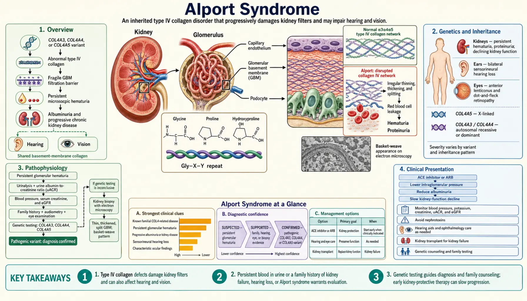
Alport syndrome is a hereditary nephritis caused by mutations in COL4A3, COL4A4, or COL4A5 genes encoding type IV collagen α-chains, components of the glomerular basement membrane (GBM). The disease produces a characteristic triad of progressive glomerulonephritis leading to end-stage renal disease, sensorineural hearing loss, and ocular abnormalities including anterior lenticonus. It accounts for approximately 2–3% of pediatric ESRD cases worldwide.
Table of Contents
1. Overview
Alport syndrome is a progressive hereditary nephritis first described by Cecil Alport in 1927 as a familial nephritis with associated hearing loss. It is caused by mutations in genes encoding type IV collagen α3, α4, or α5 chains (COL4A3, COL4A4, COL4A5), which are critical structural components of the glomerular basement membrane (GBM), cochlear basement membrane, and lens capsule. Three inheritance patterns exist: X-linked (XLAS, ~85% of cases, COL4A5 mutations), autosomal recessive (ARAS, ~15%, COL4A3 or COL4A4 mutations), and autosomal dominant (ADAS, ~5%, heterozygous COL4A3/A4, milder). XLAS affects males more severely than females. Alport syndrome accounts for approximately 0.6% of adults on renal replacement therapy but 2–3% of pediatric ESRD. Early angiotensin-converting enzyme inhibitor (ACE inhibitor) therapy delays kidney failure: in a study of three generations of Alport families (Gross et al., 2012), younger siblings treated earlier started dialysis 13 years later than older siblings treated later or not at all.
2. Genetics and Inheritance
Type IV collagen is the major structural collagen of basement membranes. The GBM contains predominantly the α3α4α5(IV) heterotrimer encoded by COL4A3 (chromosome 2q36), COL4A4 (chromosome 2q36), and COL4A5 (chromosome Xq22). In early fetal GBM, α1α2(IV) predominates; during nephrogenesis, the switch to α3α4α5(IV) occurs (the “isoform switch”). Loss-of-function mutations prevent this switch or disrupt heterotrimer assembly, resulting in persistence of fetal-type α1α2(IV) GBM that is prone to mechanical degradation.
X-linked Alport syndrome (XLAS): ~85% of Alport families. COL4A5 mutation on X chromosome. Hemizygous males: in a United States study of 681 affected males from 175 families (Bekheirnia 2010), ESRD began at an average age of 25 years with truncating mutations, 28 years with splice-site mutations and 37 years with missense mutations. Heterozygous females: in a European study of girls and women from 195 families with a proven COL4A5 mutation (Jais 2003), 95% had hematuria, 75% developed proteinuria, 28% hearing loss and 15% eye changes; the chance of reaching ESRD before age 40 was 12% (versus 90% in boys and men), and the risk appears to rise after age 60. Female carriers have variable expression due to X-inactivation.
Autosomal recessive ARAS: ~15% of Alport cases. Biallelic mutations in COL4A3 or COL4A4. Males and females equally affected. ESRD by 20s–30s, similar severity to X-linked males.
Autosomal dominant ADAS: Heterozygous COL4A3/A4 mutations. Milder phenotype; ESRD in 50s–70s if it occurs. Overlaps with thin basement membrane nephropathy (TBMN); ADAS may represent ~1% of the general population (previously called “benign familial hematuria”).
De novo mutations account for ~10–15% of XLAS cases (no family history).
3. Pathophysiology
Loss of the α3α4α5(IV) heterotrimer in the GBM disrupts the normal network of type IV collagen crosslinks, reducing GBM tensile strength. The resulting structurally fragile GBM allows passage of erythrocytes (hematuria) and plasma proteins (proteinuria). Podocyte foot process effacement develops as the diseased GBM fails to support podocyte attachment.
Mechanistically, the absence of the α3/α4/α5 network also exposes the persistent fetal α1α2(IV) chains to injury, and activates mesangial cells and tubular cells via mechanical and cytokine-mediated pathways. TGF-β1 drives progressive interstitial fibrosis. The same α3α4α5(IV) network is critical in the cochlear basilar membrane (explaining sensorineural hearing loss) and the lens capsule (explaining anterior lenticonus and macular lesions).
4. Clinical Presentation
The classic triad of Alport syndrome consists of:
1. Progressive glomerulonephritis: Persistent microscopic hematuria from infancy (universal in affected males and female carriers). Episodic macroscopic hematuria, particularly with upper respiratory infections or exercise, in childhood. Proteinuria begins in the second decade in X-linked males. Chronic kidney disease (CKD) progresses to ESRD in X-linked males at an average age of 25 (truncating), 28 (splice-site) or 37 years (missense mutations) in the Bekheirnia 2010 cohort.
2. Sensorineural hearing loss (SNHL): Bilateral, high-frequency SNHL (2,000–8,000 Hz range). Present in 67% of the 681 XLAS males in the Bekheirnia 2010 cohort; a European study (Jais 2003) put the chance of deafness before age 40 at 80% in males and 10% in females. Typically begins in teens. Hearing loss is not congenital — audiogram may be normal in young children. Also present in ~60% of ARAS patients. Often the second symptom to develop after hematuria.
3. Ocular abnormalities: Anterior lenticonus (protrusion of the lens nucleus anteriorly through the lens capsule) — pathognomonic but found in only 20–30% of XLAS males; confirmed by slit-lamp examination showing oil-droplet reflex. Macular flecks (perimacular yellowish-white dot pattern); posterior polymorphous corneal dystrophy. Vision is usually not severely impaired.
Additional features: recurrent corneal erosions; aortic aneurysm (rare, XLAS); diffuse leiomyomatosis (rare, contiguous COL4A5/COL4A6 deletion).
5. Diagnosis
Clinical diagnosis is suspected in: persistent microscopic hematuria in a child or young adult with family history of nephritis, ESRD, or hearing loss. Confirmed by:
Genetic testing: Next-generation sequencing (NGS) panel for COL4A3/A4/A5 is first-line. Pathogenic variants identified in 90–95% of clinically diagnosed Alport patients. Interpretation: truncating (nonsense/frameshift/splice) mutations are more severe in XLAS males; missense variants are assessed for pathogenicity (ClinVar, Alport Online database). Variants of uncertain significance (VUS) are re-reviewed over time, because their classification can change as databases grow.
Skin biopsy (for XLAS): Immunofluorescence staining for α5(IV) collagen. In XLAS males: absent or reduced α5(IV) in epidermal basement membrane and GBM. In XLAS females: mosaic staining (alternating positive/negative segments due to X-inactivation). Skin biopsy is less invasive than renal biopsy and ~80% sensitive for XLAS diagnosis.
Kidney biopsy: Light microscopy: initially normal or focal mesangial proliferation; later FSGS, tubular atrophy, interstitial fibrosis. Immunofluorescence: absent α3/α4/α5(IV) in GBM (in ARAS and XLAS males); mosaic in XLAS females. Electron microscopy (EM): pathognomonic — GBM shows diffuse irregular thickening, splitting, and lamellation (“basket-weave” or “moth-eaten” pattern) in advanced disease; diffuse thinning in early childhood disease. EM findings evolve with age from thin GBM to lamellated GBM.
Additional workup: Audiogram in all diagnosed patients (annual monitoring thereafter). Ophthalmology slit-lamp examination for lenticonus (annual after diagnosis). Laboratory: urinalysis with microscopy (dysmorphic RBCs, RBC casts), spot urine protein-to-creatinine ratio (PCR) to quantify proteinuria, estimated glomerular filtration rate (eGFR) trend, complement levels (normal — distinguishes from immune complex GN).
6. Histopathology
The ultrastructural changes on electron microscopy are central to Alport diagnosis and staging:
Early disease (children): Diffuse thinning of the GBM (normal adult GBM thickness ~300–400 nm; thin = <200 nm). May overlap with thin basement membrane nephropathy. Light microscopy and immunofluorescence may be normal.
Established disease: Irregular GBM thickness — areas of thinning interspersed with thickening. Splitting and lamellation of the lamina densa: the dense central layer breaks up into multiple irregular strands enclosing rounded electron-lucent areas (“basket-weave” pattern). This ultrastructural appearance is pathognomonic for Alport syndrome.
Advanced disease: Widespread GBM thickening and lamellation, foot process effacement, mesangial hypercellularity, segmental sclerosis, interstitial fibrosis, and tubular atrophy.
Immunofluorescence with α-chain specific antibodies: In ARAS and XLAS males — complete absence of α3/α4/α5(IV) in GBM and Bowman’s capsule; absent α5(IV) in distal tubular basement membrane and skin. In XLAS females — segmental/mosaic expression. Enables genotype prediction from biopsy in ~80% of cases.
7. Treatment
RAAS Blockade (cornerstone of treatment)
Angiotensin-converting enzyme inhibitors (e.g., ramipril, benazepril) or angiotensin receptor blockers (ARBs) are first-line treatment to reduce proteinuria and delay chronic kidney disease progression. A study of three generations of Alport families (Gross et al., 2012) found that untreated relatives started dialysis at a median age of 22 years; angiotensin-converting enzyme inhibitor (ACEi) therapy started once kidney function was already impaired delayed dialysis to a median age of 25, therapy started at the proteinuria stage delayed it to a median age of 40, and no patient treated at the hematuria or microalbuminuria stage had reached kidney failure at the time of the report. In sibling pairs, earlier therapy in the younger sibling delayed dialysis by 13 years compared with later or no therapy in the older sibling. Guidelines recommend initiating angiotensin-converting enzyme inhibitor in all males with XLAS and a known pathogenic variant, even before proteinuria develops (preventive treatment). Target: proteinuria <300 mg/day; blood pressure <125/75 mmHg.
The first placebo-controlled trial in children, EARLY PRO-TECT (Gross 2020), followed 66 children with few symptoms at 14 German centres on ramipril for three to six years (22 randomized to ramipril or placebo, 44 in an open comparison arm). Ramipril showed no safety problems, and it lowered the risk of disease progression by about half (combined hazard ratio 0.52), although this result was not statistically significant.
Sparsentan
A dual endothelin/angiotensin receptor antagonist approved by the U.S. Food and Drug Administration for IgA nephropathy (IgAN; accelerated approval in February 2023, converted to traditional approval in September 2024) and, in April 2026, for focal segmental glomerulosclerosis (FSGS) in patients without nephrotic syndrome; it is not approved for Alport syndrome (see the Sparsentan page). The Phase 3 DUPLEX trial was a trial in FSGS (biopsy-proven or genetic), compared with the angiotensin receptor blocker (ARB) irbesartan. A 2026 exploratory analysis of the DUPLEX patients with genetic FSGS, 25 of whom carried variants in the Alport genes COL4A3–COL4A5, found substantial and sustained reductions in proteinuria with sparsentan compared with irbesartan, and fewer reached the combined kidney endpoint.
Emerging Therapies
Newer drugs tested specifically in Alport syndrome have so far disappointed: the benefit of bardoxolone methyl was not clearly sustained after the drug was stopped, and the phase 2 trial of lademirsen was stopped for futility, with the same yearly loss of kidney function as placebo.
Bardoxolone methyl (NRF2 activator): the Phase 2/3 CARDINAL trial (Warady 2022) enrolled only people with Alport syndrome — 157 patients aged 12–70. Estimated glomerular filtration rate (eGFR) was preserved relative to placebo at 48 and 100 weeks (by 9.2 and 7.4 ml/min per 1.73 m²), but after 4 weeks off the drug the week-104 difference was not statistically significant in an analysis using only measured values (1.5 ml/min per 1.73 m²). Stopping treatment was more frequent with bardoxolone, mostly because of protocol-defined rises in liver enzymes (transaminases).
Atrasentan (endothelin-A antagonist): selective endothelin-A blockade reduces proteinuria. Its Phase 3 ALIGN trial was a trial in IgA nephropathy, not Alport syndrome, and it is finished: the final 2.5-year results (Heerspink 2026) reported that atrasentan reduced proteinuria and preserved kidney function in IgA nephropathy.
Lademirsen (anti-miR-21): microRNA-21 drives fibrosis downstream of TGF-β1 in Alport mouse models. In the Phase 2 randomized trial in 43 adults with Alport syndrome at risk of rapid decline (Gale 2024; weekly injections or placebo for 48 weeks), the trial was stopped for futility after a planned interim analysis: eGFR fell by about 5 ml/min per 1.73 m² a year in both the lademirsen and placebo groups.
Gene therapy: In vivo and ex vivo strategies for COL4A3/A5 restoration remain in preclinical development.
Anti-TGF-β therapy: Pirfenidone and losartan (with TGF-β pathway effects) have been investigated in murine Alport models.
Symptomatic Management
Hearing aids for SNHL; ophthalmology monitoring and lens management for lenticonus; strict blood pressure control; dietary sodium restriction; avoidance of nephrotoxins (nonsteroidal anti-inflammatory drugs (NSAIDs), aminoglycosides, contrast agents).
8. Prognosis and ESRD Risk
XLAS males: In the Bekheirnia 2010 study of 681 United States males, truncating COL4A5 mutations (nonsense, frameshift, large deletions) → ESRD at an average age of 25; splice-site mutations → 28; missense mutations → 37. Deletions involving COL4A5 + COL4A6 → leiomyomatosis (rare). Hearing loss was present in 67% and eye changes in 30% of the males in that study.
With early angiotensin-converting enzyme inhibitor: the delay depends on when treatment starts. In Gross 2012, untreated relatives began dialysis at a median age of 22; treatment started at the impaired-function stage moved this to 25 and treatment at the proteinuria stage to 40, and sibling pairs showed a 13-year delay with earlier therapy.
XLAS females: In Jais 2003 (girls and women from 195 European families), the chance of ESRD before age 40 was 12%, and the risk appears to rise after age 60. That study identified proteinuria that appears and increases over time, and the development of hearing loss, as risk factors for kidney failure in females; it found no clear link between the type of mutation and the outcome in females.
ARAS: ESRD by 20s–30s in both sexes if untreated; clinical severity similar to X-linked males.
ADAS: Variable. A subset reaches ESRD in 50s–70s; a significant proportion maintain normal kidney function into old age. Overlaps clinically with thin basement membrane nephropathy.
Genetic modifier effects: Variants in UMOD, MUC1, APOL1, and other genes may modulate the rate of chronic kidney disease progression independently of the primary COL4A mutation.
9. Transplantation and Anti-GBM Disease
Kidney transplantation is the treatment of choice for Alport ESRD, with outcomes comparable to other hereditary nephritides. However, a rare but serious complication is post-transplant anti-GBM nephritis (Alport anti-GBM disease):
Mechanism: Alport patients who lacked α3/α4/α5(IV) chains throughout their lives are immunologically naive to these antigens. The transplanted kidney introduces normal GBM antigens as neo-antigens. The immune system may recognize α3(IV) NC1 or α5(IV) NC1 as foreign and mount an anti-GBM antibody response, causing crescentic GN in the graft.
Incidence: Approximately 3–5% of Alport males post-transplant (higher in those with complete absence of α3/α4/α5 chains, i.e., ARAS or XLAS with truncating mutations). Onset typically within 1 year of transplant. In one United States series of 52 kidneys transplanted into 41 Alport patients (Byrne 2002), only 1 patient (2.4%; 1 of 32 men, 3.1%) developed anti-GBM disease after transplant; most graft losses came from chronic allograft nephropathy (69% of grafts) and acute rejection (22%).
Management: High-dose immunosuppression (cyclophosphamide + steroids), plasmapheresis (as for Goodpasture syndrome). Graft loss is common despite treatment. Screening: anti-GBM antibody titers post-transplant, especially in ARAS patients.
Living-related donor evaluation: Female XLAS carriers can donate; careful evaluation of glomerular filtration rate (GFR) trend, proteinuria, and genetic variant severity is required. ARAS heterozygote relatives have increased lifetime chronic kidney disease risk — carrier status alone does not preclude donation but warrants careful assessment and long-term follow-up.
10. Genetic Counseling
Formal genetic counseling explains to patients and families how Alport syndrome is passed on. Key inheritance points:
X-linked (XLAS): Sons of affected females have a 50% chance of inheriting the COL4A5 mutation. Daughters of affected females have a 50% chance of being carriers (will have hematuria; variable chronic kidney disease risk). Sons of affected males cannot inherit the X-linked variant from their father. Daughters of affected males are all obligate carriers.
Autosomal recessive (ARAS): Parents are obligate heterozygotes (TBMN carriers); siblings have a 25% chance of ARAS. Prenatal testing and preimplantation genetic diagnosis (PGD) are available options for affected families.
Autosomal dominant (ADAS): 50% transmission probability per pregnancy.
De novo mutations (~10–15%): Molecular testing can confirm de novo status. Genetic testing of first-degree relatives is commonly offered even without symptoms, as early identification enables early angiotensin-converting enzyme inhibitor therapy.
Variant reclassification: As ClinVar and disease-specific databases expand, variant classifications change over time. Genetic reports that list variants of uncertain significance (VUS) are therefore re-reviewed over time.
Key Research Papers
- Savige J, et al. Expert guidelines for the management of Alport syndrome and thin basement membrane nephropathy. J Am Soc Nephrol. 2013;24(3):364–375. — PubMed PMID: 23349312 · doi:10.1681/ASN.2012020148
- Gross O, et al. Early angiotensin-converting enzyme inhibition in Alport syndrome delays renal failure and improves life expectancy. Kidney Int. 2012;81(5):494–501. — PubMed PMID: 22166847 · doi:10.1038/ki.2011.407
- Kashtan CE, et al. Alport syndrome: a unified classification of genetic disorders of collagen IV α345: a position paper of the Alport Syndrome Classification Working Group. Kidney Int. 2018;93(5):1045–1051. — PubMed PMID: 29551517 · doi:10.1016/j.kint.2017.12.018
- Heidet L, et al. A human-mouse chimera of the alpha3alpha4alpha5(IV) collagen protomer rescues the renal phenotype in Col4a3-/- Alport mice. Am J Pathol. 2003;163(4):1633–1644. — PubMed PMID: 14507670
- Byrne MC, Budisavljevic MN, Fan Z, Self SE, Ploth DW. Renal transplant in patients with Alport's syndrome. Am J Kidney Dis. 2002;39(4):769–775. — PubMed PMID: 11920343
- Temme J, et al. Incidence of renal failure and nephroprotection by RAAS inhibition in heterozygous carriers of X-chromosomal and autosomal recessive Alport mutations. Kidney Int. 2012;81(8):779–783. — PubMed PMID: 22237748 · doi:10.1038/ki.2011.452
- van der Loop FT, et al. Autosomal dominant Alport syndrome caused by a COL4A3 splice site mutation. Kidney Int. 2000;58(5):1870–1875. — PubMed PMID: 11044206 · doi:10.1046/j.1523-1755.2000.00358.x
- Kamiyoshi N, et al. Genetic, clinical, and pathologic backgrounds of patients with autosomal dominant Alport syndrome. Clin J Am Soc Nephrol. 2016;11(8):1441–1449. — PubMed PMID: 27281700 · doi:10.2215/cjn.01000116
- Jais JP, et al. X-linked Alport syndrome: natural history and genotype-phenotype correlations in girls and women belonging to 195 families: a “European Community Alport Syndrome Concerted Action” study. J Am Soc Nephrol. 2003;14(10):2603–2610. — PubMed PMID: 14514738 · doi:10.1097/01.asn.0000090034.71205.74
- Bekheirnia MR, et al. Genotype-phenotype correlation in X-linked Alport syndrome. J Am Soc Nephrol. 2010;21(5):876–883. — PubMed PMID: 20378821 · doi:10.1681/asn.2009070784
- Kashtan CE. Alport syndrome: achieving early diagnosis and treatment. Am J Kidney Dis. 2021;77(2):272–279. — Search PubMed · doi:10.1053/j.ajkd.2020.03.026
- Gross O, Tönshoff B, Weber LT, et al. A multicenter, randomized, placebo-controlled, double-blind phase 3 trial with open-arm comparison indicates safety and efficacy of nephroprotective therapy with ramipril in children with Alport's syndrome. Kidney Int. 2020;97(6):1275–1286. — PubMed PMID: 32299679
- Warady BA, Pergola PE, Agarwal R, et al. Effects of Bardoxolone Methyl in Alport Syndrome. Clin J Am Soc Nephrol. 2022;17(12):1763–1774. — PubMed PMID: 36411058
- Gale DP, Gross O, Wang F, et al. A Randomized Controlled Clinical Trial Testing Effects of Lademirsen on Kidney Function Decline in Adults with Alport Syndrome. Clin J Am Soc Nephrol. 2024;19(8):995–1004. — PubMed PMID: 38829703
- Yee J, Gong W, Inrig J, et al. Antiproteinuric Effect of Sparsentan in Patients with Genetic-Associated FSGS Enrolled in the DUPLEX Trial. Clin J Am Soc Nephrol. 2026;21(4):605–614. — PubMed PMID: 41433094
- Heerspink HJL, Jardine MJ, Kohan DE, et al. Atrasentan in patients with IgA nephropathy (ALIGN): final 2·5-year results from a randomised, double-blind, placebo-controlled, phase 3 trial. Lancet. 2026;407(10546):2387–2401. — PubMed PMID: 42242268
PubMed Topic Searches
- PubMed: Alport syndrome COL4A5 mutation
- PubMed: Alport syndrome ACE inhibitor treatment delay ESRD
- PubMed: Alport syndrome glomerular basement membrane electron microscopy
- PubMed: Alport syndrome hearing loss genetics
- PubMed: Alport syndrome post-transplant anti-GBM nephritis
- PubMed: Thin basement membrane nephropathy COL4A3