Erysipelas
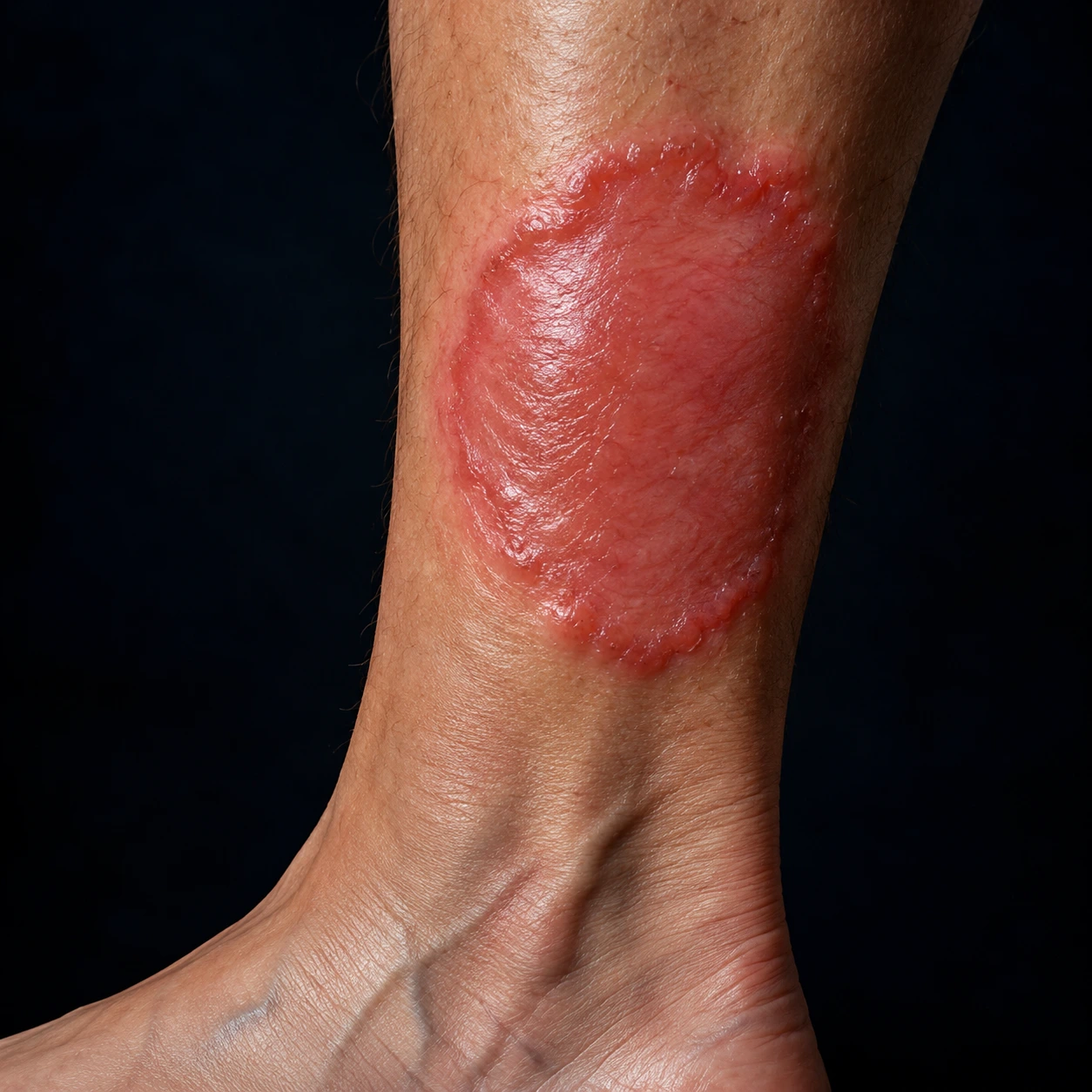
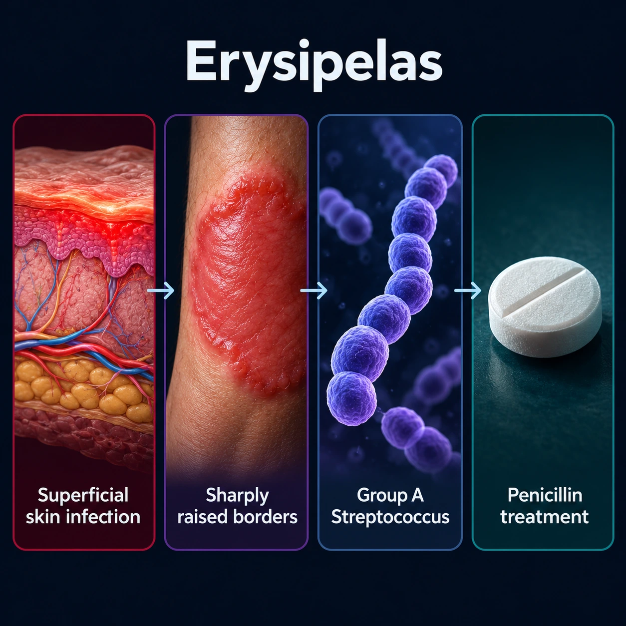
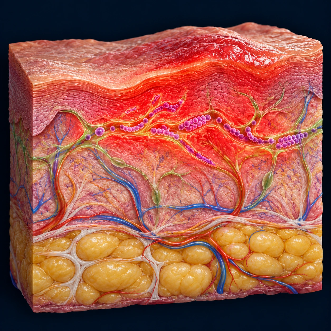
Table of Contents
- What Erysipelas Is
- Pathophysiology: Streptococcal Superficial Lymphatics
- Clinical Features: The Well-Demarcated Red Plaque
- Anatomical Distribution: Lower Limbs and Face
- Risk Factors and Predisposing Conditions
- Diagnosis and Differential Diagnosis
- Distinguishing Erysipelas from Necrotizing Fasciitis
- Treatment: Penicillin as First-Line
- Recurrence and Long-Term Prophylaxis
- Complications and Prognosis
- Key Research Papers
- Connections
- Featured Videos
What Erysipelas Is
Erysipelas is an acute bacterial infection of the upper dermis and superficial lymphatic vessels, distinguished from deeper cellulitis by its sharply demarcated, raised, advancing borders. These well-defined edges are the hallmark of the condition and reflect the infection's confinement to the superficial lymphatic network of the skin. Erysipelas is predominantly caused by Streptococcus pyogenes (Group A Streptococcus, group A Streptococcus, GAS), accounting for approximately 80% of cases.
Historically called "St. Anthony's Fire" due to the intense red color, burning pain, and the appearance of spreading flames across the skin, erysipelas has been recognized as a distinct clinical entity since the Middle Ages. The name itself derives from the Greek erythros (red) and pella (skin).
Annual incidence ranges from approximately 10 to 100 cases per 100,000 population, varying by geographic region and population characteristics. The condition affects all ages, but shows a bimodal distribution: young children tend to develop facial erysipelas (often following streptococcal pharyngitis or minor abrasions), while older adults predominantly develop lower limb erysipelas. Adults are more commonly affected than children overall.
Erysipelas carries significant morbidity primarily from its tendency to recur — approximately 30% of patients experience recurrence within 3 years, rising to 50% over 5 years. Each recurrence damages the lymphatic vasculature, creating a self-perpetuating cycle of increasing susceptibility. Chronic lymphedema is the most important long-term sequela. The condition is rarely fatal in immunocompetent hosts but can be potentially serious in elderly or immunocompromised individuals.
It is essential to distinguish erysipelas from related conditions. Impetigo affects only the superficial epidermis (not dermis or lymphatics), presenting with honey-crusted lesions without systemic symptoms. Cellulitis involves the deep dermis and subcutaneous fat, presenting with poorly defined, blending borders (not the raised, sharply demarcated plaque of erysipelas). Necrotizing fasciitis is a deeper, life-threatening infection of the fascial planes that can superficially mimic erysipelas in early stages — distinguishing these is a clinical emergency.
Pathophysiology: Streptococcal Superficial Lymphatics
The primary organism in erysipelas is Streptococcus pyogenes (Group A Streptococcus, group A Streptococcus) in approximately 80% of cases. Groups C and G beta-hemolytic streptococci account for most of the remaining cases, particularly in immunocompromised patients and the elderly. Staphylococcus aureus is a rare cause — true erysipelas with its superficial lymphatic spread pattern is predominantly a streptococcal disease. Specific M-types of group A Streptococcus, notably M-types 2, 28, and 49, are particularly associated with erysipelas.
Several virulence factors of S. pyogenes contribute to its ability to establish and spread infection:
- M protein: A major surface antiphagocytic protein that resists opsonization and complement deposition, allowing the organism to evade neutrophil killing. M-protein antibodies provide type-specific immunity — explaining susceptibility to different M-type strains.
- Hyaluronic acid capsule: Mimics host connective tissue, helping the organism avoid immune recognition.
- Streptokinase (fibrinolysin): Cleaves fibrin, breaking down tissue barriers and facilitating lateral spread through the dermis and lymphatics.
- Hyaluronidase: Degrades hyaluronic acid in the extracellular matrix, further aiding tissue penetration.
- DNases (streptodornases): Break down DNA in necrotic debris, reducing viscosity of pus and aiding spread.
- Pyrogenic exotoxins (streptococcal superantigens): Responsible for systemic symptoms including fever, chills, and in severe cases toxic shock-like syndrome.
The infection begins when the organism enters via a break in the skin barrier — most commonly interdigital tinea pedis fissures, leg ulcers, dermatitis excoriations, lymphedema-associated skin cracks, or minor trauma. Once introduced, S. pyogenes spreads laterally within the upper dermis and superficial lymphatic vessels. This specific anatomical compartment — above the lymphatic plexus that separates the upper dermis from the deep dermis — is why erysipelas has its characteristic superficial, well-demarcated nature. The infection spreads along the lymphatic channels but cannot easily penetrate the deeper fascial compartments that characterize cellulitis.
The intense erythema, heat, and induration result from a robust host inflammatory response: release of cytokines including tumor necrosis factor alpha (TNF-α), interleukin-1 beta (IL-1β), and interleukin-6 (IL-6) causes profound vasodilatation, increased vascular permeability, and massive neutrophil recruitment. The "peau d'orange" (orange-peel) appearance results from dermal edema with lymphatic obstruction causing accentuation of the dimples around hair follicles. The sharp, raised border reflects the physical boundary between infected and uninfected tissue within the superficial lymphatic compartment — below this border, deeper tissues remain uninvolved, explaining the raised edge.
Clinical Features: The Well-Demarcated Red Plaque
The cardinal clinical feature of erysipelas is a well-demarcated, sharply defined, raised, fiery-red plaque with advancing edges. This sharp border is the KEY distinguishing feature from cellulitis, which has poorly defined, gradually blending borders with surrounding normal skin. In erysipelas, one can draw a precise line between affected and unaffected skin — the edge is palpably raised above the surrounding skin level.
The surface of the plaque has a characteristic shiny, tense, orange-peel texture ("peau d'orange") resulting from dermal edema and lymphatic obstruction that causes dimpling around hair follicles. The plaque is intensely warm and exquisitely tender to touch. The color ranges from bright red to deep crimson, and the plaque expands progressively over days if untreated.
Systemic symptoms are prominent and often precede or accompany the skin findings:
- Fever: typically 38–40°C, often with rigors
- Chills and shaking
- Malaise and fatigue
- Headache
- Systemic symptoms may precede skin signs by 24–48 hours, leading to initial diagnostic uncertainty
Regional lymphadenopathy in the draining lymph node basin is common. Lymphangitic streaks — red lines extending proximally along lymphatic channels — indicate active lymphatic spread and are a hallmark of streptococcal infection in general.
Bullous erysipelas is a severe variant in which blisters form within the plaque from intense dermal edema. These bullae contain clear or serosanguineous fluid and arise from the epidermis being lifted off by subepidermal edema — this is distinct from the toxin-mediated flaccid bullae of bullous impetigo. Petechiae (pinpoint hemorrhages) within the plaque may also be present in more aggressive cases.
The lesion expands centrifugally over days if untreated, and the center may eventually appear darker, violaceous, or develop a woody induration as the acute inflammation subsides but edema persists. Without treatment, the infection can spread to involve large areas of the affected limb or face.
Anatomical Distribution: Lower Limbs and Face
Lower limbs account for 70–80% of erysipelas cases in adults, with the legs and feet being most common. The primary entry portal is interdigital tinea pedis (athlete's foot) with maceration and fissuring of the skin between the toes — this is the single most important predisposing skin breach and the most modifiable risk factor. In a case-control study of leg erysipelas (Dupuy 1999), a broken skin barrier carried an odds ratio of 23.8, and an estimated 61% of leg erysipelas in the population was attributable to toe-web intertrigo (the cracked, soggy skin between the toes); the authors proposed that finding and treating it be evaluated as a way to prevent repeat episodes. Other lower limb portals include venous leg ulcers, diabetic foot ulcers, dermatitis excoriations, lymphedema-associated skin cracks, and surgical incision sites.
Facial erysipelas has a classic clinical presentation: a butterfly distribution across the nose and cheeks with sharply demarcated erythema — this distribution is important to distinguish from the lupus malar rash (which is non-tender, non-advancing, and without fever). Facial erysipelas is now less common in developed countries than historical accounts suggest, but remains an important diagnosis particularly in children. Children more commonly develop facial erysipelas (compared to adults), with portals of entry including streptococcal pharyngitis (organisms spread from throat to facial skin) and minor facial abrasions.
Facial erysipelas carries special clinical significance due to proximity to the orbit and cavernous sinus. The valveless facial veins allow infection to spread intracranially — cavernous sinus thrombosis is a rare but potentially fatal complication requiring urgent intravenous (IV) antibiotics and anticoagulation. Periorbital involvement is one of the usual reasons for hospital admission (see Treatment).
Upper limb erysipelas is classically associated with post-mastectomy lymphedema — women who have undergone axillary lymph node dissection for breast cancer have impaired arm lymphatic drainage, making the arm highly susceptible. This is an important consideration in breast cancer survivors presenting with arm erythema and swelling.
Trunk involvement is rare but can occur in patients with severe immunosuppression or following abdominal surgery. Perianal and genital erysipelas are uncommon but recognized presentations.
Risk Factors and Predisposing Conditions
Skin barrier disruption is the most important and modifiable risk factor category:
- Tinea pedis/athlete's foot (interdigital fissures) — the single most modifiable risk factor for lower limb erysipelas; treated with topical antifungals (and maintenance therapy) as part of recurrence prevention
- Leg ulcers: venous ulcers, diabetic foot ulcers, pressure ulcers
- Dermatitis and eczema with excoriation
- Lymphedema-associated skin cracks and weeping
- Surgical wounds and incision sites
- Insect bites, minor trauma, abrasions
Lymphatic and venous insufficiency impairs the host's first-line defense against bacterial dissemination:
- Lymphedema (primary or secondary post-surgery/radiation) — impaired lymphatic drainage allows streptococci to establish and spread without being cleared
- Chronic venous insufficiency — venous hypertension leads to skin fragility, lipodermatosclerosis, and impaired local immunity
- Saphenous vein harvest for coronary artery bypass grafting — a classic iatrogenic cause of lower limb lymphedema predisposing to recurrent erysipelas
Systemic and metabolic risk factors:
- Obesity: increases lymphatic congestion, causes skin-to-skin friction in folds, and impairs lymphatic function; one of the strongest independent risk factors
- Prior ipsilateral erysipelas: the strongest single predictor of recurrence — each episode causes progressive lymphatic damage, creating a self-reinforcing vulnerability cycle
- Diabetes mellitus: impaired neutrophil function, peripheral neuropathy (reduces awareness of skin injury), microangiopathy
- Immunosuppression: human immunodeficiency virus (HIV) infection, malignancy, systemic corticosteroids, biologic agents, organ transplantation
- Age >60 years: impaired immune defenses, higher burden of comorbidities, reduced skin integrity
- Radiation therapy: causes lymphatic obliteration and skin fibrosis in the treatment field
- Alcoholism and liver disease: impaired immune function, nutritional deficiencies affecting skin integrity
Diagnosis and Differential Diagnosis
Erysipelas is a clinical diagnosis in the vast majority of cases, based on the characteristic appearance of a well-demarcated, raised, warm, tender erythematous plaque with systemic symptoms. Laboratory investigations play a supporting role:
- Blood cultures: positive in only 2–4% of erysipelas cases — bacteremia is rare, and routine blood cultures have very low yield. However, they are usually taken when systemic toxicity, immunosuppression, or diagnostic uncertainty about necrotizing infection is present.
- Inflammatory markers: elevated WBC (neutrophilia), C-reactive protein (CRP), and erythrocyte sedimentation rate (ESR) are present in the majority of cases and support the diagnosis of active bacterial infection.
- Skin swabs: surface swabs are not useful (skin surface organisms do not reflect the infecting organism in the dermis).
- Skin biopsy: rarely needed clinically; shows dense neutrophilic infiltrate in the upper dermis with dilated lymphatic vessels — useful when the diagnosis is genuinely uncertain.
- LRINEC score: calculated from CRP, WBC, hemoglobin, sodium, creatinine, and glucose — used specifically to risk-stratify for necrotizing fasciitis (see dedicated section).
The differential diagnosis of erysipelas includes several important conditions:
- Cellulitis — the most common misdiagnosis in both directions. Deep dermis/subcutaneous infection with poorly defined, blending borders and no raised plaque edge. Fever and systemic symptoms are common to both. The border distinction is the critical clinical differentiator.
- Contact dermatitis — absent fever and systemic symptoms; clear history of allergen or irritant exposure; not tender in the same way; may have vesicles and oozing.
- Superficial thrombophlebitis — follows the course of a superficial vein; cord-like tender induration; ultrasound confirms.
- Deep vein thrombosis — leg swelling and erythema but without the sharply demarcated plaque; ultrasound distinguishes.
- Drug reaction/fixed drug eruption — history of medication; may be sharply demarcated; typically non-tender without systemic symptoms.
- Tinea faciei/corporis — superficial, fine scaling at active border, central clearing; KOH preparation positive for fungal hyphae.
- Dermatomyositis — heliotrope (violaceous) periorbital rash; associated with proximal muscle weakness.
- Facial lupus erythematosus — malar (butterfly) rash is non-tender, non-advancing, non-warm; associated with other lupus features; antinuclear antibody (ANA) positive.
- Angioedema — non-tender, non-erythematous (pale/skin-colored), typically recurrent, rapid onset; no fever; associated with triggers (angiotensin-converting enzyme inhibitors (ACE inhibitors), foods, allergens).
- Necrotizing fasciitis (URGENT) — see dedicated section.
Distinguishing Erysipelas from Necrotizing Fasciitis
Necrotizing fasciitis (NF) is a surgical emergency — early NF can be clinically indistinguishable from erysipelas or cellulitis, and failure to recognize it costs lives. The diagnosis requires a high index of suspicion because the skin appearance in early NF may be deceptively benign while fascial destruction progresses rapidly beneath the surface.
Warning signs that suggest NF rather than erysipelas:
- Pain disproportionate to skin appearance: This is the most important early warning sign. Early NF may look exactly like erysipelas but cause extreme, out-of-proportion pain — then paradoxically become painless as subcutaneous nerves are destroyed (a very ominous late sign).
- Wooden or board-like induration extending beyond the visible erythema — indicates fascial involvement below the skin surface.
- Skin discoloration: Gray-brown, dusky, or violaceous color (late sign indicating skin necrosis from fascial vessel thrombosis).
- Hemorrhagic or gray bullae — distinguished from the clear/serosanguineous bullae of bullous erysipelas; gray fluid or hemorrhagic content strongly suggests deep tissue necrosis.
- Crepitus (crackling sensation on palpation, or gas on imaging) — indicates gas-producing organisms in the deep tissues; NOT present in erysipelas.
- Rapid progression — spreading centimeters per hour despite appropriate antibiotic therapy; erysipelas, while progressive, responds to penicillin within 24–48 hours.
- Systemic toxicity out of proportion to the skin appearance — septic shock, multi-organ failure, altered mental status early in the course.
The LRINEC score (Laboratory Risk Indicator for Necrotizing Fasciitis) uses six laboratory parameters: C-reactive protein (>150 mg/L = 4 points), WBC (>25 × 10⁹/L = 2 points, 15–25 = 1 point), hemoglobin (<11 g/dL = 2 points, 11–13.5 = 1 point), sodium (<135 mmol/L = 2 points), creatinine (>141 µmol/L = 2 points), and glucose (>10 mmol/L = 1 point). In the 2004 study that developed the score (Wong 2004), a cut-off of 6 points told necrotizing fasciitis apart from other soft-tissue infections with a positive predictive value of 92.0% and a negative predictive value of 96.0%, and the authors wrote that patients scoring 6 or more be carefully evaluated for necrotizing fasciitis. The LRINEC score is meant as a supplement to clinical judgment, not a replacement — a score <6 does not exclude NF.
The surgical principle commonly stated is that any diagnostic doubt about necrotizing fasciitis is settled by surgical exploration. Intraoperative findings of gray necrotic fascia, lack of tissue planes ("finger sweeps" pass through tissue without resistance), absence of bleeding from fascial edges, and "dishwater" fluid in the fascial planes confirm the diagnosis. Surgical debridement — often extensive — is the only life-saving intervention. Antibiotics alone cannot treat NF; they are an adjunct to surgery, not an alternative.
Treatment: Penicillin as First-Line
Penicillin is the drug of choice for erysipelas. Streptococcus pyogenes has never developed resistance to penicillin in over 80 years of clinical use — one of the most consistent and reassuring findings in clinical microbiology. There is no indication for broader-spectrum antibiotics in straightforward erysipelas unless methicillin-resistant Staphylococcus aureus (MRSA) or polymicrobial infection is genuinely suspected.
Outpatient oral therapy (mild-moderate erysipelas):
- Amoxicillin 500 mg three times daily × 10–14 days — first-line oral agent
- Phenoxymethylpenicillin (Penicillin V) 500 mg four times daily × 10–14 days — equally effective, widely used in Europe
- Treatment duration of 10–14 days is common practice. A randomized, double-blind trial in uncomplicated cellulitis (Hepburn 2004; 87 people given levofloxacin) found 5 days as effective as 10 days: success in 43 of 44 (98%) after 5 days and 42 of 43 (98%) after 10 days, with no difference at 14 or 28 days. That trial did not test penicillin or erysipelas specifically.
Penicillin allergy:
- Cephalexin 500 mg four times daily — whether a cephalosporin cross-reacts with penicillin depends on its side chain. A 2019 meta-analysis of 21 studies with 1,269 people whose penicillin allergy was confirmed by testing (Picard 2019) found cross-reactivity in 16.45% for aminocephalosporins (the group that includes cephalexin), which share an identical side chain with a penicillin, 5.60% for cephalosporins with an intermediate side-chain similarity, and 2.11% for those with low similarity. The higher risk with aminocephalosporins was seen whether the penicillin allergy was immunoglobulin E-mediated or T-cell-mediated.
- Clindamycin 300–450 mg three times daily — effective alternative with good tissue penetration
- Erythromycin — an option but resistance rates for streptococci are increasing in many regions
Indications for intravenous therapy and hospital admission:
- Systemic toxicity: high fever (>39.5°C), hypotension, tachycardia, altered mental status
- Facial or periorbital erysipelas — proximity to cavernous sinus; risk of intracranial spread
- Immunosuppression (diabetes with poor control, human immunodeficiency virus, malignancy, immunosuppressive therapy)
- Failure to improve on oral therapy within 48–72 hours
- Inability to tolerate oral medications
- Extreme age (young infants, frail elderly)
Intravenous regimens: Benzylpenicillin (penicillin G) 1.2–2.4 g intravenous every 4–6 hours; or ampicillin 1–2 g intravenous every 4–6 hours. Step-down to oral therapy after 48–72 hours of clinical improvement (defervescence, cessation of lesion expansion) is standard practice. Empiric methicillin-resistant Staphylococcus aureus coverage (vancomycin, daptomycin) is appropriate only when methicillin-resistant Staphylococcus aureus is specifically suspected — purulent cellulitis, abscess, known methicillin-resistant Staphylococcus aureus carriage, or failure of beta-lactam therapy.
Adjunctive measures:
- Limb elevation — reduces edema and improves lymphatic and venous drainage; important adjunct that speeds resolution
- Cool, moist compresses for symptomatic relief of burning pain
- Nonsteroidal anti-inflammatory drugs (NSAIDs) or acetaminophen for pain and fever control — however, symptom control can mask signs of progression toward NF
- Incision and drainage is not used — erysipelas has no pus pocket (abscess cavity); I&D is not indicated and may worsen the infection
- Compression garments after complete resolution to address underlying venous/lymphatic insufficiency
- Tinea pedis treated concurrently — topical antifungals (terbinafine, clotrimazole) applied to interdigital spaces throughout and after antibiotic therapy
Recurrence and Long-Term Prophylaxis
Recurrent erysipelas is a major clinical problem — approximately 30% of patients experience at least one recurrence within 3 years, rising to approximately 50% over 5 years. The mechanism of recurrence is a self-reinforcing cycle: each episode of erysipelas causes further damage to the superficial lymphatic vessels, leading to worsening lymphatic insufficiency, which in turn increases susceptibility to the next episode. Breaking this cycle requires both antibiotic prophylaxis and aggressive management of predisposing factors.
Key risk factors for recurrence: prior ipsilateral erysipelas (strongest predictor), lymphedema (primary or secondary), chronic venous insufficiency, obesity, persistent tinea pedis.
Long-term antibiotic prophylaxis is indicated for patients with 2 or more episodes per year:
- Benzathine penicillin G 1.2 million units intramuscular injection monthly — an injected regimen used in practice, reported to reduce recurrence while it is maintained
- Oral phenoxymethylpenicillin (Penicillin V) 250 mg twice daily (continuous) — the oral penicillin approach tested in the PATCH trials (Thomas et al., 2013); requires good patient adherence
- Erythromycin 250 mg twice daily — for documented penicillin allergy
What the largest trials found: in PATCH I (274 people with two or more episodes of leg cellulitis), cheap oral penicillin 250 mg twice a day for 12 months cut recurrences while it was being taken: 22% vs 37% on placebo (hazard ratio 0.55), about one recurrence prevented for every 5 people treated. After the tablets were stopped the protection faded — during the follow-up period without treatment, recurrence was 27% in both groups — and the number of people with side effects did not differ from placebo (37 vs 48). PATCH II (123 people, most after a first episode) gave oral penicillin V for 6 months: 20% vs 33% had a repeat episode (hazard ratio 0.53), a result that did not reach statistical significance because recruitment was slow.
Duration of prophylaxis: continued as long as predisposing risk factors persist, typically for years in patients with lymphedema. Recurrence rates return to pre-prophylaxis levels when antibiotics are discontinued if underlying risk factors have not been addressed. Prophylaxis reduces but does not eliminate recurrences — it is usually combined with non-antibiotic prevention strategies.
Non-antibiotic recurrence prevention (often more important for long-term control):
- Aggressive treatment of tinea pedis — topical antifungal therapy (terbinafine, clotrimazole, miconazole applied twice daily for 4 weeks) plus maintenance therapy (weekly application) indefinitely; attention to foot hygiene, moisture control, and appropriate footwear
- Compression stockings/bandaging for chronic venous insufficiency and lymphedema — reduces edema and improves lymphatic function; crucial for breaking the recurrence cycle
- Skin care and moisturization — regular emollient application to restore skin barrier integrity; particularly important in chronic venous insufficiency with lipodermatosclerosis
- Weight loss — reduces lymphatic congestion, improves lower limb circulation, and reduces skin fold moisture
- Manual lymphatic drainage (physiotherapy-guided) in patients with established lymphedema
- Protection of the affected limb from trauma, insect bites, and skin breaks
Complications and Prognosis
The prognosis of erysipelas is excellent with prompt treatment in immunocompetent adults. Mortality is less than 1% for uncomplicated cases. Response to penicillin is typically evident within 24–48 hours (defervescence, cessation of lesion expansion), with full resolution of the plaque over 1–2 weeks. The skin may remain mildly hyperpigmented or induration may persist for weeks after resolution.
Complications:
- Lymphedema — the most important long-term complication. Repeated episodes progressively destroy lymphatic vessels, causing permanent impairment of lymphatic drainage. This creates chronic swelling, skin thickening and fibrosis, and increasing susceptibility to further infections — the vicious cycle described in the Recurrence section. In advanced cases, the limb develops woody, non-pitting induration ("brawny edema") and eventual "elephantiasis nostras verrucosa" — extreme fibrotic skin thickening with verrucous (wart-like) changes.
- Bacteremia/septicemia: rare (2–4% of cases), but when it occurs, requires intravenous antibiotics and potentially intensive care unit (ICU) care. More common in immunocompromised patients and the elderly.
- Abscess formation: uncommon in typical streptococcal erysipelas; when present, suggests S. aureus co-infection or superinfection.
- Post-streptococcal glomerulonephritis: very rare complication of skin group A Streptococcus infection (though far more commonly associated with impetigo than erysipelas). Presents 1–3 weeks after skin infection with hematuria, proteinuria, and hypertension.
- Necrotizing fasciitis: rare evolution in immunocompromised patients or with particularly virulent strains; requires immediate surgical intervention.
- Cavernous sinus thrombosis (facial erysipelas complication): presents with proptosis, periorbital edema, ophthalmoplegia, and high fever. Requires intravenous antibiotics plus anticoagulation. Serious risk of visual loss or death.
- Endocarditis: extremely rare complication; consider in patients with prosthetic heart valves or native valve disease with bacteremia.
Prognostic factors for worse outcomes:
- Immunocompromised state (diabetes, human immunodeficiency virus, malignancy, immunosuppressive therapy)
- Elderly age with multiple comorbidities
- Facial or periorbital location (proximity to cavernous sinus)
- Delayed presentation or treatment
- Presence of bacteremia
- Evolution toward necrotizing fasciitis
Key Research Papers
- Celestin R, Brown J, Kihiczak G, Schwartz RA. Erysipelas: a common potentially dangerous infection. Acta Dermatovenerol Alp Pannonica Adriat. 2007;16(3):123–127. — PubMed PMID: 17994173
- McNamara DR, Tleyjeh IM, Berbari EF, et al. Incidence of lower-extremity cellulitis: a population-based study in Olmsted county, Minnesota. Mayo Clin Proc. 2007;82(7):817–821. — PubMed PMID: 17605961
- Bonnetblanc JM, Bédane C. Erysipelas: recognition and management. Am J Clin Dermatol. 2003;4(3):157–163. — PubMed PMID: 12627991
- Dupuy A et al. Risk factors for erysipelas of the leg (cellulitis): case-control study. BMJ. 1999. — PubMed PMID: 10364117
- Thomas KS et al. Penicillin to prevent recurrent leg cellulitis. N Engl J Med. 2013. — PubMed PMID: 23635049
- Jorup-Rönström C. Epidemiological, bacteriological and complicating features of erysipelas. Scand J Infect Dis. 1986;18(6):519–524. — PubMed PMID: 3810046
- Wong CH et al. Necrotizing fasciitis: clinical presentation, microbiology, and determinants of mortality. J Bone Joint Surg Am. 2003. — PubMed PMID: 12925624
- Cox NH. Oedema as a risk factor for multiple episodes of cellulitis/erysipelas of the lower leg: a series with community follow-up. Br J Dermatol. 2006. — PubMed PMID: 17034523
- Chartier C, Grosshans E. Erysipelas: an update. Int J Dermatol. 1996;35(11):779–781. — PubMed PMID: 8915727
- Swartz MN. Clinical practice. Cellulitis. N Engl J Med. 2004. — PubMed PMID: 14985488
- UK Dermatology Clinical Trials Network's PATCH Trial Team; Thomas K, Crook A, Foster K, et al. Prophylactic antibiotics for the prevention of cellulitis (erysipelas) of the leg: results of the UK Dermatology Clinical Trials Network's PATCH II trial. Br J Dermatol. 2012;166(1):169–178. — PubMed PMID: 21910701
- Picard M, Robitaille G, Karam F, et al. Cross-reactivity to cephalosporins and carbapenems in penicillin-allergic patients: two systematic reviews and meta-analyses. J Allergy Clin Immunol Pract. 2019;7(8):2722–2738.e5. — PubMed PMID: 31170539
- Hepburn MJ, Dooley DP, Skidmore PJ, et al. Comparison of short-course (5 days) and standard (10 days) treatment for uncomplicated cellulitis. Arch Intern Med. 2004;164(15):1669–1674. — PubMed PMID: 15302637
- Wong CH, Khin LW, Heng KS, Tan KC, Low CO. The LRINEC (Laboratory Risk Indicator for Necrotizing Fasciitis) score: a tool for distinguishing necrotizing fasciitis from other soft tissue infections. Crit Care Med. 2004;32(7):1535–1541. — PubMed PMID: 15241098
PubMed Topic Searches
- PubMed: Erysipelas streptococcal skin infection pathogenesis
- PubMed: Erysipelas cellulitis differential diagnosis clinical features
- PubMed: Erysipelas penicillin treatment lower limb
- PubMed: Erysipelas recurrence prophylaxis penicillin prevention
- PubMed: Necrotizing fasciitis erysipelas diagnosis LRINEC
- PubMed: Erysipelas lymphedema risk factor predisposing
- PubMed: Erysipelas epidemiology incidence adults elderly
- PubMed: Erysipelas facial streptococcus group A treatment
Connections
- Dermatology
- Cellulitis
- Impetigo
- Scabies
- Eczema
- Contact Dermatitis
- Stevens-Johnson Syndrome
- Psoriasis
- Sepsis
- Streptococcal Infection
- Lichen Planus
- Seborrheic Dermatitis
- Cellulitis: History and Discovery — Hippocrates to Fehleisen, the history both infections share.
- Lymphedema — the most important long-term sequela and, once established, the strongest risk factor for the next attack; compression and lymphatic drainage break the cycle.