Stevens-Johnson Syndrome
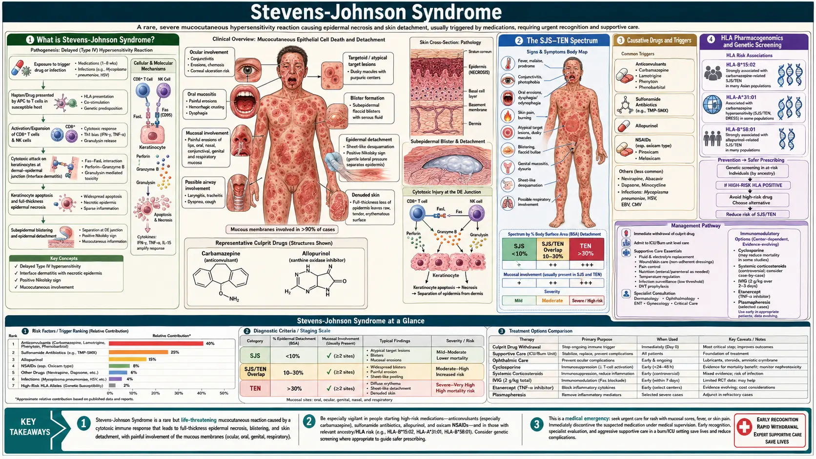
Table of Contents
- What Stevens-Johnson Syndrome Is
- The SJS–TEN Spectrum
- Causative Drugs and Triggers
- Human Leukocyte Antigen Pharmacogenomics and Genetic Screening
- Pathophysiology: Granulysin and Keratinocyte Apoptosis
- Clinical Features and Prodrome
- Diagnosis: Nikolsky Sign and Skin Biopsy
- SCORTEN Prognostic Score
- Treatment: Supportive Care and Immunomodulation
- Ophthalmological Emergency
- Key Research Papers
- Connections
- Featured Videos
What Stevens-Johnson Syndrome Is
Stevens-Johnson Syndrome (SJS) is a rare but potentially fatal mucocutaneous hypersensitivity reaction characterized by widespread keratinocyte death, skin detachment, and severe erosions of multiple mucous membranes. It was first described by Albert Mason Stevens and Frank Chambliss Johnson in 1922 in two children with fever, stomatitis, and conjunctivitis — a clinical picture now recognized as the mucocutaneous inflammatory syndrome we understand today.
SJS belongs to a continuum of escalating severity: SJS (epidermal detachment less than 10% of body surface area), SJS/TEN overlap (10–30% detachment), and toxic epidermal necrolysis (TEN or Lyell syndrome) (greater than 30% detachment). The distinction is prognostic, not mechanistic — all represent the same immune-mediated keratinocyte destruction driven by cytotoxic lymphocytes and natural killer cells. TEN, at its most severe, resembles a full-thickness burn covering the majority of the body surface.
Incidence is low — approximately 1–6 cases per million people per year for SJS, 0.4–1.2 per million for TEN — but mortality is high: roughly 5–10% for SJS and 25–35% for TEN, reaching over 90% in patients with the highest SCORTEN prognostic scores. Drug reactions account for approximately 70–80% of adult SJS/TEN cases; infection (primarily Mycoplasma pneumoniae) accounts for most pediatric SJS cases.
The SJS–TEN Spectrum
The International Consensus Classification (Bastuji-Garin, 1993) defines the spectrum by the percentage of total body surface area (BSA) showing epidermal detachment — meaning frank denuded skin or skin that detaches with lateral pressure:
- SJS: Detachment of less than 10% BSA. Widespread dusky erythematous or purpuric macules, typically with atypical target lesions and prominent mucous membrane erosions. Mortality approximately 5–10%.
- SJS/TEN overlap: Detachment 10–30% BSA. Intermediate severity; prognosis between the two poles.
- TEN (Lyell syndrome): Detachment greater than 30% BSA. Large sheets of epidermis slough off, leaving raw, bleeding dermis resembling a severe burn. Without specialized burn-unit care, death from sepsis, fluid loss, and multi-organ failure is likely. Mortality approximately 25–35%, rising sharply with the SCORTEN score.
An important pediatric subset is SJS due to Mycoplasma pneumoniae (MP-SJS or MIRM — Mycoplasma-Induced Rash and Mucositis). Children with this form have prominent mucous membrane erosions (oral, ocular, urogenital) with minimal or absent skin detachment. Some experts propose separating MIRM from drug-induced SJS given the more benign course and different trigger.
Erythema multiforme (EM) is a related but distinct entity — typically self-limited, recurrent, predominantly caused by herpes simplex virus, with classic "true" target lesions (three zones: central dusky or necrotic core, pale edematous ring, erythematous periphery) and less severe mucous membrane involvement than SJS. The old term "EM major" for severe EM with mucosal involvement has been largely abandoned in favor of the SJS classification to avoid confusion.
Causative Drugs and Triggers
Drug reactions are the predominant cause of SJS and TEN in adults. The interval from first drug exposure to SJS onset is typically 1–3 weeks (the time needed for sensitization and immune expansion). On rechallenge or with an already-sensitized patient, onset may occur within 3–10 days. Recognizing the responsible drug and stopping it immediately is the single most important therapeutic intervention.
High-Risk Drug Classes
- Allopurinol: The most common cause of SJS/TEN in Asia and increasingly in Western countries. Risk is highest in the first 2 months of use. In the EuroSCAR case-control study (379 patients, 1,505 controls), 17.4% of patients and 1.9% of controls had taken it. Risk was higher at 200 mg a day or more (adjusted odds ratio 36) than at lower doses (3.0), and was confined to the first 8 weeks of use. Strong human leukocyte antigen-B*58:01 association (see below).
- Sulfonamide antibiotics: Sulfamethoxazole (in co-trimoxazole, also called trimethoprim-sulfamethoxazole), sulfadiazine, sulfasalazine. Among the originally identified SJS triggers and remain high-risk.
- Aromatic anticonvulsants: Carbamazepine (highest risk, especially with human leukocyte antigen-B*15:02 in Han Chinese and Southeast Asian populations), lamotrigine, phenytoin, phenobarbital. Carbamazepine-induced SJS risk rises dramatically if human leukocyte antigen-B*15:02 is present — a 2013 pooled analysis put the odds at about 80 times higher (odds ratio 79.84).
- Oxicam nonsteroidal anti-inflammatory drugs (NSAIDs): Piroxicam, meloxicam. Implicated in disproportionate SJS/TEN cases relative to their use prevalence.
- Nevirapine: Non-nucleoside reverse transcriptase inhibitor (antiretroviral). High rates of SJS especially in women and patients with higher CD4 counts at initiation.
- Other anticonvulsants: Oxcarbazepine, zonisamide, levetiracetam (lower risk than aromatic anticonvulsants).
- Other antibiotics: Aminopenicillins (ampicillin, amoxicillin), cephalosporins, fluoroquinolones, tetracyclines.
Infectious Triggers
Mycoplasma pneumoniae is the dominant infectious trigger, predominantly in children and young adults. SJS following Mycoplasma tends to have less skin detachment with more severe mucosal involvement. Herpes simplex virus (HSV) triggers erythema multiforme more commonly than SJS. Histoplasma, cytomegalovirus (CMV), and human immunodeficiency virus (HIV) infection have also been associated with SJS/TEN.
Human Leukocyte Antigen Pharmacogenomics and Genetic Screening
One of the most striking advances in understanding SJS/TEN has been the discovery of specific human leukocyte antigen (HLA) alleles that confer extremely high risk for drug-specific reactions. These associations reflect the fact that the drug or its reactive metabolite must be presented by a specific major histocompatibility complex molecule to activate cytotoxic T cells.
Key Human Leukocyte Antigen–Drug Associations
- Human leukocyte antigen-B*15:02 — Carbamazepine (Han Chinese, Thai, Vietnamese, Filipino, South Asian populations): A 2013 pooled analysis of 16 studies gave an overall odds ratio for SJS/TEN of 79.84 (95% confidence interval 28.45–224.06): 115.32 in Han Chinese, 54.43 in Thais and 221.00 in Malaysians. No white or Japanese patients with SJS/TEN carried the allele. The US Food and Drug Administration (FDA) carbamazepine label carries a boxed warning stating that patients with ancestry in genetically at-risk populations "should be screened" before starting, and that those testing positive "should not be treated with carbamazepine unless the benefit clearly outweighs the risk." The label reports the allele in greater than 15% of people in Hong Kong, Thailand, Malaysia and parts of the Philippines, about 10% in Taiwan, 4% in North China, 2–4% on average in South Asians and less than 1% in Japan and Korea; it is largely absent in people not of Asian origin. A 2011 screening study in Taiwan tested 4,877 people: 7.7% carried the variant and were advised to avoid carbamazepine. None of the non-carriers who took carbamazepine developed SJS/TEN, against about 10 cases expected from the historical rate of 0.23%. Oxcarbazepine carries partial cross-reactivity.
- Human leukocyte antigen-B*57:01 — Abacavir: This human immunodeficiency virus antiretroviral causes a severe hypersensitivity reaction (including skin manifestations) almost exclusively in human leukocyte antigen-B*57:01 carriers. Prospective screening and exclusion of human leukocyte antigen-B*57:01-positive patients has essentially eliminated abacavir hypersensitivity reactions. This was the first pharmacogenomic screening to be routinely implemented in clinical practice and represents a proof-of-concept for the field.
- Human leukocyte antigen-B*58:01 — Allopurinol: Strongly associated with allopurinol-induced SJS/TEN in Han Chinese, Thai, Korean, and other Asian populations. Allele frequency approximately 6–8% in these populations versus 0.1% in Europeans. Some guidelines recommend screening in at-risk Asian populations before allopurinol initiation.
- Human leukocyte antigen-A*31:01 — Carbamazepine (European and Japanese populations): Associated with multiple carbamazepine hypersensitivity syndromes (SJS, drug reaction with eosinophilia and systemic symptoms, maculopapular exanthema) in Europeans and Japanese — a population with low human leukocyte antigen-B*15:02 prevalence. Some European guidelines recommend this screen before carbamazepine initiation.
The mechanistic basis is direct: the drug (or a reactive metabolite) binds non-covalently to the peptide-binding groove of the specific human leukocyte antigen molecule, forming a pharmacological interaction (the "p-i concept") that mimics antigen presentation and activates cytotoxic T cells bearing specific T-cell receptors. This is distinct from classical drug haptenation.
Pathophysiology: Granulysin and Keratinocyte Apoptosis
The cellular mechanism of keratinocyte death in SJS/TEN has been substantially elucidated over the past two decades. Activated drug-specific cytotoxic CD8+ T lymphocytes (CTLs) and natural killer (NK) cells accumulate in the skin in response to the drug-HLA interaction. These effector cells kill keratinocytes through multiple pathways:
Granulysin — The Key Mediator
The landmark 2008 study by Chung and colleagues identified granulysin as the dominant soluble cytotoxin responsible for widespread keratinocyte death in SJS/TEN. Granulysin is a cationic cytolytic protein stored in secretory lysosomes of CTLs and natural killer cells. It is released upon target cell recognition and penetrates the keratinocyte membrane, triggering caspase-independent apoptosis via mitochondrial pathway disruption. Critically, granulysin was found in blister fluid at concentrations two to four orders of magnitude (100 to 10,000 times) higher than perforin, granzyme B or soluble Fas ligand, and its concentration correlates directly with disease severity. In mouse models, injection of recombinant granulysin alone reproduced the skin detachment of SJS/TEN.
Additional Apoptotic Pathways
- Fas–FasL pathway: Keratinocytes in SJS/TEN upregulate Fas (CD95); blister-fluid lymphocytes express FasL. Fas–FasL interaction triggers keratinocyte apoptosis. This pathway was initially thought to be primary but is now understood as contributory alongside granulysin.
- Perforin and granzyme B: CTLs release perforin pores in the keratinocyte membrane through which granzyme B (a serine protease) enters and cleaves caspases, triggering apoptosis.
- Tumor necrosis factor alpha (TNF-α): Elevated in blister fluid; activates NF-κB and JNK pathways leading to keratinocyte apoptosis and dermal inflammation.
- Annexin A1: A 2014 study identified annexin A1, found in the fluid from drug-exposed blood mononuclear cells of SJS/TEN patients, as a key mediator of keratinocyte death. SJS/TEN keratinocytes carry abundant FPR1, its receptor, and the death pathway described is necroptosis, which depends on the RIP1 and RIP3 proteins.
The end result — massive synchronous keratinocyte apoptosis across large areas of skin — is histologically visible as full-thickness epidermal necrosis with a clean dermal-epidermal separation, seen on skin biopsy as necrotic epidermis separating above an otherwise relatively normal dermis (in contrast to burns, where the dermis is also damaged).
Clinical Features and Prodrome
Prodromal Phase (Days 1–3)
SJS/TEN characteristically begins with a nonspecific flu-like prodrome that may be mistaken for a viral illness or drug side effect: fever (often greater than 39°C), malaise, sore throat, photophobia, burning eyes, and skin tenderness or dysesthesia. This prodrome precedes the skin eruption by 1–3 days. The early mucosal burning and photophobia are important warning signs — mucous membrane involvement in SJS/TEN nearly always precedes or accompanies the rash, unlike most other drug eruptions.
Cutaneous Eruption
The rash typically begins on the trunk and face, then spreads to involve the extremities. Initial lesions are dusky erythematous macules or atypical target lesions — flat, lacking the raised edematous ring of true erythema multiforme targets. These macules coalesce rapidly, and the centers of the lesions become purpuric or necrotic. Within 1–3 days, the epidermis begins to detach, starting with blister formation within the macules. The skin may feel painful and tender before visible blistering — patients often describe it as a "burning" or "raw" sensation under the skin. The Nikolsky sign is positive: lateral pressure on erythematous skin causes the epidermis to shear and separate laterally.
Mucous Membrane Involvement — Mandatory for SJS Diagnosis
Mucous membrane erosions are a defining feature of SJS and are required for the diagnosis. Involvement includes:
- Oral mucosa: Hemorrhagic crusting of the lips (pathognomonic when paired with skin lesions), erosions of the buccal mucosa, tongue, and palate. Severe pain limits eating and drinking — nutritional support is essential.
- Ocular mucosa: Conjunctival hyperemia, chemosis, pseudomembrane formation, eyelid edema. This is the most feared mucosal site — see Ophthalmological Emergency section.
- Urogenital mucosa: Erosions of the glans penis or vaginal mucosa, dysuria, urinary retention. Urethral stricture is a long-term complication.
- Respiratory mucosa: Tracheobronchial involvement with hemorrhagic mucositis in severe TEN can cause respiratory failure.
Diagnosis: Nikolsky Sign and Skin Biopsy
Clinical Assessment
SJS/TEN is primarily a clinical diagnosis. The key diagnostic features are:
- Recent (within 1–3 weeks) drug exposure (or Mycoplasma infection in children)
- Prodromal fever and mucosal burning
- Dusky/purpuric flat macules or atypical targets — not raised urticarial targets
- Mandatory mucous membrane erosions (at least two sites)
- Positive Nikolsky sign in erythematous skin
- Measured or estimated BSA detachment to classify SJS vs. overlap vs. TEN
The RegiSCAR (Registry of Severe Cutaneous Adverse Reactions) group developed validated scoring criteria using the above features to confirm SJS/TEN diagnosis and exclude mimics (e.g., staphylococcal scalded skin syndrome, which also shows Nikolsky positivity but is caused by exfoliatin toxin rather than immune-mediated keratinocyte death, and which spares mucous membranes).
Skin Biopsy
Biopsy of an early lesion (ideally within 24–48 hours) shows full-thickness epidermal necrosis with keratinocyte apoptosis at all levels of the epidermis, a sparse lymphocytic infiltrate at the dermal-epidermal junction, and subepidermal blister formation with little dermal inflammation. The necrotic epidermis rests on an otherwise relatively intact dermis — this contrasts with staphylococcal scalded skin syndrome, where the split is superficial (subcorneal) and epidermal necrosis is absent. Frozen sections of the detached "roof" can rapidly confirm full-thickness vs. superficial split.
Laboratory Evaluation
No blood test is diagnostic of SJS/TEN, but laboratory evaluation guides severity assessment and management:
- Complete blood count (lymphopenia is common; eosinophilia may suggest drug reaction with eosinophilia and systemic symptoms rather than SJS/TEN)
- Comprehensive metabolic panel (electrolytes, renal function — fluid losses cause electrolyte disturbances)
- Liver function tests (hepatitis in some drug reactions; drugs eliminated hepatically)
- Blood cultures (fever may reflect secondary sepsis)
- Mycoplasma pneumoniae serology and polymerase chain reaction (PCR) (especially in children)
SCORTEN Prognostic Score
SCORTEN (SCORe of Toxic Epidermal Necrolysis) is a validated prognostic scoring system developed by Bastuji-Garin and colleagues (2000) to predict in-hospital mortality for SJS/TEN. It is calculated from seven binary variables assessed within the first 24 hours of hospital admission:
- Age >40 years (1 point)
- Presence of malignancy (1 point)
- Heart rate >120 bpm (1 point)
- Initial BSA epidermal detachment >10% (1 point)
- Serum blood urea nitrogen (BUN) >28 mg/dL (urea >10 mmol/L) (1 point)
- Serum bicarbonate <20 mEq/L (1 point)
- Serum glucose >252 mg/dL (14 mmol/L) (1 point)
SCORTEN score and associated predicted mortality:
- 0–1 points: ~3.2% mortality
- 2 points: ~12.1%
- 3 points: ~35.3%
- 4 points: ~58.3%
- 5+ points: >90% mortality
A study of 144 patients found SCORTEN performed well on each of the first 5 days and best on day 3, and its authors recommended computing it again on day 3. Recalculating around days 3 and 5 is also described, as BSA detachment typically peaks during this period and the metabolic variables evolve. It is used to guide triage decisions (transfer to burn unit), inform family communication, and stratify patients in clinical trials.
Treatment: Supportive Care and Immunomodulation
Step 1 — Immediate Drug Withdrawal
The single most impactful intervention is immediate identification and discontinuation of the causative drug. Each day of continued drug exposure is associated with increased BSA detachment and higher mortality. Where multiple drugs are implicated, usual practice is to stop all non-essential drugs, starting with the drug begun most recently or most strongly associated with SJS/TEN (using tools like the ALDEN algorithm — Algorithm of Drug Causality in Epidermal Necrolysis). The half-life of the offending drug also predicts how quickly the reaction will plateau after withdrawal.
Step 2 — Specialized Wound Care (Burn Unit)
Patients with SJS/TEN detachment greater than 10% are typically transferred to a burn center or specialized dermatology intensive care unit. Management parallels burn care:
- Fluid resuscitation: Significant fluid, protein, and electrolyte losses through denuded skin — aggressive intravenous (IV) fluid replacement, typically with Ringer's lactate, tailored to urine output.
- Temperature regulation: Environmental temperature set to 30–32°C to minimize the thermogenic cost of evaporative water loss through denuded skin.
- Wound management: Gentle non-adhesive dressings (petrolatum gauze, biologic membranes, or biosynthetic dressings). Intact necrotic epidermis is left in place rather than forcibly debrided, because it provides a temporary biologic barrier. Early ophthalmology consultation.
- Pain management: Intravenous opioid analgesia for wound care procedures; anxiolytics for dressing changes.
- Nutritional support: Early nasogastric or nasojejunal tube feeding (oral intake is impaired by mucosal erosions).
- Infection surveillance: Surveillance cultures (wounds, blood, urine); systemic antibiotics only for documented infection (prophylactic antibiotics are not part of usual guidance and may select resistant organisms).
Immunomodulatory Therapies
Evidence for specific immunosuppressive therapies in SJS/TEN is limited by the rarity of the disease and by the small number of small randomized controlled trials. A 2022 Cochrane review found three randomized trials among nine studies (308 participants), with very low certainty for several comparisons. A 2026 review of 43 studies concluded that there is no clear evidence that systemic therapies beat supportive care, which remains the primary treatment; compared with supportive care alone, only etanercept showed a significant reduction in deaths (risk ratio 0.32, 95% confidence interval 0.11–0.93, with imprecision and unclear clinical importance noted by the authors), and cyclosporine did better than intravenous immunoglobulin (risk ratio 0.18). The most studied options:
- Cyclosporine: Often described as the immunomodulatory agent with the best evidence base in SJS/TEN, though the Cochrane review rates the certainty for cyclosporine versus intravenous immunoglobulin as very low, and a single-center review of 64 patients found a standardized mortality ratio of 0.43 for cyclosporine and 1.43 for intravenous immunoglobulin. Mechanistically, cyclosporine inhibits calcineurin → blocks T-cell activation and IL-2 production → suppresses CTL-mediated keratinocyte killing. Multiple retrospective series and a meta-analysis suggest cyclosporine reduces mortality and may halt disease progression. Typical dosing: 3–5 mg/kg/day for 10–14 days with rapid taper. Renal function monitoring is essential. Cyclosporine also decreases tumor necrosis factor alpha and Fas-FasL-mediated apoptosis.
- Intravenous immunoglobulin (IVIg): Proposed mechanisms include Fas pathway blockade and granulysin neutralization. Early retrospective reports suggested benefit; however, a systematic review and a retrospective analysis of patients in the prospective EuroSCAR study (odds ratio for death 1.4 in France and 1.5 in Germany, neither significant) did not demonstrate a survival advantage over supportive care alone at standard doses. High-dose IVIg (2–3 g/kg) combined with cyclosporine is used in some centers.
- Etanercept: tumor necrosis factor alpha inhibitor (soluble receptor). Several prospective series from Taiwan suggest etanercept 50 mg subcutaneous ×2 doses significantly reduces mortality compared to historical controls (SCORTEN-predicted vs. actual mortality). A randomized trial from Taiwan published in 2018 (96 patients, etanercept versus corticosteroids) reported SCORTEN-predicted mortality of 17.7% against 8.3% observed on etanercept, skin healing in 14 versus 19 days in moderate-to-severe cases (P = 0.010), and gastrointestinal bleeding in 2.6% versus 18.2%. The Cochrane review concluded that etanercept may reduce deaths compared with corticosteroids, although its confidence interval was consistent with possible benefit and possible harm. Etanercept has an excellent safety profile in the short term (no immunosuppression-related infections in short courses).
- Corticosteroids: Controversial and not listed as primary therapy. Early retrospective data suggested harm (increased infection, delayed wound healing); however, very early (pre-detachment) short-course systemic corticosteroids may abort the reaction in selected cases. Current guidelines do not support routine use.
- Thalidomide: Formally studied and found to be harmful — a randomized, placebo-controlled trial was stopped because of excess deaths, with 10 of 12 patients dying on thalidomide against 3 of 10 on placebo. The authors concluded that thalidomide is detrimental in toxic epidermal necrolysis.
- Janus kinase (JAK) inhibitors (research stage): A 2024 study found interferon signalling and JAK/STAT activation in toxic epidermal necrolysis skin. JAK inhibitor pills eased the disease in two mouse models, and treatment was reported safe and followed by rapid skin regrowth in seven patients. The abstract reports these seven people without a comparison group.
Ophthalmological Emergency
Ocular involvement in SJS/TEN is an ophthalmological emergency — the most feared long-term complication is permanent blindness from progressive conjunctival cicatrization (scarring), and the window for intervention to preserve vision is narrow.
Acute Ocular Manifestations
- Conjunctival hyperemia, chemosis (edema), photophobia, purulent discharge
- Pseudomembrane formation on the palpebral conjunctiva (fibrinous membranes that, if not removed, cause severe symblepharon)
- Eyelid margin erosions and trichiasis (lashes turning inward, abrading the cornea)
- Corneal epithelial defects and ulceration — risk of corneal perforation in severe cases
Symblepharon Formation
The most catastrophic acute complication is symblepharon — adhesion of the bulbar conjunctiva (over the eyeball) to the palpebral conjunctiva (lining the eyelids). Symblephara form rapidly as the inflamed conjunctival surfaces make contact and scar. Once established, symblephara restrict eye movement, distort lid anatomy, cause chronic dry eye, and expose the cornea to persistent abrasion. Aggressive early intervention is required to prevent their formation.
Ophthalmological Management
- Daily ophthalmology assessment from day 1 of hospitalization, regardless of symptom severity at presentation (ocular involvement may evolve rapidly)
- Pseudomembrane removal by the ophthalmologist daily or twice daily using a glass rod or moist cotton-tipped applicator — prevents the symblepharon adhesions from forming
- Amniotic membrane transplantation (AMT): Fresh or cryopreserved amniotic membrane can be placed as an ocular surface overlay within the first 72–96 hours in patients with severe conjunctival involvement. AMT provides an anti-inflammatory, anti-fibrotic substrate that dramatically reduces the risk of symblepharon and long-term visual impairment
- Preservative-free lubricating eye drops every 1–2 hours; topical antibiotic drops for corneal defects; topical cyclosporine A for chronic ocular surface disease
- Long-term: Scleral contact lenses for dry eye and irregular astigmatism; surgical management of established symblephara; mucous membrane grafting in severe cases
Even with optimal acute management, approximately 35% of SJS/TEN survivors have significant long-term ocular sequelae, and 3–10% are legally blind. Patients who survived SJS/TEN are typically followed by an ophthalmologist long-term, as dry eye disease and conjunctival fibrosis can progress for years after the acute episode.
Key Research Papers
Key peer-reviewed studies on SJS/TEN pathogenesis, pharmacogenomics, and treatment. Each PMID link opens the study on PubMed.
- Chung WH, et al. Medical genetics: a marker for Stevens-Johnson syndrome. Nature. 2004;428(6982):486. — PubMed PMID: 15057820
- Chung WH, et al. Granulysin is a key mediator for disseminated keratinocyte death in Stevens-Johnson syndrome and toxic epidermal necrolysis. Nat Med. 2008;14(12):1343-1350. — PubMed PMID: 19029983
- Bastuji-Garin S, et al. SCORTEN: a severity-of-illness score for toxic epidermal necrolysis. J Invest Dermatol. 2000;115(2):149-153. — PubMed PMID: 10951229
- Mockenhaupt M, et al. Stevens-Johnson syndrome and toxic epidermal necrolysis: assessment of medication risks with emphasis on recently marketed drugs. The EuroSCAR-study. J Invest Dermatol. 2008;128(1):35-44. — PubMed PMID: 17805350
- Halevy S, et al. Allopurinol is the most common cause of Stevens-Johnson syndrome and toxic epidermal necrolysis in Europe and Israel. J Am Acad Dermatol. 2008;58(1):25-32. — PubMed PMID: 17919772
- Nassif A, et al. Evaluation of the potential role of cytokines in toxic epidermal necrolysis. J Invest Dermatol. 2004;123(5):850-855. — PubMed PMID: 15482470
- Nassif A, et al. Toxic epidermal necrolysis: effector cells are drug-specific cytotoxic T cells. J Allergy Clin Immunol. 2004;114(5):1209-1215. — PubMed PMID: 15536433
- Schneck J, et al. Effects of treatments on the mortality of Stevens-Johnson syndrome and toxic epidermal necrolysis: a retrospective study on patients included in the prospective EuroSCAR Study. J Am Acad Dermatol. 2008;58(1):33-40. — PubMed PMID: 17919775
- Kirchhof MG, et al. Retrospective review of Stevens-Johnson syndrome/toxic epidermal necrolysis treatment comparing intravenous immunoglobulin with cyclosporine. J Am Acad Dermatol. 2014;71(5):941-947. — PubMed PMID: 25087214
- Chen P, et al. Carbamazepine-induced toxic effects and HLA-B*1502 screening in Taiwan. N Engl J Med. 2011;364(12):1126-1133. — PubMed PMID: 21428768
- Dodiuk-Gad RP, et al. Stevens-Johnson syndrome and toxic epidermal necrolysis: an update. Am J Clin Dermatol. 2015;16(6):475-493. — PubMed PMID: 26481651
- Saito N, et al. An annexin A1-FPR1 interaction contributes to necroptosis of keratinocytes in severe cutaneous adverse drug reactions. Sci Transl Med. 2014;6(245):245ra95. — PubMed PMID: 25031270
- Wang CW, et al. Randomized, controlled trial of TNF-α antagonist in CTL-mediated severe cutaneous adverse reactions. J Clin Invest. 2018;128(3):985-996. — PubMed PMID: 29400697
- Jacobsen A, et al. Systemic interventions for treatment of Stevens-Johnson syndrome (SJS), toxic epidermal necrolysis (TEN), and SJS/TEN overlap syndrome. Cochrane Database Syst Rev. 2022;3(3):CD013130. — PubMed PMID: 35274741
- Heuer R, Paulmann M, Mockenhaupt M, Nast A. Systemic immunomodulating therapies for epidermal necrolysis (Stevens-Johnson syndrome/toxic epidermal necrolysis): a systematic review and meta-analysis. J Dtsch Dermatol Ges. 2026;24(1):34-42. — PubMed PMID: 40905522
- Nordmann TM, et al. Spatial proteomics identifies JAKi as treatment for a lethal skin disease. Nature. 2024;635(8040):1001-1009. — PubMed PMID: 39415009
- Tangamornsuksan W, et al. Relationship between the HLA-B*1502 allele and carbamazepine-induced Stevens-Johnson syndrome and toxic epidermal necrolysis: a systematic review and meta-analysis. JAMA Dermatol. 2013;149(9):1025-1032. — PubMed PMID: 23884208
- Guégan S, et al. Performance of the SCORTEN during the first five days of hospitalization to predict the prognosis of epidermal necrolysis. J Invest Dermatol. 2006;126(2):272-276. — PubMed PMID: 16374461
- Wolkenstein P, et al. Randomised comparison of thalidomide versus placebo in toxic epidermal necrolysis. Lancet. 1998;352(9140):1586-1589. — PubMed PMID: 9843104
Curated PubMed topic searches: