⚠ Evidence warning: Clinical trials or other evidence do not support the claims that doxycycline is an important added treatment for filarial lymphedema and that long-term antibiotics slow lymphatic deterioration. This page keeps it as a report of what people, websites or other media say.
Lymphedema
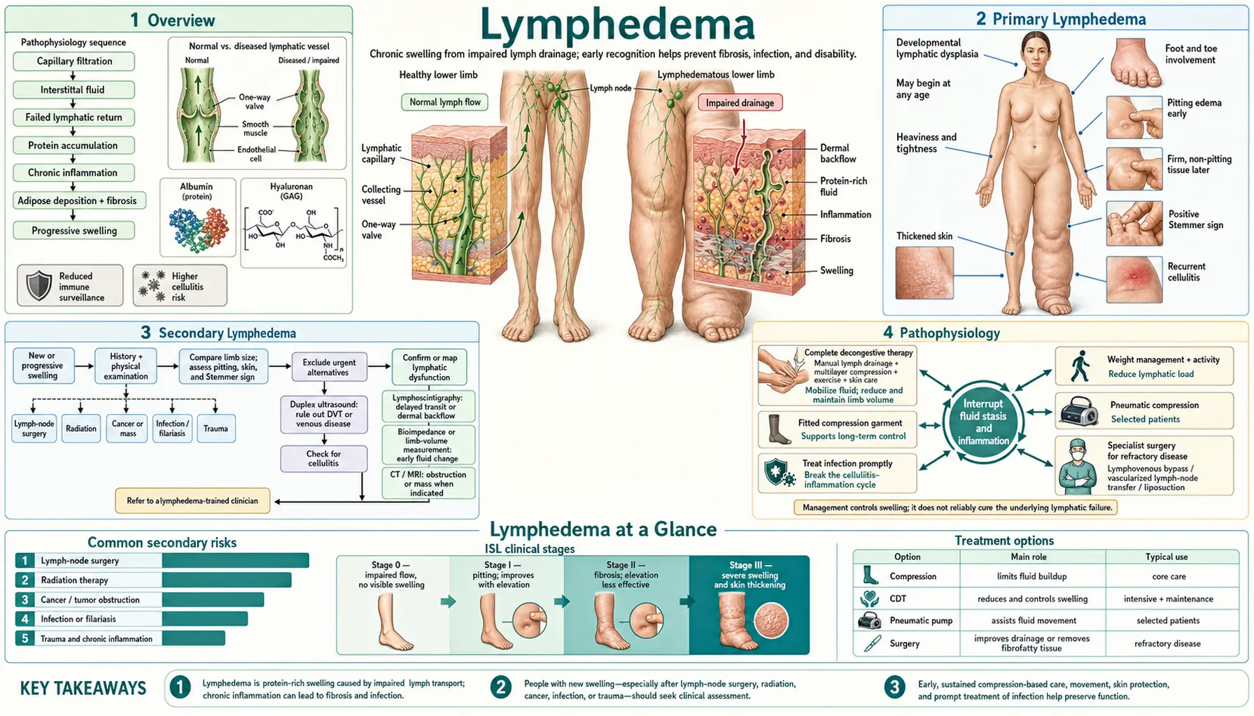
Table of Contents
1. Overview
Lymphedema is a chronic, progressive condition caused by impairment of the lymphatic system, resulting in the accumulation of protein-rich interstitial fluid within the tissues. Unlike simple venous edema — which consists of low-protein transudates — lymphedema fluid is protein-rich, a distinction with profound clinical consequences. The elevated protein concentration drives a secondary inflammatory response mediated by macrophages and mast cells, leading over time to adipose deposition, fibrosis, and irreversible tissue remodeling if the condition remains untreated or poorly managed.
Globally, the most common cause of lymphedema is filariasis — a parasitic infection caused mainly by Wuchereria bancrofti, and also by Brugia species, and transmitted by Culex, Anopheles and Aedes mosquitoes. The World Health Organization reported 51 million people infected as of 2018, a 74% decline since 2000, with 657 million people in 39 countries still living in areas that need preventive chemotherapy in 2023 and at least 36 million living with chronic disease. Older sources give an estimate of 120 million people in endemic tropical regions and over 200 million affected individuals worldwide when all causes are included. In the United States and other high-income countries, the leading cause is breast cancer treatment: axillary lymph node dissection combined with radiation therapy. Secondary lymphedema following cancer treatment profoundly affects quality of life, functional capacity, and psychological well-being for millions of cancer survivors.
Lymphedema is considered incurable with current therapies. There is no pharmacological treatment that reverses lymphatic damage or restores normal lymphatic architecture. However, with consistent, lifelong application of Complete Decongestive Therapy (CDT) — the gold standard of care — the condition is highly manageable. Limb volumes can be substantially reduced, complications prevented, and quality of life preserved. Early diagnosis and early initiation of treatment are critical to preventing progression to irreversible fibrofatty tissue changes.
2. Primary Lymphedema
Primary lymphedema arises from hereditary or congenital malformations of the lymphatic system, occurring in the absence of external injury or disease. It is considerably rarer than secondary lymphedema and is classified by age of onset into three main subtypes:
Congenital Lymphedema (Milroy Disease)
Present at birth or manifesting within the first year of life, congenital lymphedema accounts for approximately 10–15% of primary cases. The best-characterized genetic form is Milroy disease, caused by loss-of-function mutations in the FLT4 gene encoding vascular endothelial growth factor receptor 3, the principal receptor for lymphangiogenic growth factors, vascular endothelial growth factors C and D. Inheritance is autosomal dominant with variable penetrance. Presentation is typically bilateral lower extremity edema, often involving the dorsum of the feet at birth. Males with Milroy disease frequently have hydroceles. Lymphoscintigraphy demonstrates aplastic or hypoplastic lymphatics.
Lymphedema Praecox (Meige Disease)
This is the most common form of primary lymphedema, accounting for 65–80% of cases. Onset occurs between puberty and age 35, most commonly around menarche in females. Presentation is typically unilateral lower extremity edema beginning in the foot and ankle, gradually progressing proximally. The genetic basis is heterogeneous; many cases are sporadic. FLT4 mutations account for fewer cases than in congenital lymphedema. The condition predominantly affects females (approximately 9:1 female-to-male ratio).
Lymphedema Tarda
Lymphedema tarda designates primary lymphedema with onset after age 35. It is rare and may represent late-manifesting lymphatic hypoplasia that becomes clinically apparent when lymphatic reserve is challenged by additional insults such as infection, pregnancy, or weight gain.
Other Primary Lymphedema Syndromes
- Lymphedema-distichiasis syndrome: caused by FOXC2 mutations (a forkhead transcription factor); characterized by double row of eyelashes (distichiasis) plus lower extremity lymphedema; autosomal dominant; lymphatic valves are abnormal rather than absent
- GATA2 deficiency: mutations in GATA2 cause a complex syndrome including lymphedema, myelodysplastic syndrome, deafness, and immunodeficiency (MonoMAC syndrome)
- Hennekam syndrome: intestinal lymphangiectasia, peripheral lymphedema, facial anomalies, and intellectual disability; CCBE1 gene mutations
- Nonne-Milroy-Meige variants: historically used clinical descriptors now partially replaced by genotype-based classification
3. Secondary Lymphedema
Secondary lymphedema results from damage to a previously normal lymphatic system. It is far more common than primary lymphedema and spans a wide range of causes depending on geographic context and individual medical history.
Cancer Treatment (Leading Cause in Developed Countries)
Surgical removal and/or radiation of lymph nodes — the primary routes of lymphatic drainage — causes secondary lymphedema in a significant proportion of cancer survivors:
- Breast cancer: a 2013 pooled analysis of 72 studies put arm lymphedema at 16.6% overall and 21.4% in prospective cohort studies, with 19.9% after axillary lymph node dissection (ALND) and 5.6% after sentinel lymph node biopsy (SLNB); adding radiation to either procedure substantially increases risk
- Gynecologic cancers (cervical, endometrial, vulvar): pelvic and inguinal lymph node dissection produces lower extremity and genital lymphedema
- Prostate cancer: pelvic lymph node dissection; lower extremity and scrotal lymphedema
- Melanoma: lymph node dissection of axillary or inguinal nodes
- Head and neck cancers: cervical lymph node dissection and radiation → facial and cervical lymphedema
Filariasis (Leading Cause Worldwide)
Lymphatic filariasis is caused by parasitic nematodes transmitted by mosquito bites. Wuchereria bancrofti accounts for 90% of cases; Brugia malayi and B. timori account for the remainder. Adult worms inhabit the lymphatic vessels and lymph nodes, causing obstruction, inflammation, and progressive destruction of lymphatic architecture. Over years, this leads to the dramatic tissue hypertrophy characteristic of elephantiasis — massive enlargement of limbs and genitalia. Older sources give 120+ million people in endemic regions of sub-Saharan Africa, South Asia, and Southeast Asia; the World Health Organization reported 51 million infected as of 2018. Its Global Programme to Eliminate Lymphatic Filariasis (GPELF) uses mass drug administration, and the regimen depends on which other filarial diseases are present: albendazole alone twice a year where loiasis occurs; ivermectin with albendazole in countries with onchocerciasis; diethylcarbamazine with albendazole in countries without onchocerciasis; and all three together in countries without onchocerciasis where other programme conditions are met.
Recurrent Skin Infections
Recurrent bacterial cellulitis or erysipelas — most commonly caused by Group A Streptococcus — scars lymphatic channels with each episode. This creates a vicious cycle: existing lymphedema impairs skin integrity and immune surveillance, making infection more likely; each infection causes further lymphatic damage, worsening the underlying lymphedema. Compression, skin care, and antibiotic prophylaxis are the measures described for breaking this cycle; the trial results are under Complications.
Other Causes
- Obesity: a strong, independent risk factor; adipose tissue compresses lymphatics and impairs the intrinsic lymphatic pumping mechanism; substantially increases cancer treatment-related lymphedema risk
- Chronic venous insufficiency: chronically elevated venous hydrostatic pressure overloads lymphatic drainage capacity, eventually producing a combined phlebolymphostatic edema
- Trauma and burns: disruption of lymphatic channels
- Malignant obstruction: tumor invasion of lymph nodes or lymphatic channels producing obstructive lymphedema
- Radiation fibrosis: even without surgery, high-dose radiation causes progressive fibrosis of lymphatic vessels and nodes
4. Pathophysiology
Under normal conditions, the lymphatic system absorbs approximately 2–4 liters of interstitial fluid per day — the excess produced by capillary filtration that the venous system cannot recapture. Critically, this fluid contains proteins, lipids, cellular debris, and immune cells that would be pathologic if allowed to accumulate in tissue. Intrinsic contractions of lymphangions (the functional units of collecting lymphatics, each bounded by smooth muscle and intraluminal valves) propel lymph centrally toward the thoracic duct. Extrinsic forces — muscle contraction, respiratory pressure changes, arterial pulsation — augment lymphatic flow.
When lymphatic transport capacity is exceeded or the system is damaged, protein-rich fluid accumulates in the interstitium. The protein content is the key pathophysiologic driver distinguishing lymphedema from other forms of edema. High interstitial protein concentrations:
- Activate local macrophages and mast cells, initiating a chronic inflammatory response
- Drive secretion of transforming growth factor-beta (TGF-β) and other profibrotic cytokines
- Stimulate adipocyte proliferation and hypertrophy, producing the adipose component of chronic lymphedema
- Promote extracellular matrix deposition and progressive tissue fibrosis
Once significant adipose deposition and fibrosis occur — roughly corresponding to ISL Stage II–III — the tissue changes become irreversible. This is why elevation alone (effective in venous edema) fails to reduce advanced lymphedema: the volume is no longer primarily fluid but fibrofatty tissue.
Upstream of the obstruction, collecting lymphatics dilate and their smooth muscle walls hypertrophy initially, but eventually become flaccid and lose intrinsic pumping function (lymphangion failure). Increased lymphatic pressure drives retrograde flow into the superficial dermal lymphatic plexus — dermal backflow — creating characteristic skin changes including lymphatic vesicles, lymphorrhea (leakage of lymph through the skin surface), and the dermal fibrosis that produces the positive Stemmer sign.
In filariasis, adult worm metabolites and the bacteria Wolbachia (an obligate endosymbiont of filarial nematodes) both contribute to the inflammatory lymphatic damage. Targeting Wolbachia with doxycycline is described as producing significant anti-filarial effects and as an important adjunct to standard antiparasitic therapy. Three placebo-controlled trials published in 2024 tested 6 weeks of doxycycline added to hygiene care in filarial lymphedema. In Sri Lanka it “did not significantly impact lymphedema progression”; in southern India there was no difference between the groups; in Tanzania limbs improved in 14.4% and 15.2% on the two doxycycline doses against 23.4% on placebo, though fewer people had an acute attack on doxycycline in the first 6 months.
5. Clinical Presentation and Staging
The clinical presentation of lymphedema varies considerably by cause, duration, and stage. Common symptoms include a sense of limb heaviness or fullness, tightness, aching, reduced flexibility of joints, and recurrent skin infections. Cosmetic disfigurement and functional impairment contribute substantially to psychological morbidity.
Physical Examination Findings
- Early (Stage I): soft pitting edema that reduces with elevation; no skin changes
- Intermediate (Stage II): pitting edema that does not reduce reliably with elevation; skin begins to thicken; early fibrosis palpable
- Advanced (Stage III): non-pitting edema due to fibrosis and adipose; skin hyperkeratosis; papillomatosis (cobblestone skin); verrucous (wart-like) changes; massive limb deformity (elephantiasis)
The Stemmer Sign
The Stemmer sign is the most clinically useful physical examination finding specific to lymphedema. It is elicited by attempting to pinch and lift a fold of skin at the base of the second toe (lower extremity) or the base of the second finger (upper extremity). A positive Stemmer sign — inability to pick up a skin fold due to dermal fibrosis and thickening — is essentially pathognomonic for lymphedema. A negative Stemmer sign does not exclude early lymphedema, but a positive sign in the appropriate clinical context confirms the diagnosis. Importantly, venous edema alone does not produce a positive Stemmer sign, making it a useful differentiating feature.
International Society of Lymphology (ISL) Staging
- Stage 0 (Latent/Subclinical): lymphatic transport capacity is impaired and measurable by lymphoscintigraphy or bioimpedance, but clinical edema is not yet apparent; tissue at risk following lymph node surgery may be in this stage for months to years
- Stage I (Spontaneously Reversible): visible pitting edema; soft tissue; reduces substantially with limb elevation overnight; no fibrosis; skin is normal
- Stage II (Spontaneously Irreversible): edema may or may not pit; elevation alone does not substantially reduce volume; fibrosis is developing; skin is beginning to thicken; this is the critical stage — intervention prevents progression to Stage III
- Stage III (Elephantiasis): massive non-pitting edema; gross skin changes including papillomatosis, hyperkeratosis, acanthosis, verrucous projections; extreme functional impairment; classic presentation of Bancroftian filariasis; very rare in breast cancer-related lymphedema
Lipedema: An Important Differential Diagnosis
Lipedema is a distinct chronic condition of abnormal subcutaneous adipose tissue distribution, predominantly affecting women, that is frequently misdiagnosed as lymphedema or obesity. Key distinguishing features:
- Bilateral and symmetric distribution, typically involving the legs from the hips to the ankles, and sometimes the arms — but always sparing the feet
- Negative Stemmer sign (the feet are spared)
- Disproportionate pain and tenderness on palpation relative to visible appearance
- Easy bruising
- Does not improve with elevation or caloric restriction
- Female predominance; hormonal triggers (puberty, pregnancy, menopause)
- Advanced lipedema can develop secondary lymphedema (lipolymphedema), complicating the picture
6. Diagnosis
In typical clinical presentations — a breast cancer survivor developing ipsilateral arm swelling after axillary lymph node dissection, or a returning traveler from a filariasis-endemic region with progressive lower extremity edema — the diagnosis of lymphedema can often be made clinically. However, objective measurement and imaging studies are increasingly important for early detection, staging, surgical planning, and research.
Objective Limb Volume Assessment
- Circumferential measurements: sequential circumferential measurements at fixed intervals along the limb; compared to the contralateral limb; a difference of more than 2 cm at any point is a commonly used diagnostic threshold
- Water displacement volumetry: the gold standard for volume measurement; the limb is submerged in water and displaced volume measured; impractical for routine clinical use
- Bioelectrical impedance analysis: measures the ratio of extracellular to intracellular fluid; a bioimpedance spectroscopy device is U.S. Food and Drug Administration-cleared for surveillance of breast cancer-related lymphedema; particularly useful for detecting Stage 0 subclinical lymphedema before clinical edema appears; allows intervention before fibrosis develops. In the PREVENT randomized trial of breast cancer patients, early swelling triggered a 4-week compression sleeve; progression to full treatment afterward was 7.9% when bioimpedance triggered it and 19.2% when a tape measure did. In the same trial, 3-year risk of chronic lymphedema was 4.4% after sentinel node biopsy (4.2% with regional node radiation) and 25.8% after axillary dissection (26% with regional node radiation)
Imaging Studies
- Lymphoscintigraphy (radionuclide lymphoscintigraphy): the gold standard imaging modality for lymphedema; intradermal injection of technetium-99m-labeled nanocolloid is followed by dynamic and delayed imaging; visualizes lymphatic transport velocity, identifies obstruction location, and distinguishes primary from secondary lymphedema; defines collateral pathways; guides CDT planning
- Indocyanine green (ICG) near-infrared fluorescence lymphography: intradermal ICG injection with near-infrared camera imaging of superficial lymphatics; high spatial resolution; identifies dermal backflow patterns in real time; increasingly used to guide lymphatic microsurgery (lymphovenous anastomosis); not widely available outside specialized centers
- MR lymphography: high-resolution magnetic resonance imaging following intracutaneous gadolinium injection; delineates deep lymphatic anatomy; useful for surgical planning
- Duplex ultrasound: primarily used to exclude deep vein thrombosis (DVT) in acute unilateral limb swelling — an important and urgent differential diagnosis; also evaluates for venous insufficiency component
Laboratory Studies
Laboratory testing is directed at excluding alternative or concurrent diagnoses rather than confirming lymphedema itself. Serum albumin is checked to exclude hypoalbuminemia (which causes bilateral, protein-poor, pitting edema). Protein-losing enteropathy markers, thyroid function (myxedema causes non-pitting edema), and renal function (nephrotic syndrome) are relevant in appropriate clinical contexts. In endemic regions, circulating filarial antigen testing and nocturnal peripheral blood microfilaria smears diagnose active filariasis.
7. Complete Decongestive Therapy (CDT)
Complete Decongestive Therapy is the internationally recognized gold standard for lymphedema management. It is delivered by certified lymphedema therapists and consists of four components — manual lymphatic drainage, compression, therapeutic exercise, and skin care — applied across two distinct phases.
Phase I: Intensive Reduction Phase
Phase I is therapist-driven, typically conducted daily over 2–6 weeks. Its goal is maximum volume reduction before the patient transitions to self-management.
- Manual Lymphatic Drainage (MLD): a highly specialized, light-pressure massage technique developed by Emil Vodder in the 1930s. Unlike traditional deep-tissue massage, MLD uses gentle, rhythmic, circular strokes applied in the direction of lymphatic flow. The technique begins centrally (neck or axilla) to clear proximal lymphatics before working distally. Crucially, MLD redirects lymph flow around the obstructed region through collateral lymphatic pathways — for example, from the affected arm across the trunk to functioning axillary or inguinal nodes. Sessions typically last 45–60 minutes. MLD differs from conventional massage, which does not activate superficial lymphatics and may actually worsen lymphedema. How much it adds is less certain: a 2015 Cochrane review of 6 small trials after breast cancer found compression bandaging alone reduced swelling by 30–38.6%, and adding MLD gave a further 7.11% in two trials. MLD was safe in all trials, and 60–80% of participants reported feeling better for pain and heaviness whichever treatment they received.
- Multilayer Inelastic Compression Bandaging: low-elasticity (short-stretch) bandages are applied over padding in multiple layers following each MLD session. Short-stretch bandages create high working pressure during muscle contraction (supporting lymphatic propulsion) but low resting pressure (comfortable between activities). They are worn 23 hours per day during Phase I. This is distinct from elastic compression stockings, which are unsuitable for Phase I because they provide high resting pressure that obstructs lymphatics at rest.
- Decongestive Lymphatic Exercises: specific exercises performed with bandages in place to maximize the muscle-pump effect on lymphatic flow.
- Skin and Nail Care: meticulous hygiene, moisturization, and treatment of fungal infections (tinea pedis) to minimize infection entry points. A single episode of cellulitis can set back months of CDT progress.
Phase II: Maintenance Phase
Phase II is lifelong and primarily patient-managed, maintaining the volume reduction achieved in Phase I.
- Compression Garments: medical-grade flat-knit compression garments (Class II–III, typically 20–40+ mmHg) are custom measured at the end of Phase I and worn during all waking hours. Garments are replaced every 3–6 months as they lose elasticity. Proper garment fitting is critical — poorly fitted garments can create tourniquets or fail to maintain reduction. Night-time compression (bandaging or custom foam garments) is used for moderate-to-severe cases.
- Self-MLD and Self-Bandaging: patients are trained to perform simplified MLD on themselves and to apply night bandaging.
- Exercise: regular aerobic and resistance exercise is described as safe and beneficial when performed with compression garments. In the PAL trial, 141 breast cancer survivors with stable arm lymphedema did slow, progressive weight lifting twice a week: arm swelling rose 5% or more in 11% against 12% of controls, and flare-ups were 14% against 29%. Swimming, walking, cycling, and gentle resistance training all support lymphatic flow through the muscle pump. Contrary to historical fears, exercise does not worsen breast cancer-related lymphedema when appropriate compression is worn.
- Ongoing Skin Care and Precautions: long-standing precaution lists name heat, sunburn, cuts, insect bites, blood pressure measurement on the affected arm, tight jewelry, and blood draws on the affected limb, along with prompt treatment of any skin break or infection. A prospective breast cancer cohort (3,041 arm measurements) found no significant link between arm volume and blood draws, injections or flights, and concluded blood pressure readings also may not be associated; cellulitis, a body mass index of 25 or more, axillary lymph node dissection and regional node radiation were linked to arm swelling.
Adjunctive and Emerging Treatments
- Sequential Intermittent Pneumatic Compression (IPC): pneumatic pumps that sequentially inflate compartments of a sleeve from distal to proximal; evidence-based adjunct to CDT, particularly for home maintenance; described as an addition to MLD in Phase I, not a replacement
- Low-Level Laser Therapy (LLLT): photobiomodulation with near-infrared light; some evidence for tissue softening, fibrosis reduction, and pain relief in chronic upper extremity lymphedema; several small randomized controlled trials (RCTs) support benefit; mechanisms include stimulation of lymphatic endothelial cell proliferation and anti-inflammatory effects
- Pharmacologic: no drug is approved for lymphedema. Diuretics are contraindicated — they reduce intravascular volume without mobilizing interstitial protein and worsen the protein concentration in remaining tissue fluid. Benzopyrones (coumarins) showed early promise in trials but have not achieved mainstream adoption. In a 1999 placebo-controlled crossover trial of 140 women with arm lymphedema after breast cancer, coumarin 200 mg twice daily left arm volume unchanged, the authors called it not effective, and blood tests showed liver toxicity in 6%. A 2004 Cochrane review said no conclusion was possible because of poor trial quality. Ketoprofen cream has limited evidence. The human ketoprofen trials used tablets: 75 mg by mouth 3 times daily for 4 months in 21 patients (open-label) and in a placebo-controlled trial of 34, with reduced skin thickness and improved tissue findings under the microscope.
Surgical Treatments
Surgery is reserved for patients with severe or refractory lymphedema who fail optimal CDT. Three main surgical approaches exist:
- Lymphovenous Anastomosis: microsurgical connection of lymphatic vessels to small venules (venulae), creating a bypass around the obstruction. Requires supermicrosurgery techniques (vessels 0.3–0.8 mm diameter). Best results are achieved in early-to-moderate lymphedema (ISL Stage I–early II) before significant fibrosis develops. Reduces limb volumes and substantially decreases cellulitis frequency and CDT burden in appropriately selected patients.
- Vascularized Lymph Node Transfer (VLNT): healthy lymph nodes with their blood supply are transplanted from a donor site (commonly groin, axilla, or submental region) to the affected area. The mechanism is debated — may involve lymphangiogenesis from transferred nodes or physical drainage around the obstruction. Best for moderate-to-severe lymphedema. Donor-site lymphedema risk requires careful preoperative lymphatic mapping of the donor site.
- Liposuction (Lymphatic Liposuction): removes the fibrofatty component of chronic lymphedema that CDT cannot reduce. Indicated for Stage III or Stage II cases with significant adipose hypertrophy where the limb volume no longer responds to CDT. The Charles procedure (excision of all subcutaneous tissue) is reserved for extreme elephantiasis. Critical caveat: the technique relies on lifelong, full-time compression after liposuction — without it, lymphedema rapidly recurs to or beyond pre-surgical volumes.
8. Complications
Uncontrolled or poorly managed lymphedema carries significant complication risk, some of which are life-threatening.
Recurrent Cellulitis and Erysipelas
Bacterial skin infections — most commonly caused by Group A Streptococcus pyogenes — are the most frequent and clinically important complication of lymphedema. Impaired lymphatic function reduces the ability to clear pathogens from the interstitium and deliver immune cells to the site of infection. Entry points include tinea pedis (athlete's foot), fissures, minor wounds, and insect bites. Each infection episode further damages lymphatics, worsening the underlying lymphedema and increasing susceptibility to future infections.
Management of acute cellulitis requires systemic antibiotics — intravenous penicillin or cephalosporins for severe cases, oral amoxicillin-clavulanate or cephalexin for milder presentations. For repeated episodes, often quoted as two or more per year, practice includes prophylactic low-dose penicillin V (250–500 mg orally twice daily or once daily) or monthly benzathine penicillin G intramuscularly. The PATCH I trial enrolled 274 people with two or more episodes of leg cellulitis and gave penicillin 250 mg twice daily or placebo for 12 months: recurrence during prophylaxis was 22% against 37% (hazard ratio 0.55), and the protection faded progressively once the drug was stopped. Long-term antibiotic prophylaxis is also described as slowing lymphatic deterioration, which the trial’s abstract does not report.
Compression has been tested too. A 2020 randomized trial of 84 people with chronic leg swelling and repeated cellulitis was stopped early: 15% on leg compression plus education had another episode against 40% on education alone (hazard ratio 0.23). The authors note it was small, single-center and unblinded.
Lymphangitis
Infection of the lymphatic channels themselves produces red streaking extending proximally from a wound or skin break toward regional lymph nodes. Associated with fever, chills, and systemic illness. Hospitals treat this as urgent and give systemic antibiotics.
Lymphorrhea
Lymph fluid leaking through the skin surface, often through lymphatic vesicles (lymphangiomas) or fissures in advanced lymphedema. Creates an entry point for infection and requires careful wound management. Compression is central to control.
Stewart-Treves Syndrome (Lymphangiosarcoma)
Stewart-Treves syndrome is a rare but extremely aggressive angiosarcoma — a malignant tumor of vascular endothelial cells — arising in the context of chronic lymphedema. First described in 1948 by Fred Stewart and Norman Treves in patients with post-mastectomy lymphedema, it typically appears as violaceous (purple-blue), multifocal skin lesions in a chronically edematous limb, often after 10 or more years of lymphedema. The incidence is estimated below 0.1% of post-mastectomy lymphedema patients. Despite multimodal treatment including radical surgery (forequarter or hindquarter amputation) and radiation, prognosis is dismal — median survival after diagnosis is less than 1–2 years. The pathogenesis likely involves chronic lymphedema-induced angiogenesis, local immunosuppression, and upregulation of vascular endothelial growth factor A. A new skin lesion in a chronically lymphedematous limb is a sign that hospitals treat as urgent, with a biopsy.
Functional and Psychological Disability
Chronic lymphedema impairs activities of daily living, limits employment, restricts clothing and footwear choices, and demands a lifelong daily management regimen. Rates of depression and anxiety are substantially elevated compared to cancer survivors without lymphedema. Body image distress is common. Psychosocial support, peer support groups, and lymphedema-specialized occupational therapy are important components of comprehensive care.
Filariasis-Specific Complications
- Genital lymphedema: hydrocele (scrotal fluid accumulation; the most common manifestation of Bancroftian filariasis), scrotal elephantiasis, and labial/vulvar lymphedema
- Chyluria: lymph entering the urinary system, producing milky white urine; occurs when retroperitoneal lymphatics rupture into the renal collecting system or ureter
- Chylous ascites and chylothorax: lymph accumulation in the peritoneal or pleural cavities
Cheap hygiene care is what the 2024 trials in filarial lymphedema credit. The World Health Organization’s essential package is skin hygiene, raising the affected limb, exercise, protective footwear, wound care and support during acute attacks. The southern India trial of 200 people concluded this package is of substantial benefit, reducing acute attacks and improving quality of life and clinical condition, and in the Sri Lanka trial 29–34% of 200 people improved by a lymphedema stage over 2 years, with or without doxycycline.
Key Research Papers
- Rockson SG, Rivera KK. Estimating the population burden of lymphedema. Ann N Y Acad Sci. 2008;1131:147–154. — PubMed PMID: 18519968
- Armer JM, Stewart BR. A comparison of four diagnostic criteria for lymphedema in a post-breast cancer population. Lymphat Res Biol. 2005;3(4):208–217. — PubMed PMID: 16379589
- Ridner SH, Dietrich MS, Cowher MS, et al. A randomized trial evaluating bioimpedance spectroscopy versus tape measurement for the prevention of lymphedema following treatment for breast cancer: interim analysis. Ann Surg Oncol. 2019;26(10):3250–3259. — PubMed PMID: 31054038
- Kasseroller RG. The Vodder School: the Vodder method. Cancer. 1998;83(12 Suppl American):2840–2842. — PubMed PMID: 9874409
- Chang DW, Suami H, Skoracki R. A prospective analysis of 100 consecutive lymphovenous bypass cases for treatment of extremity lymphedema. Plast Reconstr Surg. 2013;132(5):1305–1314. — PubMed PMID: 24165613
- Brorson H, Ohlin K, Olsson G, Svensson B, Svensson H. Controlled compression and liposuction treatment for lower extremity lymphedema. Lymphology. 2008;41(2):52–63. — PubMed PMID: 18720912
- Garza R 3rd, Skoracki R, Hock K, Povoski SP. A comprehensive overview on the surgical management of secondary lymphedema of the upper and lower extremities related to prior oncologic therapies. BMC Cancer. 2017;17(1):468. — PubMed PMID: 28679373
- Olszewski WL. Pathophysiological and clinical observations of obstructive lymphedema of the limbs. In: Cluzan RV, Pecking AP, Lokiec FM, eds. Progress in Lymphology. Elsevier; 1994. — Search PubMed
- Damstra RJ, Mortimer PS. Diagnosis and therapy in children with lymphoedema. Phlebology. 2008;23(6):276–286. — PubMed PMID: 19029008
- Maclellan RA, Greene AK. Lymphedema. Semin Pediatr Surg. 2014;23(4):191–197. — PubMed PMID: 25241097
- Mortimer PS, Rockson SG. New developments in clinical aspects of lymphatic disease. J Clin Invest. 2014;124(3):915–921. — PubMed PMID: 24590276
- Brayton KM, Hirsch AT, O'Brien PJ, Cheville A, Karaca-Mandic P, Rockson SG. Lymphedema prevalence and treatment benefits in cancer: impact of a therapeutic intervention on health outcomes and costs. PLoS One. 2014;9(12):e114597. — PubMed PMID: 25470383
- Ridner SH, Dietrich MS, Boyages J, et al. A comparison of bioimpedance spectroscopy or tape measure triggered compression intervention in chronic breast cancer lymphedema prevention. Lymphat Res Biol. 2022;20(6):618–628. — PubMed PMID: 35099283
- Boyages J, Vicini FA, Manavi BA, et al. Axillary treatment and chronic breast cancer-related lymphedema: implications for prospective surveillance and intervention from a randomized controlled trial. JCO Oncol Pract. 2023;19(12):1116–1124. — PubMed PMID: 37816208
- DiSipio T, Rye S, Newman B, Hayes S. Incidence of unilateral arm lymphoedema after breast cancer: a systematic review and meta-analysis. Lancet Oncol. 2013;14(6):500–515. — PubMed PMID: 23540561
- Ezzo J, Manheimer E, McNeely ML, et al. Manual lymphatic drainage for lymphedema following breast cancer treatment. Cochrane Database Syst Rev. 2015;2015(5):CD003475. — PubMed PMID: 25994425
- Schmitz KH, Ahmed RL, Troxel A, et al. Weight lifting in women with breast-cancer-related lymphedema. N Engl J Med. 2009;361(7):664–673. — PubMed PMID: 19675330
- Ferguson CM, Swaroop MN, Horick N, et al. Impact of ipsilateral blood draws, injections, blood pressure measurements, and air travel on the risk of lymphedema for patients treated for breast cancer. J Clin Oncol. 2016;34(7):691–698. — PubMed PMID: 26644530
- Thomas KS, Crook AM, Nunn AJ, et al. Penicillin to prevent recurrent leg cellulitis. N Engl J Med. 2013;368(18):1695–1703. — PubMed PMID: 23635049
- Webb E, Neeman T, Bowden FJ, Gaida J, Mumford V, Bissett B. Compression therapy to prevent recurrent cellulitis of the leg. N Engl J Med. 2020;383(7):630–639. — PubMed PMID: 32786188
- Loprinzi CL, Kugler JW, Sloan JA, et al. Lack of effect of coumarin in women with lymphedema after treatment for breast cancer. N Engl J Med. 1999;340(5):346–350. — PubMed PMID: 9929524
- Badger C, Preston N, Seers K, Mortimer P. Benzo-pyrones for reducing and controlling lymphoedema of the limbs. Cochrane Database Syst Rev. 2004;2004(2):CD003140. — PubMed PMID: 15106192
- Rockson SG, Tian W, Jiang X, et al. Pilot studies demonstrate the potential benefits of antiinflammatory therapy in human lymphedema. JCI Insight. 2018;3(20):e123775. — PubMed PMID: 30333315
- Yahathugoda TC, De Silva NL, Ruben J, et al. LEDoxy-SL: a placebo-controlled, double-blind, randomized, 24-month trial of six weeks of daily doxycycline plus hygiene-based essential care for reducing progression of filarial lymphedema in Sri Lanka. Am J Trop Med Hyg. 2024;111(4_Suppl):52–65. — PubMed PMID: 39043165
- Krishnasastry S, Ashok A, Devidas A, et al. Efficacy and safety of adding 6 weeks of doxycycline to the essential package of care to treat filarial lymphedema: a double-blind, randomized, controlled trial in southern India. Am J Trop Med Hyg. 2024;111(4_Suppl):83–93. — PubMed PMID: 39362214
- Ngenya A, Klarmann-Schulz U, John W, et al. Efficacy of intensified hygiene measures with or without the addition of doxycycline in the management of filarial lymphedema: a randomized double-blind, placebo-controlled clinical trial in Tanzania. Am J Trop Med Hyg. 2024;111(4_Suppl):33–51. — PubMed PMID: 39191236
- World Health Organization. Lymphatic filariasis (fact sheet). 21 November 2024. — who.int fact sheet
10. PubMed Research Searches
Use these curated searches to explore the current research literature on lymphedema:
- Lymphedema treatment complete decongestive therapy outcomes
- Breast cancer lymphedema prevention axillary dissection
- Lymphovenous anastomosis microsurgery lymphedema
- Lymphedema filariasis Wuchereria bancrofti
- Manual lymphatic drainage evidence review
- Lymphedema compression garments management
- Primary lymphedema FOXC2 FLT4 genetics
- Stemmer sign lymphedema clinical diagnosis
- Lymphedema cellulitis prevention prophylaxis
- Stewart-Treves angiosarcoma lymphedema
- Lymph node transfer vascularized lymphedema surgery
- Lipedema vs lymphedema differential diagnosis
Connections
- Hematology
- Deep Vein Thrombosis — deep vein thrombosis causes post-thrombotic syndrome with a secondary lymphedema component; both present with unilateral leg swelling and require careful differentiation by duplex ultrasound and clinical assessment
- Erysipelas — recurrent streptococcal skin infection both causes and is exacerbated by lymphedema; bacteria enter through compromised lymphedematous skin, and each episode of infection further scars lymphatic channels in a damaging cycle
- Oncology — axillary lymph node dissection and radiation therapy for breast cancer is the leading cause of lymphedema in the United States; pelvic lymph node dissection for gynecologic and prostate cancers causes lower extremity lymphedema
- Thrombocytopenia — shares the Hematology specialty section; both involve vascular and lymphatic system pathology with significant implications for tissue integrity and infection risk
- Endocrinology — obesity is a strong independent risk factor for secondary lymphedema; adipose tissue compresses lymphatic channels and impairs intrinsic lymphatic pumping; weight management is an important adjunct to lymphedema therapy
- Manual Lymphatic Drainage — the light, rhythmic skin-stretching technique that forms one of the four pillars of complete decongestive therapy, with an honest account of how much it adds beyond compression
- Discussed in depth on these pages: Cellulitis