⚠ Evidence warning: Clinical trials or other evidence do not support the claim that the chance of reversing type 2 diabetes ends about 10 years after diagnosis. This page keeps it as a report of what people, websites or other media say.
Insulin Resistance
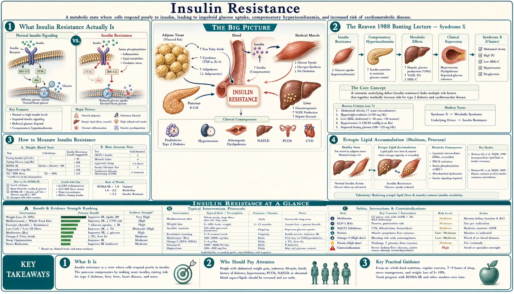
Insulin resistance is the single most important upstream cause of metabolic disease in the developed world. It is the underlying physiologic abnormality in Type 2 diabetes, fatty liver disease, polycystic ovary syndrome, gestational diabetes, and the constellation of cardiovascular risk Gerald Reaven named "Syndrome X" in his 1988 Banting Lecture. It precedes overt Type 2 diabetes by 10-20 years — meaning a person can have decades of escalating fasting insulin, advancing visceral and ectopic lipid accumulation, and progressing vascular damage while their fasting glucose and hemoglobin A1c (HbA1c) remain in the normal range. Standard primary-care screening misses this because most clinicians do not order fasting insulin, the test that would reveal the upstream problem. This deep-dive covers the Reaven framework, the practical lab tests (homeostatic model assessment of insulin resistance (HOMA-IR), fasting insulin), the Roger Unger / Gerald Shulman ectopic lipid model, the differential effect of fructose vs glucose on hepatic insulin signaling, the Roy Taylor "personal fat threshold" concept, and the reversibility window.
Table of Contents
- What Insulin Resistance Actually Is
- The Reaven 1988 Banting Lecture — Syndrome X
- How to Measure Insulin Resistance
- Ectopic Lipid Accumulation (Shulman, Petersen)
- Fructose, the Liver, and Hepatic Insulin Resistance
- The Taylor Personal Fat Threshold
- Downstream Conditions (Metabolic Dysfunction-Associated Steatotic Liver Disease, Polycystic Ovary Syndrome, Cardiovascular Disease, Cancer)
- The Reversibility Window (DiRECT, Continuous-Care Ketogenic Program, Counterpoint)
- Practical Reversal Protocols
- Key Research Papers
- Connections
- Featured Videos
What Insulin Resistance Actually Is
Insulin is the master regulator of glucose disposal. After a meal, pancreatic beta cells release insulin, which signals three tissues to dispose of the incoming glucose: skeletal muscle takes up glucose for storage as glycogen, the liver shuts down its own glucose production (hepatic gluconeogenesis suppression), and adipose tissue takes up glucose for storage as fat. The insulin receptor on each of these tissues triggers an intracellular signaling cascade (IRS-1 → PI3K → Akt → GLUT4 translocation in muscle and fat; FOXO1 inhibition in liver) that physically moves glucose transporters to the cell membrane and switches off glucose production.
Insulin resistance is the failure of this signaling cascade. The insulin molecule still binds to its receptor, but the downstream intracellular signaling is blunted. Each of the three target tissues requires more insulin than normal to achieve the same glucose-disposal effect. The pancreas compensates by secreting more insulin (hyperinsulinemia). Fasting and post-meal insulin levels rise, sometimes to 3-10× normal values, while fasting glucose initially stays in the normal range because the compensatory hyperinsulinemia is sufficient to maintain control.
This compensation can continue for years to decades. The transition to Type 2 diabetes occurs when the pancreatic beta cells fail to keep up with the rising insulin demand — either because of progressive beta-cell exhaustion, lipotoxicity from intra-pancreatic fat accumulation, or both. At that point, post-meal and then fasting glucose begin to rise, hemoglobin A1c crosses the diabetic threshold, and the diagnosis is finally made. The fundamental defect was present for 10-20 years before the standard diagnostic test caught it.
The Reaven 1988 Banting Lecture — Syndrome X
Gerald Reaven's 1988 American Diabetes Association Banting Lecture was the unification event for what became metabolic syndrome. Reaven, working at Stanford in the 1970s and 1980s with the steady-state insulin suppression test his own group devised (Shen, Reaven, Farquhar 1970), observed that insulin resistance clustered consistently with five other findings: hyperinsulinemia, glucose intolerance, hypertriglyceridemia, low high-density lipoprotein (HDL) cholesterol, and hypertension. He proposed that insulin resistance was the upstream cause of all five, and that the cluster — which he called "Syndrome X" predicted cardiovascular disease independent of any single component.
The original Reaven Syndrome X criteria:
- Insulin resistance / hyperinsulinemia
- Glucose intolerance (impaired fasting glucose or impaired glucose tolerance)
- Hypertriglyceridemia (typically >150 mg/dL)
- Low high-density lipoprotein cholesterol (<40 mg/dL men, <50 mg/dL women)
- Hypertension (≥130/85 mmHg)
Later iterations (Adult Treatment Panel III (ATP III), International Diabetes Federation (IDF), American Heart Association / National Heart, Lung, and Blood Institute (AHA/NHLBI)) added central adiposity (waist circumference >102 cm men, >88 cm women) as a sixth criterion, and the diagnostic threshold became 3 of 5. The International Diabetes Federation 2005 criteria made central adiposity mandatory, recognizing it as the strongest single predictor.
The Reaven framework reframed cardiovascular risk: it was not low-density lipoprotein (LDL) cholesterol alone driving atherosclerosis, but a metabolic milieu of hyperinsulinemia + atherogenic lipid pattern (high triglycerides, low high-density lipoprotein, small dense low-density lipoprotein) + hypertension, all driven by the upstream insulin resistance. The framework has been validated by 35 years of subsequent epidemiology and is now the dominant model for explaining the rising tide of cardiovascular and metabolic disease in the developed world.
How to Measure Insulin Resistance
The gold standard is the euglycemic-hyperinsulinemic clamp (DeFronzo, Tobin, Andres 1979). A controlled insulin infusion is given while glucose is infused at the rate needed to keep blood glucose constant. The glucose-infusion rate required equals the rate of glucose disposal, which is a direct measure of insulin sensitivity. The clamp is the research gold standard but takes 3-4 hours and is not done in clinical practice.
The clinically usable surrogates:
- Homeostatic Model Assessment of Insulin Resistance (HOMA-IR) — Matthews 1985. The simplest practical measure:
- Homeostatic model assessment of insulin resistance = (fasting insulin in microunits per milliliter (µU/mL) × fasting glucose mg/dL) / 405
- (Or with glucose in mmol/L: fasting insulin µU/mL × fasting glucose mmol/L / 22.5. The insulin figure stays in µU/mL; entering insulin in pmol/L gives a falsely high result.)
- Reference values:
- < 1.0 — optimal insulin sensitivity
- 1.0 – 1.9 — normal
- 2.0 – 2.9 — early insulin resistance
- ≥ 3.0 — significant insulin resistance
- Fasting insulin alone — even simpler. Fasting insulin > 10 µU/mL is suggestive of insulin resistance in a non-diabetic; > 15 µU/mL is clearly abnormal. The optimal range is roughly 2-5 µU/mL.
- Fasting triglyceride-to-HDL ratio — a useful no-extra-test inference from the standard lipid panel. A triglyceride-to-HDL ratio > 2.0 (US units, mg/dL) or > 0.87 (SI units, mmol/L) is used by some practitioners as a marker of insulin resistance in non-diabetics. The study-derived cut-point (McLaughlin 2003, 258 overweight non-diabetic volunteers) was 3.0 in US units or 1.8 in SI units, with sensitivity 64% and specificity 68%. The ratio is particularly useful in Caucasian populations. In 98 African Americans (Sumner 2005) the triglyceride-to-HDL ratio was among the "not reliable markers" (lipid criteria sensitivity 17%, area under the curve 0.56); different baseline lipid distributions are the explanation usually offered.
- QUICKI (Quantitative Insulin Sensitivity Check Index) — another fasting-insulin-and-glucose-based formula, less commonly used than homeostatic model assessment of insulin resistance.
- 2-hour insulin during oral glucose tolerance test — the Joseph Kraft 5-hour insulin assay (using 5 timepoints during a standard 75 g oral glucose tolerance test) is the most sensitive non-clamp test for early insulin resistance. Kraft Pattern III-V indicate insulin resistance even when glucose tolerance is normal.
The limits of these tests are measured. Matthews 1985 reported low precision for homeostatic model assessment (coefficient of variation 31% for insulin resistance), which the authors said "limits its use". Insulin assays do not fully agree with each other: an American Diabetes Association work group found a bias of more than 15.5% for 7 of 10 commercial methods in 36-100% of individual samples (Miller 2009). McLaughlin's team reported an insulin cut-point of 109 pmol/L, but "in the absence of a standardized insulin assay" suggested the triglyceride or triglyceride-to-HDL cut-points as the most practical approach. The reference bands listed above and the 2-5 µU/mL "optimal" range are practitioners' working ranges; none of the papers listed on this page sets them.
Practitioners who promote this test describe a fasting insulin as most useful for people with central adiposity, hypertension, low high-density lipoprotein, elevated triglycerides, fatty liver, polycystic ovary syndrome (PCOS), gestational diabetes history, or family history of Type 2 diabetes. They describe this single test, costing under $20, as identifying the upstream metabolic defect 10-20 years before hemoglobin A1c becomes diagnostic. Most primary-care clinicians do not order it unless a patient asks for it by name.
Ectopic Lipid Accumulation (Shulman, Petersen)
Gerald Shulman and Kitt Petersen at Yale developed the most mechanistically detailed model of how insulin resistance physically arises in muscle and liver tissue. Their key insight: insulin resistance is driven not by the total amount of body fat but by where the fat is stored. Adipose tissue (subcutaneous fat) is the metabolically appropriate fat depot. When the storage capacity of subcutaneous adipose tissue is exceeded, fat spills over into ectopic locations — skeletal muscle (intramyocellular lipid, IMCL), liver (intrahepatic triglyceride, IHTG), and the pancreas itself (intra-pancreatic fat).
The mechanism in muscle:
- Excess fatty acid uptake into myocyte exceeds β-oxidation capacity.
- Accumulation of diacylglycerol (DAG) and ceramide as intermediates.
- DAG activates protein kinase C theta (PKCθ), which phosphorylates insulin receptor substrate 1 (IRS-1) on a serine residue, blocking the normal tyrosine phosphorylation by insulin.
- Blocked IRS-1 signaling means PI3K is not activated, Akt is not phosphorylated, GLUT4 is not translocated to the cell membrane, and glucose uptake is impaired.
The mechanism in liver is parallel but uses a different protein kinase C isoform (PKCε) and produces hepatic insulin resistance, manifesting as failure to suppress gluconeogenesis and continuing glucose output even in the fed state.
This model explains otherwise puzzling observations: some lean individuals have severe insulin resistance (the "thin outside, fat inside" phenotype with high liver fat despite normal body mass index (BMI)) and some obese individuals are metabolically healthy (the "metabolically healthy obese" phenotype with predominantly subcutaneous fat storage and low ectopic fat). Body mass index is a crude proxy; the actual determinant is liver and muscle lipid content.
Fructose, the Liver, and Hepatic Insulin Resistance
Fructose is metabolically distinct from glucose. Glucose can be metabolized by virtually every cell in the body via the glycolytic pathway. Fructose is metabolized almost exclusively in the liver (with small contributions from the small intestine and kidney), entering the metabolism via fructokinase (KHK), bypassing the rate-limiting phosphofructokinase step that controls glycolytic flux.
The unregulated entry of fructose into hepatic metabolism has three consequences:
- De novo lipogenesis — fructose carbons are preferentially converted to fatty acids, increasing hepatic triglyceride synthesis. This is the primary mechanism by which fructose drives fatty liver disease.
- Hepatic insulin resistance — the accumulated hepatic DAG activates PKCε, blocking insulin signaling at the liver (the Shulman mechanism). The liver fails to suppress gluconeogenesis and continues glucose output despite high insulin.
- Uric acid production — the rapid fructokinase reaction depletes hepatic adenosine triphosphate (ATP), generating adenosine diphosphate (ADP) and adenosine monophosphate (AMP), which is then degraded to uric acid. Elevated uric acid further impairs endothelial function and is itself associated with insulin resistance.
The Stanhope 2009 Journal of Clinical Investigation study gave 32 overweight or obese adults, in matched groups, glucose-sweetened or fructose-sweetened beverages providing 25% of energy requirements for 10 weeks. For the 8 outpatient weeks they ate their usual diets as they chose; only the inpatient weeks were energy-balanced. Both groups gained similar weight, but the fructose group showed significant increases in visceral adiposity, hepatic de novo lipogenesis, fasting low-density lipoprotein and ApoB, postprandial triglycerides, and reduced whole-body insulin sensitivity. The glucose group did not show these changes, although its fasting triglycerides rose by about 10%, which the fructose group's did not. This is the cleanest demonstration that fructose and glucose are not metabolically equivalent.
The clinical implication: the form of carbohydrate matters. Sucrose (table sugar, 50% fructose), high-fructose corn syrup (55% fructose), agave nectar (90% fructose), and fruit juice all deliver concentrated fructose to the liver. Whole fruit is generally fine — the fiber matrix slows absorption and the total fructose dose per serving is modest. Fruit juice removes the fiber and delivers fructose at a rate that overwhelms hepatic processing.
The Taylor Personal Fat Threshold
Roy Taylor at Newcastle University developed the "personal fat threshold" (PFT) model to explain why some lean individuals develop Type 2 diabetes while some obese individuals do not. The PFT model proposes that each person has an individual threshold of fat accumulation above which their subcutaneous adipose tissue storage capacity is exceeded and ectopic lipid begins to accumulate in liver, muscle, and pancreas. This threshold varies dramatically between individuals.
Some people have a high PFT — they can accumulate substantial subcutaneous fat without exceeding their threshold, retain insulin sensitivity, and remain metabolically healthy even at high body mass index. Other people have a low PFT — they exceed their threshold at modest body mass index, develop ectopic lipid, and progress to insulin resistance and Type 2 diabetes despite remaining lean by conventional standards. Taylor's data suggest the PFT may be set by genetics and is approximately fixed for any individual.
The clinical implication is profound: Type 2 diabetes "reversal" in any individual requires loss of fat below their personal threshold, regardless of where that threshold sits on the body mass index scale. Some patients with longstanding Type 2 diabetes can achieve full remission by losing only 10-15 kg, restoring liver and pancreatic fat content to below their individual threshold. Other patients require larger losses, and some — those whose beta cells have suffered irreversible damage during the prolonged hyperinsulinemic phase — cannot achieve remission regardless of weight loss.
The Taylor "twin cycle hypothesis" pulls all of this together: hepatic insulin resistance increases hepatic very-low-density lipoprotein (VLDL) output → pancreatic lipid accumulation → beta cell lipotoxicity → impaired insulin secretion → hyperglycemia → further hepatic lipogenesis. Breaking this cycle requires sustained negative energy balance until both liver and pancreatic fat are reduced below the personal threshold.
Downstream Conditions (Metabolic Dysfunction-Associated Steatotic Liver Disease, Polycystic Ovary Syndrome, Cardiovascular Disease, Cancer)
Insulin resistance is the upstream defect in a large fraction of clinical conditions, often unrecognized:
- Type 2 diabetes — the obvious endpoint, but only after 10-20 years of compensated hyperinsulinemia. See our Endocrinology section.
- Metabolic dysfunction-associated steatotic liver disease (called nonalcoholic fatty liver disease until a 2023 multisociety rename) — now the most common chronic liver disease in the developed world. A 2023 pooled analysis put global prevalence at 30.05% across 1990-2019, rising from 25.26% in 1990-2006 to 38.00% in 2016-2019. The liver fat accumulation is both consequence and driver of hepatic insulin resistance.
- Polycystic ovary syndrome (PCOS) — insulin resistance is the unifying mechanism. Hyperinsulinemia stimulates ovarian theca cell androgen production, suppresses sex hormone binding globulin, and amplifies luteinizing hormone-driven testosterone synthesis. Metformin is effective in polycystic ovary syndrome precisely because it addresses the upstream insulin resistance.
- Gestational diabetes — pregnancy physiologically induces insulin resistance to ensure fetal glucose supply. Women with pre-existing borderline insulin resistance cross the gestational diabetes threshold; the diagnosis identifies the underlying chronic insulin resistance. A commonly quoted figure is Type 2 diabetes in about 50% of affected women within 10 years; a systematic review of 28 studies found cumulative incidence ranging from 2.6% to over 70% across follow-up from 6 weeks to 28 years, rising most in the first 5 years and appearing to plateau after 10.
- Cardiovascular disease — the Reaven Syndrome X cluster (high triglycerides, low high-density lipoprotein, small dense low-density lipoprotein, hypertension) is fundamentally driven by hepatic insulin resistance. The atherogenic lipoprotein profile (high apoB, high triglyceride-to-HDL ratio, predominance of small dense low-density lipoprotein particles) is more predictive of cardiovascular events than low-density lipoprotein cholesterol (LDL-C) alone.
- Cancer — hyperinsulinemia and elevated insulin-like growth factor 1 (IGF-1) stimulate proliferation of multiple cancer cell lines. Epidemiologically, insulin resistance is associated with elevated risk of colorectal, breast (postmenopausal), endometrial, and pancreatic cancer.
- Alzheimer's disease — sometimes called "Type 3 diabetes" for the strong association with insulin resistance. Brain insulin signaling impairment may contribute to the pathology, though the causal direction remains debated.
- Erectile dysfunction — endothelial dysfunction driven by insulin resistance affects penile arteries early in the disease course. Erectile dysfunction (ED) is often the first symptom of underlying cardiovascular insulin resistance in middle-aged men.
- Acanthosis nigricans — the velvety dark patches in the neck, axillae, and groin folds are a direct skin manifestation of hyperinsulinemia stimulating keratinocyte and fibroblast insulin-like growth factor 1 receptors.
Fatty liver disease now has drug trials behind it. In a 2024 trial of 966 people with biopsy-confirmed steatohepatitis and scarring, resmetirom pills cleared steatohepatitis at 52 weeks in 25.9% (80 mg) and 29.9% (100 mg), against 9.7% on placebo; diarrhea and nausea were more frequent. In a 2025 trial, a planned 72-week analysis of 800 people, weekly semaglutide injections (2.4 mg) resolved steatohepatitis in 62.9% against 34.3% on placebo and reduced scarring in 36.8% against 22.4%; gastrointestinal side effects were more common.
The Reversibility Window (DiRECT, Continuous-Care Ketogenic Program, Counterpoint)
For decades, Type 2 diabetes was characterized as a chronic, progressive disease requiring lifelong medication. Three influential studies over the past 15 years challenged this framing; only DiRECT was randomized:
- Lim et al. Counterpoint study (2011) — a single-group study of 11 patients with Type 2 diabetes (duration under 4 years), compared with 8 matched non-diabetic people, who underwent an 8-week 600 kcal/day liquid diet. Within 7 days, hepatic insulin sensitivity normalized and fasting glucose returned to normal. By 8 weeks, pancreatic beta cell function had also recovered. The paper does not use the word "remission" or give a count of 7 of 11. It reports that 12 weeks after the diet ended, one participant was unavailable for retesting and three had a recurrence of diabetes on a 2-hour glucose test, which leaves seven of the 11 without a recurrence at that point; the diabetes drugs had been stopped before the study.
- DiRECT trial (Lean et al. Lancet 2018) — the definitive remission trial. 306 Type 2 diabetes patients in UK primary care randomized to weight management intervention (Total Diet Replacement 825-853 kcal/day for 12-20 weeks, then stepped food reintroduction) vs standard care. At 12 months, 46% of the intervention group achieved diabetes remission off all medication, compared to 4% of controls. Remission rates correlated linearly with weight loss: 86% remission with >15 kg loss, 57% with 10-15 kg, 34% with 5-10 kg.
- A continuous-care ketogenic program (Hallberg et al. 2018) — an alternative path to the same endpoint via sustained nutritional ketosis rather than caloric restriction, in an open-label, non-randomized study funded by the company whose care model it tested. Of 262 Type 2 diabetes patients enrolled in the program, 218 (83%) remained at 1 year. Of the 204 with a 1-year result, 60.3% (123) had hemoglobin A1c < 6.5% while taking no diabetes medication or only metformin. Insulin therapy was reduced or eliminated in 94% of insulin users: 40% stopped, and the rest went from 105.2 to 53.8 units a day.
The unifying mechanism across all three studies: reducing liver and pancreatic fat below the personal fat threshold, by whatever means (caloric restriction, ketogenic diet, or bariatric surgery), reverses both hepatic insulin resistance and pancreatic beta cell dysfunction. Type 2 diabetes is a metabolic state, not a permanent disease, in most patients whose beta cells have not been irreversibly damaged.
The reversibility window is often described as closing approximately 10 years after diagnosis. The studies measure it differently. DiRECT enrolled only people diagnosed within the past 6 years. In Steven and Taylor 2015, 29 people completed an 8-week very-low-calorie diet with similar weight loss (about 14-15%): 87% of those with diabetes under 4 years and 50% of those with diabetes over 8 years reached non-diabetic fasting glucose at week 8, and in the long-duration group some responded quickly, some slowly and some not at all. The authors of the Counterpoint study wrote that the extent of reversibility with longer duration still needed study. Patients with diabetes duration < 6 years have the highest remission rates. Patients with diabetes duration > 10 years have meaningful but lower remission rates, and patients with severe insulin deficiency (very low fasting C-peptide) may not be able to achieve remission regardless of weight loss because their beta cells are irretrievably damaged.
Remission often fades as weight returns. In DiRECT, 46% of the intervention group were in remission at 1 year and 36% at 2 years, and the authors linked sustained remission to sustained weight loss. At 5 years, 13% (11 of 85) of those given continued low-intensity support were in remission, and 26% of people in remission at year 2 still were.
Practical Reversal Protocols
Approaches studied or used for confirmed insulin resistance or early Type 2 diabetes:
- Very-low-carbohydrate ketogenic diet — the continuous-care ketogenic approach, < 30 g net carbohydrate per day, with adequate protein (1.2-1.5 g/kg ideal body weight) and unlimited natural fats. Restores insulin sensitivity through both calorie reduction and substrate switching to ketosis. Hallberg 2018 states that carbohydrate restriction "necessitates prompt medication changes": unchanged doses of insulin and sulfonylureas on this diet carry a risk of low blood sugar (hypoglycemia). See our Ketogenic Diet page.
- Total diet replacement (DiRECT protocol) — 825-853 kcal/day liquid meal replacement for 12-20 weeks, then stepped reintroduction of solid food. Requires structured clinical support. The most rapid and reliable path to substantial weight loss and diabetes remission in patients who can tolerate the restriction.
- Time-restricted eating — eating window of 6-10 hours per day as practitioners describe it, with the eating window in the early/mid day (early time-restricted feeding). Sutton 2018 tested a 6-hour window with dinner before 3 p.m., in men with prediabetes, for 5 weeks, with food set to hold their weight, and reported improved insulin sensitivity even without weight loss. See our Intermittent Fasting page.
- Mediterranean-style diet plus exercise — often grouped with the Diabetes Prevention Program approach. Less aggressive than the ketogenic or total diet replacement approaches but with strong long-term evidence. The Diabetes Prevention Program was not itself described as a Mediterranean diet program: its two goals were at least 7% weight loss and at least 150 minutes per week of physical activity similar in intensity to brisk walking. Against placebo over an average of 2.8 years it showed a 58% reduction in progression from elevated glucose to diabetes.
- Bariatric surgery — for severely insulin-resistant patients with body mass index > 35 and Type 2 diabetes, Roux-en-Y gastric bypass and vertical sleeve gastrectomy produce diabetes remission rates of 60-80% at 1 year. The mechanism is partly weight loss but also direct effects on gut hormone (glucagon-like peptide-1 (GLP-1), PYY) secretion.
- Metformin — an oral medication that reduces the liver's glucose output. A 2017 review says the explanation has moved from "a simple picture", action on the liver via AMP-activated protein kinase (AMPK) activation, to "a much more complex picture", with AMPK-dependent and AMPK-independent mechanisms and a key role for the gut. 500-2000 mg daily. Modest weight neutrality or slight loss, low hypoglycemia risk, cheap. The standard first-line pharmacologic agent.
- Pioglitazone — another oral medication, which improves insulin sensitivity. In the IRIS trial, 3,876 people without diabetes, with insulin resistance (homeostatic model assessment score above 3.0) and a recent stroke or mini-stroke, took pioglitazone or placebo. By 4.8 years, stroke or heart attack occurred in 9.0% vs 11.8% and new diabetes in 3.8% vs 7.7%, with no significant difference in deaths. Pioglitazone also brought more weight gain over 4.5 kg (52.2% vs 33.7%), swelling (35.6% vs 24.9%) and bone fractures requiring surgery or hospital care (5.1% vs 3.2%).
- Tirzepatide — a weekly injection tested for 176 weeks in 1,032 people with obesity and prediabetes: type 2 diabetes was diagnosed in 1.3% vs 13.3% on placebo, with weight falling 12.3% to 19.7% depending on dose (1.3% on placebo). After 17 weeks off treatment the diabetes figures were 2.4% vs 13.7%. Apart from coronavirus disease 2019, the most common side effects were gastrointestinal, mostly mild to moderate.
- Berberine — a plant alkaloid with metformin-like mechanism (AMP-activated protein kinase activation). In one pilot study, 36 adults with newly diagnosed Type 2 diabetes were assigned to berberine or metformin, 500 mg three times daily, for 3 months; the authors report a similar glucose-lowering effect, with hemoglobin A1c falling from 9.5% to 7.5% on berberine. Across that study, 34.5% of patients had transient gastrointestinal side effects. See our Berberine page.
- Resistance training — increases muscle mass and GLUT4 expression. The Sigal 2007 trial of 251 adults with Type 2 diabetes showed combined aerobic + resistance training produced larger hemoglobin A1c reductions than either modality alone: an additional 0.46 percentage point over aerobic training and 0.59 over resistance training.
Key Research Papers
- Reaven GM (1988). Role of insulin resistance in human disease. Banting Lecture. Diabetes. — PubMed PMID: 3056758
- DeFronzo RA, Tobin JD, Andres R (1979). Glucose clamp technique: a method for quantifying insulin secretion and resistance. Am J Physiol. — PubMed PMID: 382871
- Matthews DR et al. (1985). Homeostasis model assessment: insulin resistance and beta-cell function from fasting plasma glucose and insulin concentrations in man. Diabetologia. — PubMed PMID: 3899825
- Petersen KF, Shulman GI (2006). Etiology of insulin resistance. Am J Med. — PubMed PMID: 16563942
- Samuel VT, Shulman GI (2012). Mechanisms for insulin resistance: common threads and missing links. Cell. — PubMed PMID: 22385956
- Stanhope KL et al. (2009). Consuming fructose-sweetened, not glucose-sweetened, beverages increases visceral adiposity and lipids and decreases insulin sensitivity in overweight/obese humans. J Clin Invest. — PubMed PMID: 19381015
- Taylor R (2013). Type 2 diabetes: etiology and reversibility. Diabetes Care. — PubMed PMID: 23520370
- Taylor R, Holman RR (2015). Normal weight individuals who develop type 2 diabetes: the personal fat threshold. Clin Sci. — PubMed PMID: 25515001
- Lim EL et al. (Counterpoint, 2011). Reversal of type 2 diabetes: normalisation of beta cell function in association with decreased pancreas and liver triacylglycerol. Diabetologia. — PubMed PMID: 21656330
- Lean ME et al. (DiRECT, 2018). Primary care-led weight management for remission of type 2 diabetes (DiRECT): an open-label, cluster-randomised trial. Lancet. — PubMed PMID: 29221645
- Hallberg SJ et al. (2018). Effectiveness and Safety of a Novel Care Model for the Management of Type 2 Diabetes at 1 Year: An Open-Label, Non-Randomized, Controlled Study. Diabetes Ther. Erratum: 2018;9(2):613-621. — PubMed PMID: 29417495
- Knowler WC et al. (Diabetes Prevention Program Research Group, 2002). Reduction in the incidence of type 2 diabetes with lifestyle intervention or metformin. N Engl J Med. — PubMed PMID: 11832527
- Lean ME et al. (DiRECT 2-year results, 2019). Durability of a primary care-led weight-management intervention for remission of type 2 diabetes: 2-year results of the DiRECT open-label, cluster-randomised trial. Lancet Diabetes Endocrinol. — PubMed PMID: 30852132
- Lean ME et al. (DiRECT 5-year follow-up, 2024). 5-year follow-up of the randomised Diabetes Remission Clinical Trial (DiRECT) of continued support for weight loss maintenance in the UK: an extension study. Lancet Diabetes Endocrinol. — PubMed PMID: 38423026
- Steven S, Taylor R (2015). Restoring normoglycaemia by use of a very low calorie diet in long- and short-duration Type 2 diabetes. Diabet Med. — PubMed PMID: 25683066
- Sutton EF et al. (2018). Early Time-Restricted Feeding Improves Insulin Sensitivity, Blood Pressure, and Oxidative Stress Even without Weight Loss in Men with Prediabetes. Cell Metab. — PubMed PMID: 29754952
- Sigal RJ et al. (2007). Effects of aerobic training, resistance training, or both on glycemic control in type 2 diabetes: a randomized trial. Ann Intern Med. — PubMed PMID: 17876019
- Diabetes Prevention Program (DPP) Research Group (2002). The Diabetes Prevention Program (DPP): description of lifestyle intervention. Diabetes Care. — PubMed PMID: 12453955
- Kernan WN et al. (2016). Pioglitazone after Ischemic Stroke or Transient Ischemic Attack. N Engl J Med. — PubMed PMID: 26886418
- Rena G, Hardie DG, Pearson ER (2017). The mechanisms of action of metformin. Diabetologia. — PubMed PMID: 28776086
- Yin J, Xing H, Ye J (2008). Efficacy of berberine in patients with type 2 diabetes mellitus. Metabolism. — PubMed PMID: 18442638
- Jastreboff AM et al. (2025). Tirzepatide for Obesity Treatment and Diabetes Prevention. N Engl J Med. — PubMed PMID: 39536238
- Younossi ZM et al. (2023). The global epidemiology of nonalcoholic fatty liver disease (NAFLD) and nonalcoholic steatohepatitis (NASH): a systematic review. Hepatology. — PubMed PMID: 36626630
- Rinella ME et al. (2023). A multisociety Delphi consensus statement on new fatty liver disease nomenclature. Hepatology. — PubMed PMID: 37363821
- Harrison SA et al. (2024). A Phase 3, Randomized, Controlled Trial of Resmetirom in NASH with Liver Fibrosis. N Engl J Med. — PubMed PMID: 38324483
- Sanyal AJ et al. (2025). Phase 3 Trial of Semaglutide in Metabolic Dysfunction-Associated Steatohepatitis. N Engl J Med. — PubMed PMID: 40305708
- Kim C, Newton KM, Knopp RH (2002). Gestational diabetes and the incidence of type 2 diabetes: a systematic review. Diabetes Care. — PubMed PMID: 12351492
- Shen SW, Reaven GM, Farquhar JW (1970). Comparison of impedance to insulin-mediated glucose uptake in normal subjects and in subjects with latent diabetes. J Clin Invest. — PubMed PMID: 5480843
- McLaughlin T et al. (2003). Use of metabolic markers to identify overweight individuals who are insulin resistant. Ann Intern Med. — PubMed PMID: 14623617
- Sumner AE et al. (2005). Fasting triglyceride and the triglyceride-HDL cholesterol ratio are not markers of insulin resistance in African Americans. Arch Intern Med. — PubMed PMID: 15983289
- Lin JD et al. (2007). Serum uric acid and leptin levels in metabolic syndrome: a quandary over the role of uric acid. Metabolism. — PubMed PMID: 17512306
- Miller WG et al. (2009). Toward standardization of insulin immunoassays. Clin Chem. — PubMed PMID: 19325009
PubMed Topic Searches
Connections
- All Remedies
- Blood Sugar (Main)
- Glycemic Index & Load
- Continuous Glucose Monitoring
- Exercise & Meal Timing
- Endocrinology
- Metabolic Syndrome
- Insulin Resistance: History and Discovery — how the concept was actually built: Himsworth 1936, the Yalow-Berson radioimmunoassay, DeFronzo’s euglycemic clamp, homeostatic model assessment of insulin resistance, and Reaven’s Syndrome X.
- Ketogenic Diet
- Intermittent Fasting
- Berberine
- Cinnamon
- Chromium
- Magnesium
- Fasting Insulin — the lab test that detects this condition years before glucose or hemoglobin A1c turn abnormal, plus the homeostatic model assessment of insulin resistance calculation.
- Lab Tests
- Food
- Metabolic Syndrome: History and Discovery
- Type 2 Diabetes — the downstream diagnosis that arrives 10-20 years after insulin resistance begins, once beta cells stop compensating.
- Isoleucine for Glucose Uptake — the insulin-independent PI3K/GLUT4 route that still works when insulin signaling is blunted, plus the branched-chain amino acid (BCAA) paradox.
- Fasting Insulin and Homeostatic Model Assessment of Insulin Resistance: The Overlooked Early-Warning Labs — how to get the tests ordered, what they cost, and how to read the ranges and traps.
- Hemoglobin A1C — the 2-3 month average blood sugar marker that only turns abnormal after insulin resistance has been building for years.
- Polycystic Ovary Syndrome — the downstream syndrome in which hyperinsulinemia drives ovarian androgen excess and anovulation in roughly 70% of patients.
- Chromium for Blood Sugar Regulation — the chromodulin and GLUT4 mechanism, plus the supplement trial record.
- Homeostatic Model Assessment of Insulin Resistance — the two-number calculation from fasting insulin and glucose, with the formula, the unit traps, and how to read the result.
- Sleep, Stress & Cortisol in Insulin Resistance — short sleep, shift work, apnea, and chronic cortisol drive resistance independently of what you eat.
- Insulin Resistance: The Silent Upstream Driver — the disease-side overview, with testing thresholds, clinical signs, primary drivers, and the reversal framework.
- Reversal Protocol: Diet and Fasting — the DiRECT 5% weight-loss threshold, ketogenic and Mediterranean patterns, time-restricted eating, and protein-sparing modified fasts.
- Metabolic Syndrome and Cardiovascular Risk — what unaddressed insulin resistance costs downstream: the TG/HDL ratio, small-dense low-density lipoprotein, coronary artery calcium (CAC) scoring, and why a statin alone is not enough.
- Discussed in depth on these pages: Gestational Diabetes Mellitus · Berberine, AMP-Activated Protein Kinase & Metabolic Syndrome · Agaricus Blazei Mushroom for Blood Sugar Support · Inositol: The Versatile Pseudovitamin for Mental Health and Hormonal B