Taurine for Retinal and Eye Health
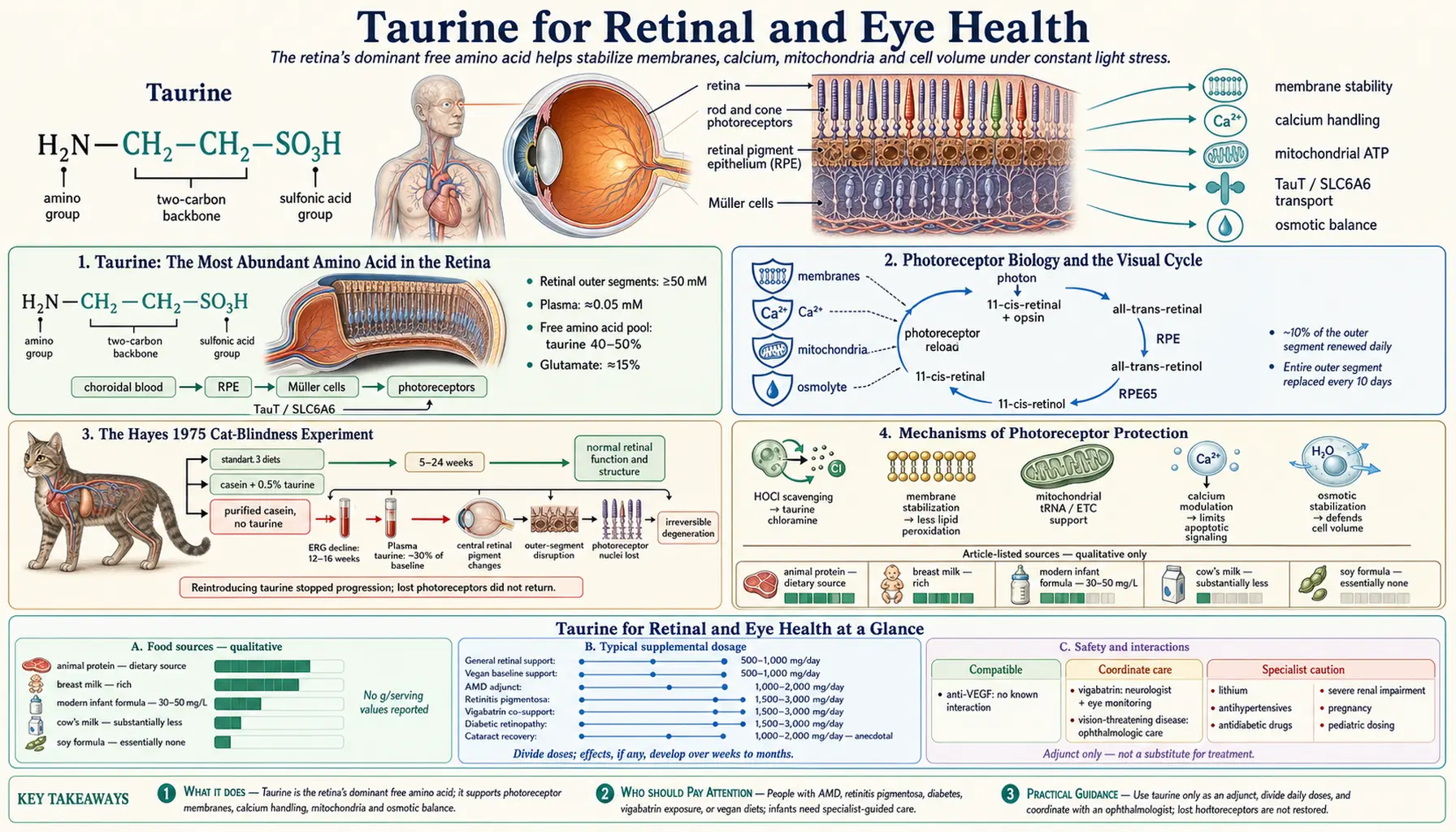
Taurine is the most abundant amino acid in the retina — comprising up to 50% of the total free amino acid pool of retinal photoreceptor cells, present at intracellular concentrations of 50 millimolar or higher in the outer segments of rods and cones. The pivotal experiment that established taurine essentiality came in 1975 when Knopf Hayes and colleagues at Harvard showed that cats fed a casein-based, taurine-free diet developed progressive irreversible photoreceptor degeneration leading to blindness within 16 weeks — a finding that earned Hayes the prestigious Bowditch Award and overturned the prevailing assumption that taurine was a biologically inert metabolic dead-end. The cat-blindness story directly motivated the addition of taurine to commercial cat food (the modern industry standard), and later to human infant formula. This deep-dive walks through the photoreceptor biology, the Hayes experiment that demonstrated essentiality, age-related decline of retinal taurine, the role of taurine in macular function, and the practical implications for adult patients with macular degeneration, retinitis pigmentosa, and vigabatrin-associated retinopathy.
Table of Contents
- Taurine: The Most Abundant Amino Acid in the Retina
- Photoreceptor Biology and the Visual Cycle
- The Hayes 1975 Cat-Blindness Experiment
- Mechanisms of Photoreceptor Protection
- Osmotic Regulation in Retinal Pigment Epithelium
- Calcium Handling in Photoreceptors
- Mitochondrial Function in the Retina
- Age-Related Macular Degeneration and Taurine
- Retinitis Pigmentosa
- Vigabatrin-Associated Retinopathy
- Diabetic Retinopathy
- Infant Formula and the Pediatric Story
- Practical Dosing for Eye Health Indications
- Cautions and Drug Interactions
- Key Research Papers
- Connections
- Featured Videos
Taurine: The Most Abundant Amino Acid in the Retina
The retina is a peculiar tissue. Layer for layer, it is the most metabolically active tissue in the body per gram of weight — the photoreceptors alone consume oxygen at a rate exceeding that of cerebral cortex. The mitochondria packed into the inner segments of rods and cones run at maximum capacity from waking to sleep, regenerating the photosensitive pigment cycle, pumping ions against gradient, and producing the enormous quantities of ATP required to maintain the dark current. And throughout this metabolic intensity, the retina maintains the highest taurine concentration of any tissue in the body.
The numbers are striking. Free taurine concentration in retinal photoreceptor outer segments reaches 50 millimolar or higher — compared to plasma concentrations of approximately 0.05 millimolar. The free amino acid pool of retinal tissue is dominated by taurine, often comprising 40 to 50% of the total free amino acid content. Glutamate (which serves as a major retinal neurotransmitter) is the next most abundant at roughly 15%. Every other amino acid — including the essential amino acids needed for protein synthesis — sits at single-digit percentages.
The retinal taurine pool is maintained by aggressive expression of the sodium-taurine cotransporter (TauT, SLC6A6) at retinal pigment epithelium (RPE) cells, Müller cells (the principal retinal glia), and photoreceptors themselves. TauT pumps taurine from the choroidal blood supply across the RPE basal membrane, then through Müller cell processes to the photoreceptor outer segments. The concentration gradient that taurine maintains across these cell layers is among the steepest of any small molecule in the body, requiring continuous ATP-driven sodium-gradient maintenance to defend.
This abundance is not coincidence or excess. The functions of retinal taurine include photoprotection, calcium modulation, osmotic regulation, mitochondrial preservation, and membrane stabilization — each of which is essential to photoreceptor survival under the extraordinary metabolic and oxidative stress of constant light exposure. The Hayes experiment of 1975 (described below) demonstrated what happens when this abundance is depleted: the photoreceptors die.
Photoreceptor Biology and the Visual Cycle
Vision begins with the absorption of a photon by a pigment molecule embedded in the disc membranes of a rod or cone photoreceptor outer segment. The pigment in rods is rhodopsin (which mediates scotopic / low-light vision), and the pigments in cones are the related photopsins (mediating color vision and high-acuity photopic vision). All visual pigments share the same fundamental architecture: a seven-transmembrane opsin protein covalently linked, via a Schiff base, to the small molecule 11-cis-retinal (the active form of Vitamin A in vision).
When a photon strikes the pigment, the 11-cis-retinal absorbs the energy and isomerizes to all-trans-retinal. This conformational change is transmitted to the opsin protein, which activates a G-protein cascade (transducin in rods) that ultimately closes cation channels in the outer segment plasma membrane, hyperpolarizing the photoreceptor and producing the electrical signal that the inner retina processes into vision.
The all-trans-retinal then dissociates from the opsin and must be regenerated to 11-cis-retinal to make the pigment receptive again. This requires the retinoid visual cycle, in which the all-trans-retinal is transported to the retinal pigment epithelium (RPE), reduced to all-trans-retinol, isomerized to 11-cis-retinol by the RPE65 enzyme, oxidized to 11-cis-retinal, and shuttled back to the photoreceptor outer segment to reload the opsin. The whole cycle takes a few seconds in the dark.
The metabolic cost of running this cycle continuously, plus the constant pumping required to maintain the dark current, plus the daily renewal of about 10% of the photoreceptor outer segment (the entire outer segment is replaced every 10 days through a process of basal addition and apical phagocytosis by the RPE), makes the retina enormously demanding. The mitochondria in the inner segments produce ATP at near-maximum rates throughout life. The byproducts of this metabolism include large quantities of reactive oxygen species, which must be neutralized continuously to prevent photoreceptor damage.
Taurine sits at the center of the defenses against this metabolic stress. It stabilizes membrane phospholipids against oxidative damage, modulates intracellular calcium to prevent calcium-driven apoptosis, supports mitochondrial efficiency through its role in tRNA modification and mPTP inhibition, and serves as an organic osmolyte to defend cell volume in the highly osmotically-stressed retinal environment. Take any of these defenses away and photoreceptors die.
The Hayes 1975 Cat-Blindness Experiment
The discovery that taurine is essential to mammalian retinal function came from one of the more elegant nutritional experiments of twentieth-century vision science. In 1975, Knopf C. Hayes, working with Robert Carey and Stephen Schmidt at Harvard Medical School and the Joslin Diabetes Center, published a paper in Science titled “Retinal degeneration associated with taurine deficiency in the cat.” The findings reshaped the understanding of taurine's biological role.
The experimental design was straightforward. Hayes fed adult cats one of three semi-purified diets: a standard diet containing taurine-rich casein and animal protein; a casein-based diet supplemented with 0.5% taurine; or a critical experimental diet using highly purified casein (which is low in taurine) with no taurine supplementation. The cats were kept on these diets for periods ranging from 5 to 24 weeks, with regular ophthalmologic examinations including electroretinography (ERG) and fundus photography.
The results were dramatic. Cats on the taurine-free diet showed:
- Progressive reduction in ERG amplitudes beginning around 12 to 16 weeks
- Visible fundus changes including granular pigmentary changes in the central retina
- Histologic examination revealing widespread photoreceptor degeneration, with disruption of outer segment architecture, loss of photoreceptor nuclei, and migration of pigment from the RPE into the neural retina
- Plasma taurine concentrations dropped to roughly 30% of baseline
- The degeneration was irreversible once established; reintroducing taurine to the diet stopped further progression but did not restore lost photoreceptors
The control groups — cats on the taurine-replete diets — showed normal retinal function and structure throughout the experiment, ruling out any non-specific effect of the casein diet itself.
The conclusion was inescapable: taurine is essential for the structural and functional integrity of the cat retina, and dietary deprivation produces irreversible blindness. Cats, unlike most mammals, have limited capacity to synthesize taurine from cysteine due to low CSAD activity, making them obligately dependent on dietary taurine. The experiment did not by itself prove that humans had the same dependency, but it raised the question urgently enough that the entire pet food industry was forced to reckon with it.
The follow-up implications cascaded:
- The cat-food industry mandated taurine supplementation in all commercial cat food, eliminating taurine-deficiency blindness as a clinical entity in housecats by the mid-1980s
- Infant formula manufacturers added taurine to formula, recognizing that human infants have limited taurine synthesis and that breast milk is rich in taurine — a change that proceeded after retrospective concerns about the developmental outcomes of pre-supplementation formula-fed infants
- The veterinary community recognized the cat-cardiomyopathy / cat-blindness pattern as parallel manifestations of taurine deficiency, leading to the Pion 1987 dilated cardiomyopathy discovery
- Vision scientists turned their attention to taurine as a potential therapeutic in human retinal degeneration, particularly in vigabatrin-induced retinopathy (described below)
The Hayes 1975 paper remains one of the most cited works in vision science. It established the principle that taurine is essential to retinal function in mammalian biology — not merely abundant, but irreplaceable.
Mechanisms of Photoreceptor Protection
The photoreceptor outer segment is exposed to a higher intensity of oxidative stress than almost any other cell in the body. Every photon absorbed by rhodopsin initiates a chemistry that produces reactive intermediates. Light energy itself drives photochemical reactions in the lipid-rich outer segment membranes, generating singlet oxygen, peroxyl radicals, and hydrogen peroxide. The mitochondria packed into the inner segments produce additional reactive oxygen species (ROS) as a byproduct of constant high-rate ATP synthesis.
The retina's antioxidant defenses include enzymatic systems (superoxide dismutase, catalase, glutathione peroxidase), small-molecule antioxidants (glutathione, ascorbate, α-tocopherol, the carotenoids lutein and zeaxanthin in the macula), and structural protection from melanin in the RPE. Taurine contributes to this defense in several ways:
- Direct hypochlorous acid scavenging. Taurine reacts with hypochlorous acid (HOCl) produced by activated immune cells in the retinal vasculature to form taurine chloramine, a much less toxic oxidant that itself has anti-inflammatory signaling properties. This protects retinal tissue from collateral damage during inflammatory responses to infection or injury.
- Membrane phospholipid stabilization. Taurine binds reversibly to phosphatidylcholine and phosphatidylethanolamine head groups in cellular membranes, reducing lipid peroxidation and stabilizing the membrane against oxidative damage. The disc membranes of photoreceptor outer segments are particularly rich in polyunsaturated fatty acids (especially DHA, the predominant fatty acid in retinal phospholipids) and therefore particularly vulnerable to peroxidation.
- Mitochondrial protection. Through its role in mitochondrial tRNA modification (forming 5-taurinomethyluridine, τm&sup5;U), taurine supports normal mitochondrial protein synthesis and electron transport chain function. Adequate taurine reduces electron leak from the ETC and lowers superoxide generation at the source. Taurine also inhibits mitochondrial permeability transition pore opening, preventing the catastrophic apoptotic cascade triggered by calcium overload or oxidative stress.
- Calcium modulation. Photoreceptor outer segments are exquisitely sensitive to intracellular calcium concentrations, which must be tightly regulated through the phototransduction cycle. Taurine modulates calcium handling in photoreceptors through effects on plasma membrane channels and intracellular calcium stores, preventing the calcium-driven cell death pathways that contribute to photoreceptor loss in various retinal degenerations.
- Osmotic stabilization. The retinal extracellular environment is osmotically stressed by the high metabolic activity of the tissue, with rapid local changes in ion concentrations as the dark and light currents shift. Taurine, as the principal organic osmolyte in retinal cells, defends cell volume against these osmotic swings.
The combined effect of these mechanisms is to provide the photoreceptor with an extraordinarily robust protective system that allows it to survive the metabolic intensity of constant light exposure for a human lifetime. When that system is compromised — by taurine deficiency, by aging, by drug exposure, by genetic mutation — the photoreceptors progressively succumb to oxidative damage and apoptotic cell death, producing the characteristic patterns of retinal degeneration.
Osmotic Regulation in Retinal Pigment Epithelium
The retinal pigment epithelium (RPE) is a single layer of cells separating the photoreceptors from the choroidal blood supply. Its functions include phagocytosis of shed photoreceptor outer segment tips, recycling of all-trans-retinal to 11-cis-retinal via the visual cycle, transport of nutrients from choroid to retina, ion transport (particularly maintenance of the subretinal space ion composition), and the blood-retina barrier function that separates immunologically privileged neural retina from circulating blood.
The RPE maintains extraordinary taurine concentrations both for its own metabolic needs and as the supplier of taurine to the photoreceptor layer. RPE cells express TauT at their basolateral membrane (facing the choroidal blood supply, importing taurine from circulation) and at their apical membrane (releasing taurine into the subretinal space for photoreceptor uptake).
The osmotic environment of the subretinal space is particularly demanding. During light adaptation, photoreceptor outer segment ion currents shift dramatically, producing local osmotic changes that the RPE must accommodate while maintaining its barrier function. Taurine, betaine, and myo-inositol serve as the principal compatible osmolytes that allow the RPE to defend cell volume through these shifts. Loss of any of these osmolytes — whether through deficient supply or genetic mutation — compromises RPE function and ultimately photoreceptor survival.
The age-related decline of RPE function is a major driver of age-related macular degeneration (AMD, see below). Aged RPE cells show reduced TauT expression, lower intracellular taurine, accumulation of lipofuscin (an oxidative-damage byproduct), and progressive loss of cellular function. This is one of the mechanistic links between cellular aging and macular degeneration: failing osmolyte regulation in the RPE compromises the support system on which photoreceptors depend.
Calcium Handling in Photoreceptors
Photoreceptors maintain among the steepest calcium gradients in the body. The outer segment cytoplasm holds calcium at submicromolar concentrations during darkness, with the gradient maintained by the plasma membrane sodium-calcium-potassium exchanger (NCKX, the principal calcium extrusion mechanism in photoreceptors). During phototransduction, calcium concentrations drop further as cyclic GMP levels fall and the cation channels close.
This calcium signaling is essential for visual processing, but it also makes photoreceptors vulnerable to calcium dysregulation. Inappropriate calcium elevation in the outer segment activates calpain proteases and triggers apoptotic cascades that kill the photoreceptor. Many forms of inherited retinal degeneration involve mutations that disturb calcium handling, either directly (mutations in NCKX, calmodulin, or calcium-binding proteins) or indirectly (mutations in phototransduction components that lead to abnormal calcium loads).
Taurine modulates photoreceptor calcium handling at several levels:
- Stabilizing NCKX function through membrane phospholipid interactions
- Modulating L-type calcium channel activity in the inner segment (where calcium-dependent neurotransmitter release occurs at the photoreceptor synaptic terminal)
- Inhibiting mitochondrial permeability transition pore opening, preventing the calcium-driven apoptotic cascade
- Supporting ATP production needed to drive the calcium pumps that maintain low cytoplasmic calcium
The protective effects of taurine on photoreceptor calcium handling are particularly relevant in retinitis pigmentosa, where many of the disease-causing mutations create dysregulated calcium states in the outer segment.
Mitochondrial Function in the Retina
Photoreceptor inner segments contain the highest density of mitochondria of any cell type in the body. This is the metabolic engine that produces the enormous ATP needed to run the dark current and the phototransduction cycle. Anything that impairs mitochondrial function impairs vision: this is the principle behind the visual loss in mitochondrial diseases (Leber hereditary optic neuropathy, MELAS, Kearns-Sayre syndrome) and in many forms of toxic optic neuropathy.
Taurine's role in mitochondrial tRNA modification (forming 5-taurinomethyluridine) makes adequate taurine essential to normal mitochondrial protein synthesis. The most affected mitochondrial proteins in taurine deficiency are subunits of the electron transport chain encoded by mitochondrial DNA. With insufficient taurine, these proteins are misassembled, ETC efficiency drops, ATP production falls, and superoxide generation increases. The photoreceptor, with its extreme metabolic demand, is among the cells most affected.
This mechanism provides a partial explanation for the irreversible nature of the cat-blindness phenotype in the Hayes experiment. Once taurine deficiency drives mitochondrial dysfunction in photoreceptors past a certain threshold, the cells enter the apoptotic cascade and die. Restoring taurine after the fact prevents further loss but does not regenerate the dead cells — photoreceptors do not divide and are not replaced.
Age-Related Macular Degeneration and Taurine
Age-related macular degeneration (AMD) is the leading cause of irreversible vision loss in adults over 60 in the developed world. The disease involves progressive degeneration of the macula — the central retina responsible for high-acuity central vision — through a complex pathology involving RPE dysfunction, drusen accumulation (subretinal deposits of lipid, protein, and oxidative damage products), choroidal vascular changes, and ultimately photoreceptor loss.
The pathogenesis of AMD involves multiple converging factors:
- Chronic oxidative stress in the macula (driven by light exposure, mitochondrial activity, and inflammation)
- RPE dysfunction and lipofuscin accumulation
- Inflammation (the complement pathway, particularly factor H polymorphisms, plays a major role in genetic susceptibility)
- Choroidal vascular changes (reduced choroidal blood flow with aging)
- Genetic predisposition (CFH and ARMS2 polymorphisms are the strongest risk factors)
- Modifiable risk factors: smoking, hypertension, obesity, diet poor in antioxidants and omega-3s
The landmark AREDS (Age-Related Eye Disease Study) and AREDS2 trials established the role of nutritional supplementation in AMD. The AREDS2 formula — vitamins C and E, zinc, copper, lutein, zeaxanthin — reduces progression of intermediate AMD to advanced disease by approximately 25% over 5 years. Notably, neither AREDS nor AREDS2 included taurine in their formulations.
The case for adding taurine to AMD support is mechanistic rather than trial-proven:
- Taurine levels in the retina decline with age (paralleling the broader age-related decline of taurine documented by Yadav et al. 2023)
- Taurine's photoprotective, calcium-modulating, mitochondrial-supporting, and membrane-stabilizing functions all address mechanisms implicated in AMD pathogenesis
- Animal models of light-induced retinal damage consistently show protective effects of taurine
- Cellular studies of RPE cells under oxidative stress show preservation by taurine supplementation
What is missing is large randomized clinical trial data specifically showing reduced AMD progression with taurine supplementation. For the patient with AMD, taurine 1 to 2 g/day is a reasonable evidence-supported adjunct to standard AREDS2 supplementation, with an excellent safety profile. It should not replace ophthalmologic follow-up, AREDS2 supplementation, anti-VEGF therapy when indicated, or modifiable risk factor management (smoking cessation, blood pressure control, healthy diet).
Retinitis Pigmentosa
Retinitis pigmentosa (RP) is a heterogeneous group of inherited retinal degenerations characterized by progressive rod photoreceptor death (causing night blindness as the initial symptom), followed by cone photoreceptor degeneration (causing peripheral vision loss then central vision loss). RP affects approximately 1 in 4,000 people and can result from mutations in more than 70 different genes. The molecular mechanisms vary, but oxidative stress, calcium dysregulation, mitochondrial dysfunction, and apoptotic photoreceptor death are common downstream pathways across most genotypes.
Taurine has been investigated as a potential neuroprotective agent in RP. Animal models of RP show progressive depletion of retinal taurine as the disease progresses, suggesting that taurine deficiency develops secondary to photoreceptor stress and may contribute to ongoing photoreceptor loss. Taurine supplementation in animal models slows the rate of photoreceptor degeneration in some genotypes.
Human trial data are limited. A few small clinical studies have evaluated taurine in RP with generally favorable but underpowered results. The most encouraging data come from combination protocols including taurine along with other antioxidants (vitamin E, lutein, omega-3 fatty acids) and adjuncts (DHA, valproic acid in some studies). The current standard of care for RP includes vitamin A supplementation (15,000 IU/day in some protocols, though this is controversial), lutein, omega-3 DHA, and where genotype-specific therapy is available, gene therapy (e.g., voretigene neparvovec for RPE65-mediated disease).
For the RP patient who is supplementing with the standard protocol and seeking additional support, taurine 1.5 to 3 g/day is reasonable, with the understanding that it represents adjunctive support rather than a disease-modifying therapy. Coordination with the retinal specialist managing the patient is appropriate.
Vigabatrin-Associated Retinopathy
Vigabatrin is an antiepileptic medication that inhibits GABA transaminase, raising central nervous system GABA levels. It is particularly effective for infantile spasms (West syndrome) and is sometimes used for refractory focal epilepsy. However, vigabatrin has a major adverse effect: it causes irreversible peripheral visual field constriction in roughly 30% of patients exposed for more than 6 months, with some patients losing peripheral vision so severely as to be functionally blind.
The mechanism of vigabatrin-induced retinopathy involves taurine. Vigabatrin and taurine share structural similarity (both contain a sulfur-related amine), and vigabatrin competes with taurine for retinal transporters and binding sites. The result is functional retinal taurine depletion, particularly affecting cone photoreceptors in the peripheral retina. The peripheral cones progressively die, producing the characteristic tunnel-vision pattern of vigabatrin retinopathy.
This mechanism makes vigabatrin-induced retinopathy one of the few clinical conditions where taurine supplementation has a clear mechanistic rationale and a body of supportive evidence. Studies including those from the Jammoul / Picaud group at Vision Institute Paris have demonstrated that taurine supplementation in animal models of vigabatrin retinopathy substantially reduces retinal damage, and small case series in patients have shown promising results.
Current best practice for patients requiring vigabatrin therapy includes:
- Careful informed consent about the retinopathy risk before initiating therapy
- Baseline ophthalmologic examination including visual fields (where age and cooperation allow)
- Limiting the duration of vigabatrin exposure to the minimum necessary
- Considering taurine co-supplementation (1.5 to 3 g/day in adults; pediatric dosing under specialist guidance) during vigabatrin therapy
- Regular ophthalmologic follow-up, with vigabatrin discontinuation at the first sign of visual field constriction
For patients with established vigabatrin-induced retinopathy, taurine supplementation may slow further progression but cannot restore lost vision — the photoreceptor death is irreversible. This is one of the strongest clinical demonstrations that retinal taurine deficiency, once severe enough to cause photoreceptor death, produces permanent blindness.
Diabetic Retinopathy
Diabetic retinopathy is the leading cause of blindness in working-age adults in the developed world. The pathology involves a complex interplay of hyperglycemia-driven microvascular damage, oxidative stress, advanced glycation endproduct (AGE) accumulation, polyol pathway flux, protein kinase C activation, and inflammatory cytokine release. The end result is microvascular damage in the retinal capillaries leading to ischemia, neovascularization, macular edema, and retinal hemorrhage.
Multiple lines of evidence connect taurine to diabetic retinopathy:
- Plasma taurine is reduced in diabetes, particularly in poorly controlled disease (the glucose-driven upregulation of TauT in non-retinal tissues effectively borrows taurine from circulation, depleting the plasma pool)
- Retinal taurine levels in diabetic animal models are reduced
- Taurine supplementation in animal models of diabetes reduces several markers of diabetic retinopathy: reduced AGE formation, reduced VEGF expression, reduced retinal microvascular damage, preserved retinal capillary endothelial function
- Small human trials of taurine in diabetic patients have shown improvements in retinal vascular function as measured by retinal blood flow studies
For the diabetic patient (particularly with longstanding poorly controlled disease, signs of early diabetic retinopathy, or markedly elevated HbA1c), taurine supplementation at 1.5 to 3 g/day is a reasonable adjunct to standard diabetic care. It does not replace the foundational priorities of glycemic control, blood pressure management, lipid management, smoking cessation, and regular ophthalmologic follow-up with appropriate intervention (laser, anti-VEGF, surgery) when retinopathy progresses.
Infant Formula and the Pediatric Story
Human infants have limited capacity to synthesize taurine from cysteine in the first months of life. CSAD activity is low in the neonatal liver and matures gradually over the first year. This means infants are largely dependent on dietary taurine.
Breast milk is rich in taurine — colostrum particularly so, and mature breast milk continues to provide substantial taurine throughout the lactation period. Breast-fed infants therefore receive adequate taurine without any supplementation. The story is different for formula-fed infants.
Until the 1980s, commercial infant formula based on cow's milk or soy was not taurine-supplemented. Cow's milk has substantially less taurine than human milk, and soy-based formulas contain essentially none. Formula-fed infants of that era developed low plasma taurine concentrations, and concerns arose about developmental implications — particularly for premature infants who had less hepatic taurine synthesis capacity than full-term infants, and even more so for infants requiring prolonged parenteral nutrition.
Studies by Jurevics, Watkins, and others in the 1980s documented that taurine-deficient formula-fed infants had:
- Lower plasma taurine concentrations
- Abnormal taurine:glycine bile acid conjugation ratios (with glycine conjugates dominating)
- Reduced fat absorption
- Some indications of suboptimal retinal development (electroretinographic differences from breast-fed controls)
The findings, combined with the cat-blindness experience, motivated the addition of taurine to all major infant formula brands beginning in the early 1980s. Modern infant formula contains taurine at concentrations approximating those found in mature human breast milk (approximately 30 to 50 mg/L). The pediatric concerns of pre-supplementation formula-fed infants have largely been resolved.
For total parenteral nutrition (TPN) in neonates and infants, taurine supplementation is now standard. Premature infants requiring prolonged TPN are particularly dependent on exogenous taurine, both for retinal development and to support bile acid conjugation (preventing TPN cholestasis).
Practical Dosing for Eye Health Indications
- General retinal support / maintenance (mild dry eye, mild age-related visual changes): 500 to 1,000 mg per day
- Adjunctive support for age-related macular degeneration: 1,000 to 2,000 mg per day, alongside AREDS2 supplementation
- Adjunctive support for retinitis pigmentosa: 1,500 to 3,000 mg per day, in coordination with retinal specialist
- Vigabatrin co-supplementation: 1,500 to 3,000 mg per day during vigabatrin therapy (adult dosing; pediatric under specialist guidance)
- Diabetic retinopathy adjunct: 1,500 to 3,000 mg per day, alongside standard diabetic care
- Cataract surgery recovery: 1,000 to 2,000 mg per day for several weeks pre- and post-operatively (anecdotal practice; not validated by large trials)
- Vegan supplementation for baseline retinal taurine support: 500 to 1,000 mg per day
Taurine for eye health indications is best taken consistently across the day in divided doses. Improvement in visual function, where it occurs, develops slowly over weeks to months as retinal taurine pools are replenished. The supplementation does not provide acute improvement in vision and should be presented to patients with appropriate expectations about its supportive rather than dramatic role.
Taurine pairs well with other retinal-protective nutrients: omega-3 fatty acids (especially DHA), lutein, zeaxanthin, zinc, vitamin C, vitamin E, and the carotenoids in colorful plant foods. The combination approach addresses multiple converging mechanisms of retinal damage.
Cautions and Drug Interactions
- Not a substitute for ophthalmologic care. Patients with diabetic retinopathy, AMD, RP, glaucoma, or any other vision-threatening condition require regular ophthalmologic follow-up and appropriate medical or surgical intervention. Taurine supplementation is an adjunct, not a primary therapy.
- Coordination with vigabatrin therapy. Taurine co-supplementation during vigabatrin therapy is mechanistically rational but should be coordinated with the prescribing neurologist. Vigabatrin requires careful neurologic monitoring for seizure control as well as ophthalmologic monitoring for retinopathy.
- Anti-VEGF therapy compatibility. Patients receiving intravitreal anti-VEGF injections (ranibizumab, aflibercept, bevacizumab) for AMD or diabetic macular edema can safely take taurine concurrently. There is no known pharmacologic interaction.
- Standard taurine cautions apply. See the main taurine overview for the full list: lithium, antihypertensives, antidiabetic drugs, severe renal impairment, pregnancy.
- Pediatric dosing. Pediatric taurine dosing for vigabatrin co-supplementation or other retinal indications should be done under specialist guidance, not extrapolated from adult doses by body weight alone.
Key Research Papers
- Hayes KC, Carey RE, Schmidt SY (1975). Retinal degeneration associated with taurine deficiency in the cat. Science. — PubMed
- Sturman JA (1993). Taurine in development. Physiological Reviews. — PubMed
- Ripps H, Shen W (2012). Review: taurine, a “very essential” amino acid. Molecular Vision. — PubMed
- Froger N, Cadetti L, Lorach H, et al. (2012). Taurine provides neuroprotection against retinal ganglion cell degeneration. PLOS ONE. — PubMed
- Jammoul F, Wang Q, Nabbout R, et al. (2009). Taurine deficiency is a cause of vigabatrin-induced retinal phototoxicity. Annals of Neurology. — PubMed
- Jammoul F, Degardin J, Pain D, et al. (2010). Taurine deficiency damages photoreceptors and retinal ganglion cells in vigabatrin-treated neonatal rats. Molecular and Cellular Neuroscience. — PubMed
- Heller-Stilb B, van Roeyen C, Rascher K, et al. (2002). Disruption of the taurine transporter gene (taut) leads to retinal degeneration in mice. FASEB Journal. — PubMed
- Lima L, Obregon F, Cubillos S, Fazzino F, Jaimes I (2001). Taurine as a micronutrient in development and regeneration of the central nervous system. Nutritional Neuroscience. — PubMed
- Yu X, Xu Z, Mi M, et al. (2008). Dietary taurine supplementation ameliorates diabetic retinopathy via anti-excitotoxicity of glutamate in streptozotocin-induced Sprague-Dawley rats. Neurochemical Research. — PubMed
- Macaione S, Ruggeri P, De Luca F, Tucci G (1974). Free amino acids in developing rat retina. Journal of Neurochemistry. — PubMed
- Pasantes-Morales H, Klethi J, Ledig M, Mandel P (1972). Free amino acids of chicken and rat retina. Brain Research. — PubMed
- Geggel HS, Ament ME, Heckenlively JR, Martin DA, Kopple JD (1985). Nutritional requirement for taurine in patients receiving long-term parenteral nutrition. New England Journal of Medicine. — PubMed
PubMed Topic Searches
- PubMed: Taurine and retinal photoreceptors
- PubMed: Taurine and age-related macular degeneration
- PubMed: Taurine and vigabatrin retinopathy
- PubMed: Taurine and retinitis pigmentosa
- PubMed: Taurine and diabetic retinopathy
- PubMed: Taurine in infant formula
- PubMed: TauT knockout and retinal degeneration
Connections
- All Amino Acids
- Taurine Overview
- Taurine Benefits Hub
- Taurine for Cardiac Function
- Taurine for Bile Acids
- Taurine for Electrolyte Balance
- Vitamin A (Retinal Substrate)
- Vitamin A for Vision
- Macular Degeneration
- Diabetes
- Omega-3 Fatty Acids (DHA)
- Zinc
- Vitamin E
- Cysteine (Taurine Precursor)
- Vitamin B6 (P5P)
- Calcium — the ion whose influx taurine modulates inside photoreceptors.