Vitamin K2 vs K1 and the Calcium Paradox
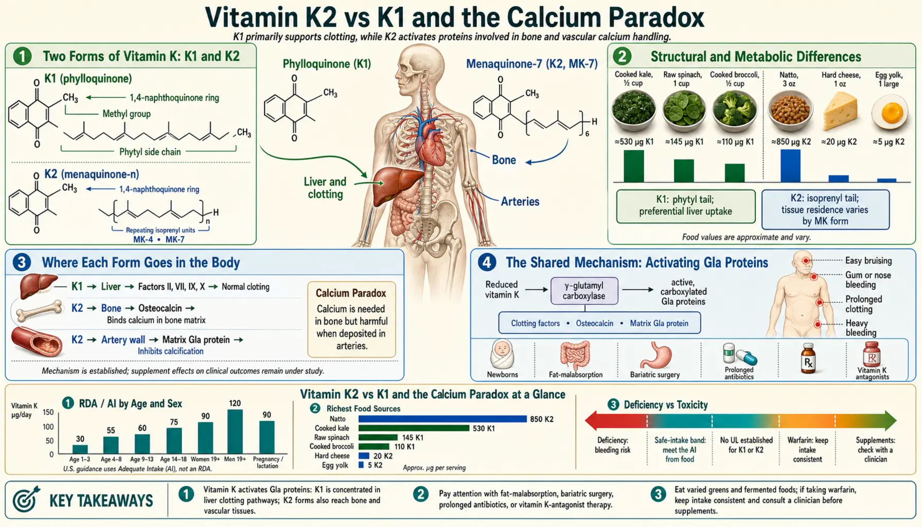
"Vitamin K" covers two related families: K1 (phylloquinone), found mainly in green vegetables and plant margarines, and K2 (the menaquinones), found in foods such as meat, liver, butter, egg yolk, natto and cheese (Schurgers 2000); menaquinones from MK-4 to MK-13 have been measured in dairy foods (Fu 2017). Both act as the cofactor that activates vitamin-K-dependent proteins, but they differ in how long they stay in the blood and where they are carried in the body, which is one reason K2 is studied mainly for bone and arteries. That difference underlies the "calcium paradox" idea: one review describes the paradox as a lack of calcium in bone alongside its storage in the vessel wall, and proposes that vitamin K2 deficiency could be responsible (Flore 2013). The idea draws on biochemistry, animal studies, biomarkers and observational cohorts; the randomized trials so far have measured calcification, bone density and blood markers rather than heart attacks or fractures, and their results are mixed. This page sets out the K1/K2 differences, then what supports the calcium-paradox hypothesis and what it still lacks.
Table of Contents
- Two Forms of Vitamin K: K1 and K2
- Structural and Metabolic Differences
- Where Each Form Goes in the Body
- The Shared Mechanism: Activating Gla Proteins
- The "Calcium Paradox" Hypothesis
- Why the Hypothesis Is Biologically Plausible
- The Gap: Association Is Not Proven Outcome
- The Vitamin D Interplay
- Summary of the Evidence
- Key Research Papers
- External Resources
- Connections
- Featured Videos
Two Forms of Vitamin K: K1 and K2
Vitamin K is a group name for K1 (phylloquinone) and K2, a series of related compounds known as menaquinones; the menaquinones differ structurally in the length of their isoprenyl side chain, from four isoprenoid residues in MK-4 up to nine in MK-9, and still longer ones have been described (Knapen 2007). Menaquinones also differ from phylloquinone in the saturation of that side chain (Fu 2017).
- Vitamin K1 (phylloquinone) is mainly present in green vegetables and plant margarines (Schurgers 2000). In a Dutch cohort of 16,057 women, K1 made up most of the vitamin K intake: mean intake was 211.7 micrograms a day of K1 and 29.1 micrograms a day of K2 (Gast 2009). See the Vitamin K overview.
- Vitamin K2 (menaquinones) is named MK-n by the number of isoprenoid units in the side chain. MK-4 is present throughout tissues outside the liver; the body makes it from phylloquinone, either directly within tissues or by way of menadione (vitamin K3), and in 2010 the human enzyme responsible, UBIAD1, was identified (Nakagawa 2010). Natto, fermented soybeans, contains a large amount of MK-7 (Kaneki 2001), and full-fat dairy products contain menaquinones mainly as MK-9, MK-10 and MK-11, with modest amounts of MK-4, MK-8 and MK-12 (Fu 2017).
Both are genuine vitamin K. K1 and K2 both act as cofactor for the enzyme gamma-glutamyl carboxylase; the difference in their structure shows up as differences in absorption, tissue distribution and bioavailability (Halder 2019), which is the starting point of the K2 story.
Structural and Metabolic Differences
The side chain affects how each form behaves once absorbed. Schurgers and colleagues (2007) compared synthetic K1 with natto-derived MK-7 in healthy volunteers:
- Half-life. Both K1 and MK-7 were absorbed well, with peak serum concentrations 4 hours after intake. The authors describe the "very long half-life time of MK-7" as the major difference, resulting in much more stable serum levels than K1 and accumulation of MK-7 to 7- to 8-fold higher levels during prolonged intake (Schurgers 2007). A Dutch trial report gives MK-7 "a halflife in the circulation of 3 days" and MK-4 a very short half-life of 1–2 hours (Knapen 2007).
- Carboxylation. In the same comparison, MK-7 induced more complete carboxylation of osteocalcin than K1 (Schurgers 2007).
The forms are also carried differently in the blood. When six healthy men were given K1, MK-4 and MK-9 together, all three were carried at first mainly in triacylglycerol-rich lipoproteins, which are cleared mainly by the liver; unlike K1, both menaquinones were also found in LDL, and MK-4 in HDL as well. The authors suggest this explains why, in their earlier studies, K1 was preferentially accumulated in the liver while menaquinones had a more widespread distribution, and they describe MK-9's half-life in the circulation as very long (Schurgers 2002). MK-7 was not tested in that study. Citing MK-7's 3-day half-life, Knapen and colleagues called it "the most logical choice for use as a low-dose food supplement" (Knapen 2007); several of the microgram-dose trials described below used MK-7.
Where Each Form Goes in the Body
When vitamin K is in short supply, the body appears to favour clotting. A 2012 review notes that the dietary vitamin K requirement for the coagulation factors is much lower than for the Gla proteins made outside the liver, and links this to the "triage theory": in the healthy population all clotting factors are made in their active form, while the synthesis of other Gla proteins is sub-optimal in people not taking supplements (Vermeer 2012). Another review describes a transport system that distributes dietary vitamin K preferentially to the liver when it is limiting, so that the first signs of insufficiency are seen as undercarboxylation of the extra-hepatic Gla proteins, which are only partly carboxylated in the healthy adult population (Theuwissen 2012). McCann and Ames (2009), who set out the triage theory for vitamin K, ranked the vitamin K-dependent proteins by how lethal their loss was in knockout mice: the coagulation proteins were essential, while mice lacking osteocalcin or matrix Gla protein survived at least through weaning.
The menaquinones' wider reach is described in the Dutch trial report: in human volunteers both K1 and K2 were transported to the liver, but only the K2 vitamins (MK-4 and MK-7) were incorporated into the LDL fraction for transport to extrahepatic tissues, and in animal experiments K1 mainly accumulated in the liver, whereas MK-4 was far more abundant in other tissues including pancreas, bone and vessel wall (Knapen 2007). This difference in distribution is the main argument for studying K2 separately for bone and arteries, even though both forms drive the same carboxylation reaction. Whether it changes outcomes is the question the trials below address.
The Shared Mechanism: Activating Gla Proteins
The calcium-paradox argument depends on what K1 and K2 have in common. Both serve as the cofactor for gamma-glutamyl carboxylase, and only the carboxylated proteins are active (Halder 2019). Seventeen Gla proteins had been discovered by 2012, among them four coagulation factors made exclusively in the liver, and osteocalcin, matrix Gla protein (MGP) and Gas6, which have key functions in maintaining bone strength, inhibiting arterial calcification and regulating cell growth respectively (Vermeer 2012). MGP is made by vascular smooth muscle cells and is described as the most important inhibitor of arterial mineralization currently known (Theuwissen 2012).
K1 can drive this carboxylation too. In a dose-finding trial in healthy adults aged 19–36, phylloquinone lowered the percentage of undercarboxylated osteocalcin dose-dependently, and the authors concluded that a daily intake of approximately 1,000 micrograms is required to carboxylate circulating osteocalcin maximally (Binkley 2002). In the head-to-head comparison above, MK-7 induced more complete osteocalcin carboxylation than K1 (Schurgers 2007).
The "Calcium Paradox" Hypothesis
"Calcium paradox" is a name reviewers use for a lack of calcium in bone alongside its storage in the vessel wall. One review proposes that vitamin K2 deficiency could be responsible, and states that large studies are needed to confirm what it calls preliminary experiences (Flore 2013). The observation it starts from has some longitudinal support: in the Framingham Heart Study, 364 women and 190 men had radiographs about 25 years apart, and women with the greatest bone loss showed the most severe progression of abdominal aortic calcification, while no association was seen in men; the authors concluded that the two processes may be related (Kiel 2001).
The vitamin K hypothesis links bone and artery through the two proteins above:
- When vitamin K is insufficient, more osteocalcin is left undercarboxylated; one review describes prolonged sub-clinical vitamin K deficiency as a risk factor for osteoporosis and atherosclerosis (Vermeer 2012).
- At the same time more matrix Gla protein is left undercarboxylated, and MGP is generally accepted to be a potent inhibitor of arterial calcification, although its mechanism at the molecular level is not completely understood (Schurgers 2008).
The popular one-line version, "vitamin K2 directs calcium to the bones and away from the arteries", is a hypothesis, not an established finding in humans. A related idea is that calcium supplements taken without enough vitamin K could add to calcium in soft tissue. A 2019 review states that it is "tempting to speculate" that vitamin K supplementation might protect against potentially untoward effects of calcium supplementation (Wasilewski 2019). No trial among the sources reviewed for this page tested whether adding vitamin K changes the cardiovascular effect of calcium supplements.
Calcium supplements and heart attacks: the meta-analyses and the debate
Separately from vitamin K, two meta-analyses by Bolland and colleagues reported more heart attacks with calcium supplements. The 2010 analysis of placebo-controlled trials of calcium supplements given without vitamin D found more myocardial infarctions with calcium: a hazard ratio of 1.31 (95% CI 1.02–1.67) in the five trials with patient-level data, and a pooled relative risk of 1.27 (1.01–1.59) in the trial-level analysis (Bolland 2010). The 2011 reanalysis added the Women's Health Initiative calcium and vitamin D trial, using the women who were not taking their own calcium supplements at randomization, and found that calcium or calcium plus vitamin D increased the risk of myocardial infarction (relative risk 1.24, 1.07–1.45) and of myocardial infarction or stroke (1.15, 1.03–1.27) (Bolland 2011).
Later analyses came to a different conclusion. A collaborative meta-analysis, which noted that concern had been raised over the design of the earlier meta-analyses, counted coronary events verified by clinical review, hospital record or death certificate; 18 trials with 63,563 participants met its criteria, and the pooled relative risk was 1.02 (0.96–1.09) for coronary heart disease in the five trials that contributed coronary events, 0.96 (0.91–1.02) for all-cause mortality in seventeen trials, and 1.08 (0.92–1.26) for myocardial infarction, and concluded that the evidence does not support an increase in coronary heart disease or mortality in elderly women (Lewis 2015). A 2016 systematic review funded by the National Osteoporosis Foundation found no statistically significant differences in cardiovascular events or mortality in the four randomized trials it included, in all of which cardiovascular outcomes were secondary end points, and concluded that calcium intake within tolerable upper intake levels (2,000 to 2,500 mg a day) is not associated with cardiovascular risk in generally healthy adults (Chung 2016). The National Osteoporosis Foundation and the American Society for Preventive Cardiology then adopted the position that there is moderate-quality evidence that calcium, with or without vitamin D, from food or supplements, has no relationship, beneficial or harmful, to the risk of cardiovascular and cerebrovascular disease or mortality in generally healthy adults (Kopecky 2016). None of these analyses involved vitamin K.
Why the Hypothesis Is Biologically Plausible
The calcium-paradox hypothesis has several lines of mechanistic support:
- The proteins are vitamin-K-dependent. Osteocalcin and MGP need vitamin K-dependent carboxylation for activity, and in the healthy adult population they are only partly carboxylated (Theuwissen 2012).
- The knockout mouse. Mice lacking MGP develop to term but die within two months as a result of arterial calcification that leads to blood-vessel rupture; the authors concluded that calcification must be actively inhibited in soft tissues (Luo 1997). This is an animal study.
- Biomarkers. A review reports that circulating desphospho-uncarboxylated MGP (dp-ucMGP) was predictive of cardiovascular risk and mortality, and that total uncarboxylated MGP was associated with the extent of existing arterial calcification (Theuwissen 2012); another describes uncarboxylated MGP as a promising biomarker for detecting cardiovascular calcification (Schurgers 2008).
- Vitamin K antagonists. In rats, high doses of warfarin caused calcification of the artery wall and aortic heart valves, first seen after 2 weeks; the authors suggest warfarin acts by inhibiting the carboxylation of MGP (Price 1998). In humans the evidence is a small cross-sectional study: 19 people under 55 who had used coumarin anticoagulants for more than 10 years had more femoral arterial calcification than 18 matched healthy controls (odds ratio 8.5, 95% CI 2.01–35.95), an association the authors suggest may act through inhibition of MGP carboxylation (Rennenberg 2010).
- Cohorts. In the Rotterdam Study, an observational cohort of 4,807 Dutch adults, the highest third of menaquinone intake had a relative risk of coronary heart disease death of 0.43 (95% CI 0.24–0.77) and an odds ratio for severe aortic calcification of 0.48 (0.32–0.71) compared with the lowest third, while phylloquinone (K1) intake was not related to any of the outcomes (Geleijnse 2004). In 16,057 Dutch women the hazard ratio for coronary heart disease was 0.91 (0.85–1.00) per 10 micrograms a day of K2 intake (Gast 2009), and in a cross-sectional study of 564 postmenopausal women higher menaquinone intake was associated with less coronary calcification (Beulens 2009). These are associations; they do not show that menaquinone caused the difference.
Together these make a coherent hypothesis. Mechanistic, animal and observational evidence does not by itself show that taking K2 changes outcomes in people; that is what the trials in the next section test.
The Gap: Association Is Not Proven Outcome
The randomized human trials have mostly measured surrogate endpoints — calcification scores, arterial stiffness, bone density and the dp-ucMGP marker — rather than heart attacks, strokes, fractures or deaths. A 2015 Cochrane review of vitamin K for the primary prevention of cardiovascular disease found only one small trial (60 participants, 3 months), which did not report cardiovascular events, and concluded that there was a lack of evidence to determine whether vitamin K supplementation is effective (Hartley 2015).
Calcification trials published since then have given mixed results:
- No effect found. In 365 older men with aortic valve calcification (AVADEC), 720 micrograms a day of MK-7 plus 25 micrograms of vitamin D for two years did not influence progression of valve calcification, and aortic and coronary calcification also did not differ from placebo, although dp-ucMGP fell (Diederichsen 2022). In 68 people with type 2 diabetes and cardiovascular disease, 360 micrograms a day of MK-7 for 6 months did not change calcification mass on CT, and active calcification on PET tended to increase (P = 0.06, not significant) (Zwakenberg 2019). In a pilot trial in 86 haemodialysis patients (iPACK-HD), 10 mg of vitamin K1 three times a week for 12 months lowered dp-ucMGP by 86% but made no difference to the progression of coronary calcification (Holden 2023).
- Slower progression reported. In the open-label VitaVasK trial in haemodialysis patients, which was stopped early because of slow recruitment and analysed 40 of 60 randomized patients, 5 mg of vitamin K1 three times weekly was followed by 56% lower progression of thoracic aortic calcification at 18 months (p = .039), while the 68% lower progression of coronary calcification was not significant (P = .072) (Saritas 2022). In the VitaK-CAC trial in patients with symptomatic coronary artery disease, 360 micrograms a day of MK-7 for two years slowed the rise in coronary calcium score compared with placebo (P = .02); the authors state that the clinical significance for plaque stability remains to be determined (Vossen 2026). In the DANCODE trial, 400 people with severe coronary calcification were randomized to 720 micrograms a day of vitamin K2 plus 25 micrograms of vitamin D3 or placebo for 24 months; the mean coronary calcium score rose by 196 units with the supplements and 248 with placebo (difference −52, 95% CI −79 to −24) (Hasific 2026).
None of these trials had heart attacks, strokes or deaths as its primary outcome; in AVADEC, deaths and cardiovascular events, recorded as safety outcomes, did not differ between the groups (Diederichsen 2022).
On the bone side, the largest trial — the OF study, which randomized 4,378 osteoporotic postmenopausal women aged 50 or older to calcium alone or calcium plus menatetrenone (pharmaceutical MK-4) — found no significant difference in new vertebral fractures over 36 months, its primary endpoint (Inoue 2009). A 2019 systematic review that identified 36 studies concluded that for postmenopausal or osteoporotic patients there is no evidence that vitamin K affects bone mineral density or vertebral fractures, and that it may reduce clinical fractures but the evidence is insufficient to confirm this (Mott 2019); a 2020 correction adjusted every estimate that had included the Yamaguchi Osteoporosis Prevention Study, after concerns were raised about that study's integrity (Mott 2020). The earlier 2006 meta-analysis of the Japanese fracture trials (Cockayne 2006) carries a 2018 notice titled "Caution of Conclusions of a Meta-analysis Including Problematic Trials" (JAMA Internal Medicine 2018), alongside a caution to readers from its senior author (Torgerson 2018). Several vitamin K trial reports in this literature have since been retracted: a 2005 fracture trial in elderly women with Alzheimer's disease, at the corresponding author's request because it contains fabricated clinical trial data (Sato 2005); a 2011 hip-fracture trial in Alzheimer's disease, because of scientific misconduct (Sato 2011); and the Yamaguchi study, whose 2025 retraction states that the evidence "strongly argues for scientific misconduct" (Ishida 2004). The bone trials are covered in more detail on the Bone Health page.
So the calcium paradox remains a hypothesis with incomplete human outcome data: the biochemistry is well documented, some trials found slower calcification on imaging and others found none, no trial cited here has shown that K2 prevents heart attacks, and the fracture evidence was judged insufficient (Mott 2019).
The Vitamin D Interplay
Vitamin K2 is often discussed with vitamin D. A 2017 review states that vitamin D promotes the production of vitamin K-dependent proteins, which require vitamin K for carboxylation in order to function properly (van Ballegooijen 2017). In a rat bone-tumour cell line, the active hormone 1,25-dihydroxyvitamin D3 increased MGP messenger RNA within 4 hours and MGP secretion 15-fold by 24 hours; the authors note an earlier finding of a 6-fold stimulation of the other vitamin K-dependent bone protein, bone Gla protein (BGP), by the same hormone in another cell line (Fraser 1988). The review concludes that current evidence supports the notion that joint supplementation of vitamins D and K might be more effective than either alone for bone and cardiovascular health, while noting that few intervention trials studied cardiovascular-related outcomes (van Ballegooijen 2017).
The concern sometimes raised — that high-dose vitamin D without enough vitamin K could favour soft-tissue calcification — comes mainly from animal work. High doses of vitamin D are known to cause calcification of the artery media in rats within 3 to 4 days, and giving warfarin at the same time dramatically increased that calcification (Price 2000). In humans the trial results differ by outcome: in a three-year trial in postmenopausal women, the elastic properties of the carotid artery stayed unchanged with a supplement of minerals, vitamin D and vitamin K1, but declined with minerals plus vitamin D alone and with placebo (Braam 2004); MK-7 combined with vitamin D did not slow aortic valve calcification in AVADEC (Diederichsen 2022), while vitamin K2 combined with vitamin D3 slowed the rise in coronary calcium score in DANCODE (Hasific 2026). See Vitamin D3 and Calcium.
Summary of the Evidence
- Both K1 and K2 are vitamin K. Both activate the same Gla proteins; K1 is preferentially retained in the liver while menaquinones are distributed more widely (Schurgers 2002), and MK-7 stays in the blood far longer than K1 or MK-4 (Schurgers 2007; Knapen 2007).
- Food sources differ. K1 comes mainly from green vegetables and plant margarines; K2 from meat, liver, butter, egg yolk, natto, cheese and curd cheese (Schurgers 2000).
- The calcium paradox is a hypothesis. It rests on biochemistry, animal studies, biomarkers and observational cohorts; the calcification trials are mixed, and none had heart attacks as its primary outcome.
- The calcium-supplement question is disputed. Two meta-analyses reported more heart attacks with calcium supplements (Bolland 2010; Bolland 2011); a later meta-analysis of verified events, a 2016 systematic review and a 2016 guideline statement found no relationship (Lewis 2015; Chung 2016; Kopecky 2016). No trial cited here tested whether vitamin K changes that effect.
- Warfarin-type anticoagulants: the authors of the K1 and MK-7 comparison noted that preparations supplying 50 micrograms a day or more of MK-7 may interfere with oral anticoagulant treatment in a clinically relevant way (Schurgers 2007), and in healthy volunteers taking acenocoumarol, MK-7 at doses as low as 10 micrograms a day significantly influenced anticoagulation in some individuals (Theuwissen 2013). Decisions about anticoagulants and supplements belong with the prescribing clinician. Details are on the Sources & MK-4 vs MK-7 page.
Key Research Papers
- Schurgers LJ et al. (2007). Vitamin K-containing dietary supplements: comparison of synthetic vitamin K1 and natto-derived menaquinone-7. Blood — PubMed PMID: 17158229
- Vermeer C (2012). Vitamin K: the effect on health beyond coagulation — an overview. Food & Nutrition Research — PubMed PMID: 22489224
- Luo G et al. (1997). Spontaneous calcification of arteries and cartilage in mice lacking matrix GLA protein. Nature — PubMed PMID: 9052783
- Schurgers LJ, Cranenburg ECM, Vermeer C (2008). Matrix Gla-protein: the calcification inhibitor in need of vitamin K. Thromb Haemost — PubMed PMID: 18841280
- Theuwissen E, Smit E, Vermeer C (2012). The role of vitamin K in soft-tissue calcification. Adv Nutr — PubMed PMID: 22516724
- van Ballegooijen AJ et al. (2017). The synergistic interplay between vitamins D and K for bone and cardiovascular health: a narrative review. Int J Endocrinol — PubMed PMID: 29138634
- Geleijnse JM et al. (2004). Dietary intake of menaquinone is associated with a reduced risk of coronary heart disease: the Rotterdam Study. J Nutr — PubMed PMID: 15514282
- Beulens JW et al. (2009). High dietary menaquinone intake is associated with reduced coronary calcification. Atherosclerosis — PubMed PMID: 18722618
- Binkley NC et al. (2002). A high phylloquinone intake is required to achieve maximal osteocalcin gamma-carboxylation. Am J Clin Nutr — PubMed PMID: 12399278
- Rennenberg RJMW et al. (2010). Chronic coumarin treatment is associated with increased extracoronary arterial calcification in humans. Blood — PubMed PMID: 20354170
- Hartley L et al. (2015). Vitamin K for the primary prevention of cardiovascular disease. Cochrane Database Syst Rev — PubMed PMID: 26389791
- Knapen MH, Schurgers LJ, Vermeer C (2007). Vitamin K2 supplementation improves hip bone geometry and bone strength indices in postmenopausal women. Osteoporos Int 18(7): 963-972 — PubMed PMID: 17287908
- Schurgers LJ, Vermeer C (2002). Differential lipoprotein transport pathways of K-vitamins in healthy subjects. Biochim Biophys Acta 1570(1): 27-32 — PubMed PMID: 11960685
- Schurgers LJ, Vermeer C (2000). Determination of phylloquinone and menaquinones in food. Effect of food matrix on circulating vitamin K concentrations. Haemostasis 30(6): 298-307 — PubMed PMID: 11356998
- Fu X, Harshman SG, Shen X, et al. (2017). Multiple vitamin K forms exist in dairy foods. Curr Dev Nutr 1(6): e000638 — PubMed PMID: 29955705
- Gast GC, de Roos NM, Sluijs I, et al. (2009). A high menaquinone intake reduces the incidence of coronary heart disease. Nutr Metab Cardiovasc Dis 19(7): 504-510 — PubMed PMID: 19179058
- Nakagawa K, Hirota Y, Sawada N, et al. (2010). Identification of UBIAD1 as a novel human menaquinone-4 biosynthetic enzyme. Nature 468(7320): 117-121 — PubMed PMID: 20953171
- Kaneki M, Hodges SJ, Hosoi T, et al. (2001). Japanese fermented soybean food as the major determinant of the large geographic difference in circulating levels of vitamin K2: possible implications for hip-fracture risk. Nutrition 17(4): 315-321 — PubMed PMID: 11369171
- Halder M, Petsophonsakul P, Akbulut AC, et al. (2019). Vitamin K: double bonds beyond coagulation insights into differences between vitamin K1 and K2 in health and disease. Int J Mol Sci 20(4): 896 — PubMed PMID: 30791399
- McCann JC, Ames BN (2009). Vitamin K, an example of triage theory: is micronutrient inadequacy linked to diseases of aging? Am J Clin Nutr 90(4): 889-907 — PubMed PMID: 19692494
- Flore R, Ponziani FR, Di Rienzo TA, et al. (2013). Something more to say about calcium homeostasis: the role of vitamin K2 in vascular calcification and osteoporosis. Eur Rev Med Pharmacol Sci 17(18): 2433-2440 — PubMed PMID: 24089220
- Kiel DP, Kauppila LI, Cupples LA, et al. (2001). Bone loss and the progression of abdominal aortic calcification over a 25 year period: the Framingham Heart Study. Calcif Tissue Int 68(5): 271-276 — PubMed PMID: 11683533
- Wasilewski GB, Vervloet MG, Schurgers LJ (2019). The bone-vasculature axis: calcium supplementation and the role of vitamin K. Front Cardiovasc Med 6: 6 — PubMed PMID: 30805347
- Bolland MJ, Avenell A, Baron JA, et al. (2010). Effect of calcium supplements on risk of myocardial infarction and cardiovascular events: meta-analysis. BMJ 341: c3691 — PubMed PMID: 20671013
- Bolland MJ, Grey A, Avenell A, Gamble GD, Reid IR (2011). Calcium supplements with or without vitamin D and risk of cardiovascular events: reanalysis of the Women's Health Initiative limited access dataset and meta-analysis. BMJ 342: d2040 — PubMed PMID: 21505219
- Lewis JR, Radavelli-Bagatini S, Rejnmark L, et al. (2015). The effects of calcium supplementation on verified coronary heart disease hospitalization and death in postmenopausal women: a collaborative meta-analysis of randomized controlled trials. J Bone Miner Res 30(1): 165-175 — PubMed PMID: 25042841
- Chung M, Tang AM, Fu Z, Wang DD, Newberry SJ (2016). Calcium intake and cardiovascular disease risk: an updated systematic review and meta-analysis. Ann Intern Med 165(12): 856-866 — PubMed PMID: 27776363
- Kopecky SL, Bauer DC, Gulati M, et al. (2016). Lack of evidence linking calcium with or without vitamin D supplementation to cardiovascular disease in generally healthy adults: a clinical guideline from the National Osteoporosis Foundation and the American Society for Preventive Cardiology. Ann Intern Med 165(12): 867-868 — PubMed PMID: 27776362
- Price PA, Faus SA, Williamson MK (1998). Warfarin causes rapid calcification of the elastic lamellae in rat arteries and heart valves. Arterioscler Thromb Vasc Biol 18(9): 1400-1407 — PubMed PMID: 9743228
- Price PA, Faus SA, Williamson MK (2000). Warfarin-induced artery calcification is accelerated by growth and vitamin D. Arterioscler Thromb Vasc Biol 20(2): 317-327 — PubMed PMID: 10669626
- Diederichsen ACP, Lindholt JS, Möller S, et al. (2022). Vitamin K2 and D in patients with aortic valve calcification: a randomized double-blinded clinical trial. Circulation 145(18): 1387-1397 — PubMed PMID: 35465686
- Zwakenberg SR, de Jong PA, Bartstra JW, et al. (2019). The effect of menaquinone-7 supplementation on vascular calcification in patients with diabetes: a randomized, double-blind, placebo-controlled trial. Am J Clin Nutr 110(4): 883-890 — PubMed PMID: 31387121
- Holden RM, Booth SL, Zimmerman D, et al. (2023). Inhibit progression of coronary artery calcification with vitamin K in hemodialysis patients (the iPACK-HD study): a randomized, placebo-controlled multi-center, pilot trial. Nephrol Dial Transplant 38(3): 746-756 — PubMed PMID: 35641194
- Saritas T, Reinartz S, Krüger T, et al. (2022). Vitamin K1 and progression of cardiovascular calcifications in hemodialysis patients: the VitaVasK randomized controlled trial. Clin Kidney J 15(12): 2300-2311 — PubMed PMID: 37216675
- Vossen LM, de Leeuw PW, Schurgers LJ, et al. (2026). Two years of menaquinone-7 supplementation and coronary artery calcification: a randomized clinical trial. JAMA Cardiol 11(8): 719-726 — PubMed PMID: 42268593
- Hasific S, Øvrehus KA, Mejldal A, et al. (2026). Vitamin K2 and D3 supplementation in patients with severe coronary artery calcification: the DANCODE trial. Circulation (published online 2026) — PubMed PMID: 42661461
- Inoue T, Fujita T, Kishimoto H, et al. (2009). Randomized controlled study on the prevention of osteoporotic fractures (OF study): a phase IV clinical study of 15-mg menatetrenone capsules. J Bone Miner Metab 27(1): 66-75 — PubMed PMID: 19082528
- Mott A, Bradley T, Wright K, et al. (2019). Effect of vitamin K on bone mineral density and fractures in adults: an updated systematic review and meta-analysis of randomised controlled trials. Osteoporos Int 30(8): 1543-1559 — PubMed PMID: 31076817
- Mott A, Bradley T, Wright K, et al. (2020). Correction to: Effect of vitamin K on bone mineral density and fractures in adults: an updated systematic review and meta-analysis of randomised controlled trials. Osteoporos Int 31(11): 2269-2270 — PubMed PMID: 32965511
- Cockayne S, Adamson J, Lanham-New S, et al. (2006). Vitamin K and the prevention of fractures: systematic review and meta-analysis of randomized controlled trials. Arch Intern Med 166(12): 1256-1261 — PubMed PMID: 16801507 — Expression of concern (notice: PubMed PMID: 29582046)
- JAMA Internal Medicine (2018). Caution of conclusions of a meta-analysis including problematic trials. JAMA Intern Med 178(6): 875-876 — PubMed PMID: 29582046
- Torgerson DJ (2018). Caution to readers about systematic review on vitamin K and prevention of fractures that included problematic trials. JAMA Intern Med 178(6): 863-864 — PubMed PMID: 29582044
- Sato Y, Kanoko T, Satoh K, Iwamoto J (2005). Menatetrenone and vitamin D2 with calcium supplements prevent nonvertebral fracture in elderly women with Alzheimer's disease. Bone 36(1): 61-68 — PubMed PMID: 15664003 — Retracted (retraction notice: PubMed PMID: 29150013)
- Sato Y, Honda Y, Umeno K, et al. (2011). The prevention of hip fracture with menatetrenone and risedronate plus calcium supplementation in elderly patients with Alzheimer disease: a randomized controlled trial. Kurume Med J 57(4): 117-124 — PubMed PMID: 21778673 — Retracted (retraction notice: PubMed PMID: 28408729)
- Ishida Y, Kawai S (2004). Comparative efficacy of hormone replacement therapy, etidronate, calcitonin, alfacalcidol, and vitamin K in postmenopausal women with osteoporosis: the Yamaguchi Osteoporosis Prevention Study. Am J Med 117(8): 549-555 — PubMed PMID: 15465502 — Retracted (retraction notice: PubMed PMID: 40222753)
- Fraser JD, Otawara Y, Price PA (1988). 1,25-Dihydroxyvitamin D3 stimulates the synthesis of matrix gamma-carboxyglutamic acid protein by osteosarcoma cells. Mutually exclusive expression of vitamin K-dependent bone proteins by clonal osteoblastic cell lines. J Biol Chem 263(2): 911-916 — PubMed PMID: 3257212
- Braam LA, Hoeks AP, Brouns F, et al. (2004). Beneficial effects of vitamins D and K on the elastic properties of the vessel wall in postmenopausal women: a follow-up study. Thromb Haemost 91(2): 373-380 — PubMed PMID: 14961167
- Theuwissen E, Teunissen KJ, Spronk HM, et al. (2013). Effect of low-dose supplements of menaquinone-7 (vitamin K2) on the stability of oral anticoagulant treatment: dose-response relationship in healthy volunteers. J Thromb Haemost 11(6): 1085-1092 — PubMed PMID: 23530987