GLP-1 Receptor Agonists — Mechanism and Insulin Response
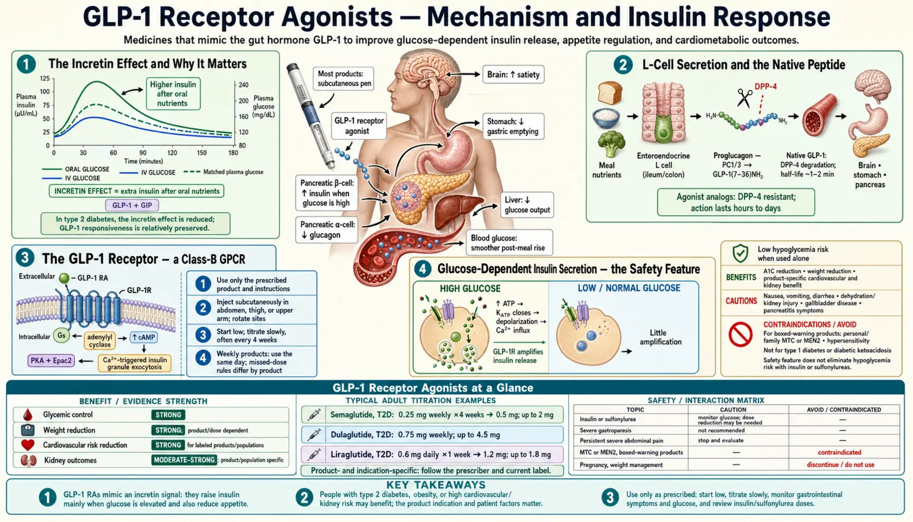
Glucagon-like peptide 1 (GLP-1) is a 30-amino-acid peptide hormone secreted by L-cells in the distal small intestine and colon in response to a meal. Its receptor — a class-B G-protein-coupled receptor on pancreatic beta cells, hypothalamic neurons, gastric smooth muscle, and a dozen other tissues — couples to Gs and elevates intracellular cyclic AMP. The clinical consequence is glucose-dependent insulin secretion (the safety-defining feature versus sulfonylureas), glucagon suppression, delayed gastric emptying, and reduced food intake. Engineered agonists (semaglutide, tirzepatide, liraglutide) extend the native 1–2 minute half-life to days by combining DPP-4 resistance with albumin binding via a fatty-acid side chain.
Interactive Visualization Hunger, Leptin & Why Diets Fight Back Watch the hunger neurons and the fullness neurons fight it out — then lose weight and see leptin fall, hunger climb and metabolism drop. It isn’t willpower. It’s a defended system. Launch →
Table of Contents
- The Incretin Effect and Why It Matters
- L-Cell Secretion and the Native Peptide
- The GLP-1 Receptor — a Class-B GPCR
- Glucose-Dependent Insulin Secretion — the Safety Feature
- Glucagon Suppression
- Gastric Emptying and Satiety Signaling
- Central Nervous System: Hypothalamus and Brainstem
- Engineering Around the Two-Minute Half-Life
- Tirzepatide and the GIP/GLP-1 Dual Agonist Concept
- Key Research Papers
- Connections
- Featured Videos
The Incretin Effect and Why It Matters
The "incretin effect" describes a long-observed paradox: the same dose of glucose delivered orally produces a much larger insulin response than the same dose delivered intravenously. The difference — roughly 50–70% of post-prandial insulin secretion in healthy adults — is accounted for by gut-derived hormones that signal to the pancreas during meal absorption. Two hormones account for the bulk of the incretin effect: glucose-dependent insulinotropic polypeptide (GIP), secreted by K-cells in the duodenum and jejunum, and glucagon-like peptide 1 (GLP-1), secreted by L-cells in the distal small intestine and colon.
Michael Nauck's 1986 Diabetologia paper documented the seminal finding that the incretin effect is dramatically reduced in patients with type 2 diabetes. This deficit became the rationale for the entire incretin therapeutic class. Restoring incretin signaling pharmacologically can be done in two ways: by blocking the enzyme that degrades native GLP-1 (DPP-4 inhibitors such as sitagliptin and linagliptin), or by administering an engineered GLP-1 analog that resists degradation (exenatide, liraglutide, semaglutide, dulaglutide). The two approaches produce different magnitudes of effect — DPP-4 inhibition raises GLP-1 to roughly twice physiological levels, while agonist injection produces concentrations 6–10 times physiological, which is why the agonists drive much larger weight loss.
L-Cell Secretion and the Native Peptide
L-cells are open-type enteroendocrine cells with a luminal surface that samples the contents of the gut. They concentrate in the distal ileum and colon, but a small population exists throughout the small intestine. L-cells secrete GLP-1 in response to multiple stimuli that all converge on intracellular calcium and cAMP elevation:
- Nutrient sensors — glucose via SGLT1 transport, long-chain fatty acids via GPR120, short-chain fatty acids via FFAR2 (these are the products of fiber fermentation by colonic bacteria), amino acids (especially glutamine and tryptophan), and bile acids via the TGR5 receptor.
- Bitter taste receptors (T2Rs) — the same family expressed on the tongue is expressed on L-cells. Bitter compounds in coffee, hops, gentian, and cruciferous vegetables stimulate GLP-1 secretion.
- Neural input — vagal cholinergic and adrenergic fibers innervate the gut and modulate L-cell output.
Native GLP-1 is released as two forms (GLP-1 7-36 amide and GLP-1 7-37), both of which are biologically active. Within 1–2 minutes of release, ~50% is cleaved at the Ala-2 / Glu-3 bond by dipeptidyl peptidase-4 (DPP-4), a serine protease found on the surface of endothelial cells and in soluble form in plasma. The cleaved fragment (GLP-1 9-36 amide) is biologically inactive at the GLP-1 receptor. This is why native GLP-1 cannot be administered as a drug at clinically useful doses — it has to be re-engineered.
The GLP-1 Receptor — a Class-B GPCR
The GLP-1 receptor is a class-B G-protein-coupled receptor (the same family that includes the receptors for glucagon, GIP, parathyroid hormone, secretin, and corticotropin-releasing hormone). Class-B receptors have a distinctive structural architecture — a large extracellular N-terminal domain that captures the peptide ligand by its C-terminus, followed by the canonical seven transmembrane helices, which the peptide's N-terminus then engages to drive conformational change.
The receptor couples primarily to Gs, activating adenylyl cyclase and raising intracellular cAMP. The cAMP signal activates two downstream effectors: protein kinase A (PKA), and Epac2 (exchange protein activated by cAMP). In pancreatic beta cells, both effectors converge to amplify glucose-stimulated insulin secretion. Secondary coupling to Gq/phospholipase C, with elevation of intracellular calcium, contributes a smaller signal.
GLP-1 receptors are expressed in pancreatic beta cells (the primary therapeutic target), pancreatic alpha cells (where activation suppresses glucagon), hypothalamic and brainstem neurons (appetite regulation), gastric smooth muscle and the pyloric sphincter (gastric emptying), cardiomyocytes and vascular endothelium (cardiovascular outcome benefits), renal tubular cells (renal outcome benefits), Brunner's glands in the duodenum, the rodent thyroid C-cell (driving the MTC warning), and the gallbladder (cholelithiasis risk). The breadth of expression is the reason GLP-1 agonists produce effects across multiple organ systems.
Glucose-Dependent Insulin Secretion — the Safety Feature
The defining feature of incretin-driven insulin secretion is that it requires elevated blood glucose. The mechanism: glucose enters the beta cell via GLUT2, is phosphorylated by glucokinase, enters glycolysis and mitochondrial oxidation, and elevates intracellular ATP. Rising ATP closes the ATP-sensitive potassium channel (KATP), depolarizes the membrane, opens voltage-gated calcium channels, and triggers exocytosis of insulin granules. This is the canonical glucose-stimulated insulin secretion (GSIS) pathway.
GLP-1 acts downstream of this pathway. PKA and Epac2 (the cAMP effectors activated by GLP-1) increase calcium release from the endoplasmic reticulum, sensitize the secretory machinery to calcium, and recruit additional insulin granules to the readily releasable pool. None of this matters unless glucose is already driving the upstream depolarization step. At fasting glucose levels of ~5 mmol/L, GLP-1 cannot drive significant insulin release. At post-prandial glucose of 8–10 mmol/L, GLP-1 amplifies the existing GSIS by 5–10 fold.
The clinical safety implication is large. Sulfonylureas close the KATP channel directly, driving insulin secretion regardless of glucose — which is why they cause hypoglycemia and why this hypoglycemia accounts for a substantial fraction of elderly diabetic emergency-department visits. GLP-1 agonist monotherapy almost never causes hypoglycemia, because the incretin amplification simply turns off when blood glucose drops. When GLP-1 agonists are combined with sulfonylureas or insulin, hypoglycemia risk is restored and dose reduction of the other agent is required.
Glucagon Suppression
The pancreatic alpha cell secretes glucagon, the counter-regulatory hormone that raises blood glucose by stimulating hepatic gluconeogenesis and glycogenolysis. In type 2 diabetes, glucagon secretion is paradoxically high during meals (when it should be suppressed) and inadequate during hypoglycemia (when it should rise). This dysregulated alpha-cell behavior is a substantial contributor to post-prandial hyperglycemia — the inappropriate hepatic glucose output during meal absorption adds to the glucose already arriving from the gut.
GLP-1 suppresses glucagon secretion in a glucose-dependent manner. The exact mechanism remains debated — direct receptor effects on alpha cells, paracrine effects via beta-cell-derived insulin and somatostatin, and indirect effects on delta cells have all been documented. The net clinical effect is a reduction in the post-prandial glucagon surge, which contributes meaningfully to the improvement in post-prandial hyperglycemia. Crucially, the suppression is glucose-dependent — at hypoglycemic levels, glucagon secretion is preserved, which means GLP-1 agonists do not abolish the body's defense against hypoglycemia in the way that high-dose insulin can.
Gastric Emptying and Satiety Signaling
GLP-1 activates receptors on gastric smooth muscle and the pyloric sphincter, slowing the rate at which a meal exits the stomach. Native GLP-1 release after a meal contributes a small physiological delay; pharmacological doses produce a much larger effect — gastric emptying half-times can extend from a normal ~90 minutes to 4–6 hours during early titration of liraglutide or semaglutide. The effect attenuates with chronic dosing (tachyphylaxis) over weeks to months, but never fully resolves.
The delayed emptying has two consequences. The intended consequence is satiety — a slowly emptying stomach signals fullness via mechanoreceptors and via the prolonged exposure of upper-gut nutrient sensors to food, suppressing the next meal. The unintended consequence is the nausea and vomiting that affect 40–75% of patients during titration — food sitting in the stomach causes the classical "food still there hours later" feeling that many patients report. The same delayed emptying creates the pre-operative aspiration risk that has led the American Society of Anesthesiologists to recommend withholding GLP-1 agonists for one week (daily) or one dosing interval (weekly) before elective surgery requiring general anesthesia.
Central Nervous System: Hypothalamus and Brainstem
GLP-1 receptors are abundant in the central nervous system, with particularly high density in the arcuate nucleus of the hypothalamus (the master appetite-regulating center) and in the nucleus tractus solitarius of the brainstem (which integrates vagal afferent input from the gut). Anders Secher's 2014 J Clin Invest paper used fluorescently labeled liraglutide to demonstrate direct binding in the arcuate nucleus and showed that selective lesioning of these neurons abolished the weight-loss effect, establishing the hypothalamus as the primary site of action for the appetite-suppressing effect.
In the arcuate nucleus, GLP-1 activates POMC/CART neurons (the anorexigenic / satiety-promoting subset) and inhibits NPY/AgRP neurons (the orexigenic / hunger-promoting subset). The downstream signal projects to the paraventricular nucleus and from there to widespread cortical and limbic circuits that govern food-related decision making. The clinical manifestation is reduced hunger, smaller meal size, reduced snacking frequency, and reduced food-related preoccupation — a constellation patients often describe as "food noise reduction."
The same central GLP-1 receptor activity has unexpected effects on reward-related behavior. Reports of reduced craving for alcohol, nicotine, opioids, and gambling in patients on semaglutide have prompted formal trials in alcohol use disorder. The mechanism appears to involve GLP-1 modulation of mesolimbic dopaminergic signaling in the ventral tegmental area and nucleus accumbens.
Engineering Around the Two-Minute Half-Life
Native GLP-1 is useless as a drug because of its sub-2-minute half-life. The therapeutic agonists all share two engineering strategies that defeat DPP-4 cleavage and renal clearance:
- Resistance to DPP-4 cleavage. The vulnerable Ala-2 / Glu-3 bond is protected by amino acid substitutions — exenatide uses the lizard exendin-4 backbone, which naturally resists DPP-4; liraglutide and semaglutide substitute Aib (alpha-aminoisobutyric acid) at position 2; dulaglutide uses a similar Gly-Gly modification.
- Albumin binding via a fatty-acid side chain. Liraglutide attaches a C16 palmitic acid via a glutamate linker at Lys-26, allowing reversible binding to circulating albumin. Bound peptide is invisible to renal filtration and to DPP-4. Semaglutide extends this with a C18 fatty diacid plus a longer linker, achieving an effective half-life of about one week and enabling once-weekly dosing. Dulaglutide fuses the GLP-1 sequence to an IgG4-Fc domain, providing albumin-like circulation time by a different mechanism.
The result is that engineered agonists circulate at concentrations 5–10 fold above native GLP-1 throughout the dosing interval, providing continuous receptor stimulation rather than the brief meal-triggered pulses of native physiology. This continuous stimulation is essential to the weight-loss effect — the central nervous system requires sustained GLP-1 receptor activity to maintain appetite suppression — but is also responsible for the persistent gastrointestinal side effects and the tachyphylaxis seen with chronic dosing.
Tirzepatide and the GIP/GLP-1 Dual Agonist Concept
Tirzepatide (Mounjaro / Zepbound) is the first "twincretin" — a single peptide engineered to activate both the GLP-1 receptor and the glucose-dependent insulinotropic polypeptide (GIP) receptor. The molecule was designed by Eli Lilly using a backbone derived from native GIP, with substitutions that confer GLP-1 receptor activity. The result is a peptide with higher affinity for GIP than GLP-1 (about 5-fold) but balanced activity at both receptors.
The clinical rationale for dual agonism was initially controversial. GIP signaling in type 2 diabetes had been considered impaired and not therapeutically useful — the GIP-driven incretin effect is reduced in T2D. The unexpected finding from SURPASS-1 through SURPASS-5 and SURMOUNT-1 was that dual GIP/GLP-1 agonism produced larger glucose reductions and substantially larger weight reductions than pure GLP-1 agonism. In SURMOUNT-1, tirzepatide 15 mg produced 22.5% mean weight loss at 72 weeks vs ~15% for semaglutide 2.4 mg in STEP-1.
The mechanism of the additional GIP contribution is incompletely understood. Candidate explanations include GIP-mediated effects on adipose tissue (where GIP receptors are expressed), CNS effects via brainstem GIP receptors, anti-emetic effects of GIP signaling that allow higher tolerable GLP-1 dosing, and synergistic insulin sensitization. The follow-on triple agonist retatrutide adds glucagon receptor activity to GIP and GLP-1 and produced 24% weight loss in phase-2 data, suggesting the multi-receptor approach has further room to run.
Key Research Papers
- Mojsov S et al., GLP-1 (7-37) as insulin-releasing hormone (J Clin Invest 1987) — Search PubMed
- Nauck MA et al., reduced incretin effect in type 2 diabetes (Diabetologia 1986) — PubMed: Nauck 1986
- Holst JV, the physiology of the incretin hormones (Physiol Rev 2007) — PubMed: Holst Physiol Rev 2007
- Baggio LL & Drucker DJ, biology of incretins (Gastroenterology 2007) — PubMed: Baggio Drucker 2007
- Drucker DJ, mechanisms of action of GLP-1 (Cell Metab 2018) — PubMed: Drucker Cell Metab 2018
- Knudsen LB & Lau J, liraglutide and semaglutide discovery (Front Endocrinol 2019) — Search PubMed
- Lau J et al., semaglutide engineering for once-weekly use (J Med Chem 2015) — PubMed: Lau J Med Chem 2015
- Secher A et al., arcuate nucleus is primary site of action of liraglutide (J Clin Invest 2014) — PubMed: Secher 2014
- Coskun T et al., tirzepatide GIP/GLP-1 dual agonist (Mol Metab 2018) — PubMed: Coskun 2018
- Frias JP et al., SURPASS-2 tirzepatide vs semaglutide (NEJM 2021) — PubMed: Frias SURPASS-2
- Jastreboff AM et al., retatrutide triple agonist phase 2 (NEJM 2023) — PubMed: Jastreboff retatrutide
- Vilsboll T et al., GLP-1 reduces post-prandial glucose (Diabetologia 2003) — Search PubMed
Connections
- All Remedies
- Hunger, Leptin & Why Diets Fight Back — interactive animation
- GLP-1 Benefits Hub
- Weight Loss Studies
- Side Effects & Cautions
- Natural GLP-1 Boosting Foods
- GLP-1 Receptor Agonists (Hub)
- Type 2 Diabetes
- Insulin Resistance
- Metabolic Syndrome
- Obesity
- Berberine
- HbA1c
- Fasting Insulin