Omega-3 Fatty Acids for Brain and Cognition
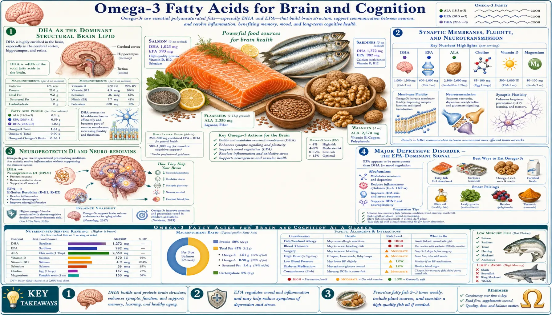
DHA (docosahexaenoic acid, 22:6n-3) is the dominant structural lipid of the human brain — comprising approximately 30% of the phospholipid in cortical gray matter, 50% of the phospholipid in retinal photoreceptor outer segments, and the bulk of the membrane lipid in synaptic terminals where neurotransmitter vesicles dock and release. EPA (eicosapentaenoic acid, 20:5n-3) plays a complementary role as the dominant substrate for the resolvin-and-protectin family of inflammation-resolving mediators that protect against neuroinflammation. The therapeutic implications cut across mood disorders (where EPA-dominant supplementation has the most consistent antidepressant signal), age-related cognitive decline (mixed trial results), ADHD in children (modest but reproducible effect), and the lifelong DHA-accretion biology that begins in the third trimester of pregnancy. This page walks through the structural, signaling, and clinical evidence for omega-3 effects on the brain.
Table of Contents
- DHA as the Dominant Structural Brain Lipid
- Synaptic Membranes, Fluidity, and Neurotransmission
- Neuroprotectin D1 and Neuro-resolvins
- Major Depressive Disorder — the EPA-Dominant Signal
- Anxiety, Bipolar Disorder, and Perinatal Mood Disorders
- Age-Related Cognitive Decline and Dementia Prevention
- ADHD in Children and Adolescents
- Omega-3 Index, Brain Volume, and MRI Findings
- Dosing for Brain and Cognitive Indications
- Cautions and Drug Interactions
- Key Research Papers
- Connections
- Featured Videos
DHA as the Dominant Structural Brain Lipid
The human brain is approximately 60% lipid by dry weight, of which roughly half is phospholipid. Within that phospholipid pool, DHA occupies the sn-2 position of phosphatidylethanolamine and phosphatidylserine at extraordinarily high enrichment — up to 30% of all fatty acid in cortical gray matter, far higher than in any other organ system. Skeletal muscle has <1% DHA in membrane fatty acids; the brain has 30%.
This enrichment is not arbitrary. DHA's 22-carbon chain with six cis double bonds creates a uniquely flexible, low-melting-point lipid — the chain folds into multiple low-energy conformations on the picosecond timescale, generating membrane fluidity that other fatty acids cannot provide. The membrane proteins embedded in DHA-rich membranes (G-protein coupled receptors, ion channels, neurotransmitter transporters, rhodopsin in photoreceptors) require this fluidity for the conformational changes that constitute their signaling function. Replacing DHA with shorter or less-unsaturated fatty acids (which happens in dietary DHA deficiency) reduces membrane fluidity and impairs these signaling functions.
The body accretes DHA into the brain across a defined developmental window. DHA accumulates in fetal brain primarily during the third trimester of pregnancy (when fetal brain growth accelerates), continues at high rates through the first two years of life (when the human brain triples in volume), and reaches roughly adult levels by school age. Adult brain DHA content is then maintained over decades through a combination of dietary intake and slow turnover.
Across the lifespan, the average daily turnover of brain DHA is small — perhaps 0.5-1% per day — so deficient intake does not produce rapid measurable cognitive change in adults. Severe deficiency over months to years can deplete brain DHA modestly, particularly in conditions of accelerated turnover (chronic inflammation, alcohol use, or repeated pregnancies in nutritionally marginal mothers).
Synaptic Membranes, Fluidity, and Neurotransmission
Within the brain, DHA is most highly concentrated in synaptic membranes — the specialized regions where presynaptic vesicles dock with the active zone and release neurotransmitter into the synaptic cleft. Synaptic vesicle fusion with the presynaptic membrane requires membrane curvature changes, lipid raft reorganization, and conformational changes in SNARE proteins, all of which depend on membrane fluidity provided by DHA-rich phospholipids.
Experimentally reducing brain DHA (in rodent models fed n-3-deficient diets across generations) produces:
- Reduced dopamine D2 receptor density in frontal cortex
- Altered serotonin receptor function
- Impaired long-term potentiation (the cellular correlate of learning)
- Reduced BDNF (brain-derived neurotrophic factor) expression
- Spatial-learning deficits in maze tasks
- Increased anxiety-like behavior
These effects can be partially reversed by reintroducing dietary DHA, though the reversal is incomplete if deficiency occurred during the developmental DHA-accretion window (third trimester through age two). The implication for human nutrition is that early-life DHA adequacy has lifetime consequences that are not fully recoverable with later supplementation — the rationale for pregnancy and lactation DHA recommendations.
Neuroprotectin D1 and Neuro-resolvins
Beyond its structural role, DHA serves as the precursor to a family of bioactive lipid mediators with specifically neuroprotective and inflammation-resolving functions. The most studied is neuroprotectin D1 (NPD1), also called protectin D1, biosynthesized in retinal pigment epithelium and brain tissue from DHA via 15-lipoxygenase.
NPD1 has been shown to:
- Protect retinal pigment epithelial cells against oxidative stress (relevant to age-related macular degeneration)
- Reduce neuronal apoptosis in cerebral ischemia models
- Modulate microglial activation toward a resolving phenotype
- Reduce beta-amyloid-induced neurotoxicity in cell culture models of Alzheimer's disease
- Suppress NF-kappaB activation and reduce neuroinflammatory cytokine production
The discovery of NPD1 (Bazan, Serhan, and colleagues, 2003-2005) reframed the understanding of why dietary DHA matters for brain health: it is not just a passive membrane component but an active substrate for endogenous neuroprotective mediators. The implication is that DHA inadequacy compromises both the brain's structural infrastructure and its capacity to resolve neuroinflammatory insults — a double hit relevant to age-related cognitive decline, stroke recovery, and neurodegenerative disease.
Companion D-series resolvins (RvD1, RvD2, etc.) are produced in the brain and CNS-adjacent tissues from DHA via similar lipoxygenase pathways, with related neuroprotective and resolution-promoting actions.
Major Depressive Disorder — the EPA-Dominant Signal
Major depressive disorder (MDD) is the clinical domain with the most consistent omega-3 antidepressant signal. Multiple meta-analyses (Liao et al. Transl Psychiatry 2019, Mocking et al. Transl Psychiatry 2016) have shown statistically and clinically meaningful antidepressant effect of omega-3 supplementation in MDD, particularly when:
- EPA-dominant formulations are used — products with EPA:DHA ratio >2:1 (or pure EPA preparations) consistently outperform DHA-dominant or pure-DHA preparations
- Total EPA dose is at least 1 g/day — lower doses show inconsistent effects
- Treatment duration is 8+ weeks — the antidepressant effect develops gradually similar to SSRI timelines
- Used as adjunct to standard antidepressant — the effect size as monotherapy is modest, but as augmentation of SSRI/SNRI it is larger
The two earliest well-known trials illustrate the EPA/DHA split directly. Marangell and colleagues (American Journal of Psychiatry, 2003, PubMed 12727707) gave 2 g/day of pure DHA or placebo as monotherapy to 36 depressed adults for 6 weeks; response rates were 27.8% on DHA versus 23.5% on placebo, and the authors reported plainly that the trial failed to show any significant effect of DHA monotherapy. Su and colleagues (European Neuropsychopharmacology, 2003, PubMed 12888186) instead gave an EPA-dominant 6.6 g/day (4.4 g EPA + 2.2 g DHA) on top of usual treatment to 28 patients for 8 weeks, and saw a significantly larger fall in the 21-item Hamilton score than placebo (P < 0.001) — though the authors themselves labelled it preliminary, and 28 patients is a very small trial. Read together they are the clearest early hint that the form and the adjunctive design matter more than the omega-3 label. Later meta-analyses put the pooled effect at roughly 0.3–0.5 standard deviations, in the range of a mild-to-moderate antidepressant effect, with wide heterogeneity between trials.
The mechanism is multifactorial: (a) anti-inflammatory effect on the cytokine-driven inflammatory hypothesis of depression, (b) restoration of membrane phospholipid composition altered by chronic stress, (c) modulation of HPA axis reactivity, and (d) BDNF upregulation. The EPA-dominance is consistent with EPA's greater role in eicosanoid competition and resolvin production versus DHA's primarily structural role.
The formal guidance that exists comes from nutritional psychiatry rather than from mainstream psychiatric bodies. The International Society for Nutritional Psychiatry Research practice guidelines (Guu and colleagues, Psychotherapy and Psychosomatics, 2019, PubMed 31480057) recommend pure EPA or an EPA-dominant blend at 1–2 g/day of net EPA as an adjunctive option in major depression. The American Psychiatric Association's own major-depression practice guideline does not list omega-3 among its recommended treatments, and no U.S. regulator has approved fish oil for depression — so this remains an adjunct that a clinician may reasonably try, not a standard of care.
Anxiety, Bipolar Disorder, and Perinatal Mood Disorders
Anxiety disorders — the evidence for omega-3 in anxiety is thinner than for depression. A 2018 meta-analysis (Su and colleagues, JAMA Network Open, PubMed 30646157) pooled 19 studies — 1,203 treated and 1,037 control participants — and found a small-to-moderate reduction in anxiety symptoms (Hedges g 0.374, 95% CI 0.081–0.666). Two limits matter: the benefit appeared only in the subgroups taking at least 2,000 mg/day and not below that, and it was concentrated in people with a diagnosed clinical condition rather than in generally anxious healthy volunteers. The authors' own conclusion was that omega-3s "might" help and that trials in populations whose main problem is anxiety are still needed.
Bipolar disorder — Stoll et al. 1999 published an early positive trial of 9.6 g/day EPA+DHA in bipolar disorder, finding significant reduction in mood episode recurrence. Subsequent trials have been mixed; current consensus is that omega-3 may be useful as adjunct to mood stabilizers in bipolar depression but not in mania.
Postpartum depression — observational studies have shown an inverse correlation between maternal seafood consumption in pregnancy and postpartum depression risk. The biological rationale is that the third-trimester fetal DHA accretion depletes maternal DHA pools, and women with marginal intake become deficient, exacerbating mood vulnerability. The trial evidence, however, points the opposite way from the popular "take fish oil in pregnancy to prevent postpartum depression" advice. A meta-analysis of 18 randomized trials in 4,052 women (Mocking and colleagues, Journal of Clinical Psychiatry, 2020, PubMed 32898343) found only a small overall benefit (standardized difference −0.24, P = .04) that was driven entirely by subgroups: supplementation during pregnancy had a negligible effect (−0.07) and no effect at all in women who were not depressed to begin with, and the authors explicitly advise against prescribing omega-3s to treat or prevent depressive symptoms during pregnancy. What signal there is sits in already depressed, postpartum women, where the point estimates were larger but the confidence intervals crossed zero — promising as an add-on treatment after delivery, not established as prevention before it.
PTSD — limited evidence; some animal data suggesting omega-3 may protect against fear-memory consolidation, but human trials are small and inconclusive.
Age-Related Cognitive Decline and Dementia Prevention
The age-related cognitive decline domain has produced mixed trial results despite strong observational evidence of association between higher fish intake / higher omega-3 index and lower dementia incidence. The mixed trials reflect difficulty of testing prevention strategies in chronic neurodegenerative disease where the brain pathology accumulates over decades.
Key trials:
- MIDAS (Yurko-Mauro 2010, Alzheimer's & Dementia) — 485 healthy adults age 55+ with age-related memory complaints randomized to 900 mg/day algal DHA versus placebo for 24 weeks. The primary endpoint, CANTAB Paired Associate Learning errors, improved modestly but significantly (difference −1.63 errors, P = .03), as did immediate and delayed verbal recognition memory (P < .02); working memory and executive function did not improve. The investigators characterized the size of the benefit as roughly the performance of someone three years younger. This is the most positive of the DHA-cognition trials — and it was designed, funded and co-authored by Martek Biosciences, the manufacturer of the algal DHA tested, which is a real reason to weight it alongside, not above, the independent null trials below.
- OmegaAD (Freund-Levi 2006, Arch Neurol) — 174 patients with mild-to-moderate Alzheimer's disease randomized to 1.7 g DHA + 0.6 g EPA daily versus placebo for 6 months. Overall no significant effect on Alzheimer's Disease Assessment Scale or MMSE, but a pre-specified subgroup with very mild AD (MMSE >27) showed cognitive stabilization versus decline in placebo
- OPAL (Dangour 2010, AJCN) — 867 cognitively healthy adults 70-79 years randomized to 700 mg/day EPA+DHA for 2 years. No significant cognitive benefit. Notably this population had relatively good baseline cognitive function and may have been below the threshold where omega-3 effects on cognition are detectable
- AREDS2 cognitive substudy — roughly 3,700 older adults at risk of macular degeneration randomized to the AREDS2 supplement with or without 1 g/day EPA+DHA. No significant cognitive benefit at 5 years
- VITAL-Cognition (Kang 2022, Alzheimer's & Dementia: TRCI) — the largest test to date: 3,424 community-dwelling adults aged 60+ in the VITAL trial randomized to 1 g/day marine omega-3 (840 mg EPA+DHA) or placebo, cognition measured over 2–3 years, plus 794 assessed in person. No cognitive benefit whatsoever — the pooled difference in annual rate of cognitive change was −0.01 standard units (95% CI −0.02 to 0.003), i.e. numerically favouring placebo and statistically indistinguishable from zero (PubMed 35415212)
The interpretation of this collection: omega-3 supplementation does not reverse established Alzheimer's disease and probably does not prevent progression once neurodegenerative pathology is well-established. It is also worth stating the balance plainly — the three large independent trials in cognitively healthy older adults (OPAL, AREDS2, VITAL-Cognition, together well over 8,000 people) were all null, and the one clearly positive trial was industry-run in a selected group with memory complaints. On current evidence, taking fish oil in later life to protect your memory is not supported. The MIDAS finding suggests a possible benefit in adults with memory complaints but normal cognitive screening — a hypothesis worth testing, not a demonstrated effect. Population epidemiology (fish consumption correlated with reduced dementia incidence over 10-30 years) suggests prevention may require lifetime intake rather than late-life supplementation.
ADHD in Children and Adolescents
Attention-deficit/hyperactivity disorder (ADHD) is associated with modestly lower red blood cell omega-3 status in cross-sectional studies. Randomized trials of omega-3 supplementation in children with ADHD have generally shown small-to-moderate improvement in attention and hyperactivity measures, with effect size approximately 0.2-0.3 standard deviations — clinically meaningful but smaller than first-line stimulant medications.
The 2011 Bloch & Qawasmi meta-analysis (JAACAP) of 10 trials with 699 children with ADHD found significant benefit, with effect size correlated with EPA content of the supplement. Subsequent trials have largely confirmed this pattern.
Practical implications: omega-3 supplementation is a reasonable adjunctive intervention in pediatric ADHD, particularly for (a) families seeking nonpharmacologic options, (b) children with partial response to stimulants, or (c) children with sub-threshold symptoms not meeting full ADHD criteria but causing functional difficulty. Typical dosing: 1-2 g/day combined EPA+DHA, with EPA-dominant formulation, for at least 3 months. It is not a replacement for evidence-based behavioral or pharmacologic treatment of full-criteria ADHD.
Omega-3 Index, Brain Volume, and MRI Findings
The Pottala et al. 2014 study (Neurology) of 1,111 postmenopausal women from the Women's Health Initiative Memory Study found that higher omega-3 index (RBC EPA+DHA) was associated with larger total brain volume and larger hippocampal volume on MRI 8 years later. The effects were small and the statistics borderline: a 1 standard deviation higher omega-3 index corresponded to a 2.1 cm³ larger total brain volume (P = 0.048) and a 50 mm³ larger hippocampus (P = 0.036). Against a total brain volume near 1,000 cm³, that is a fraction of one percent — the investigators framed it as being on the order of one to two years of normal brain aging. The same study found no relationship at all between omega-3 status and ischemic lesion volume.
Other cohorts, including the Framingham Offspring study, have reported broadly similar associations between higher omega-3 status and larger gray-matter volumes or better white-matter integrity on diffusion imaging. The white-matter picture is not consistent, however: the WHIMS-MRI analysis above found no association with ischemic lesion volume, and reports of reduced white-matter hyperintensity burden have not reproduced reliably. Treat the volumetric associations as suggestive and the vascular-lesion ones as unsettled.
These observational MRI findings are consistent with the structural role of DHA in brain phospholipids and provide neuroimaging confirmation of the broader epidemiologic association between omega-3 status and cognitive aging. They do not prove causation — people who consume more fish typically have other healthy-lifestyle correlates — but together with the membrane-structural mechanism and the trial data on cognitive function in mild memory complaints, they support adequate omega-3 intake across the lifespan as part of brain-healthy nutrition.
Dosing for Brain and Cognitive Indications
- General brain-health maintenance in adults — 500-1000 mg/day combined EPA+DHA, equivalent to 2 servings of oily fish per week. Algal DHA 300-500 mg/day is suitable for plant-based eaters
- Major depressive disorder (adjunct to standard treatment) — EPA-dominant formulation with at least 1 g/day EPA. Total EPA+DHA typically 1.5-2 g/day, EPA:DHA ratio of 2:1 or higher. Pure EPA (icosapent ethyl) is the most studied form
- Mild cognitive impairment in older adults — the MIDAS-style dose: 900 mg/day algal DHA, or equivalent EPA+DHA from fish oil (~1 g/day)
- Pediatric ADHD adjunct — 1-2 g/day combined EPA+DHA, EPA-dominant, for at least 3 months
- Pregnancy and lactation (brain development) — minimum 200 mg/day DHA per most expert consensus, with many sources recommending 300-500 mg/day DHA. Algal DHA is the cleanest source. See the pregnancy deep-dive page for detail
Form considerations for brain-health dosing: DHA-enriched formulations (algal DHA, or fish oil with higher DHA fraction) are appropriate when the primary indication is membrane-structural (cognitive aging, pregnancy/infant development). EPA-enriched formulations are appropriate when the primary indication is mood disorder, inflammation, or cardiovascular disease.
Cautions and Drug Interactions
- Bipolar mania — case reports of switch to mania with high-dose omega-3 in bipolar patients. Use with mood-stabilizer coverage; not appropriate during manic or hypomanic episodes
- Bleeding diatheses — mild antiplatelet effect; coordinate with anticoagulation provider if on warfarin, DOACs, or antiplatelet therapy. Generally well tolerated but warrants awareness for high-dose use
- SSRI/SNRI augmentation — generally well tolerated. Theoretical concern about additive serotonin effect with high-dose EPA in patients on serotonergic drugs; serotonin syndrome has not been reported clinically
- Atrial fibrillation risk at high doses — documented signal at >3 g/day combined EPA+DHA; less relevant at typical brain-health doses (<2 g/day)
- Mercury and PCB contamination — choose products with third-party purity testing (USP, ConsumerLab, IFOS). Pregnant women and children should prefer algal DHA or distilled fish oil
- Pediatric dosing caution — weight-based dosing in young children; consult pediatric provider for ADHD adjunct dosing in children <6 years
- Surgery and procedures — consider holding 7 days before major surgery; for minor procedures usually unnecessary
Key Research Papers
- Yurko-Mauro K et al. (2010). Beneficial effects of docosahexaenoic acid on cognition in age-related cognitive decline (MIDAS). Alzheimer's & Dementia. — PubMed 20434961
- Liao Y et al. (2019). Efficacy of omega-3 PUFAs in depression: A meta-analysis. Translational Psychiatry. — PubMed 31383846
- Mocking RJT et al. (2016). Meta-analysis and meta-regression of omega-3 PUFAs in major depressive disorder. Translational Psychiatry. — PubMed 26978738
- Salem N Jr et al. (2001). Mechanisms of action of docosahexaenoic acid in the nervous system. Lipids. — PubMed 11724467
- Bloch MH, Qawasmi A (2011). Omega-3 fatty acid supplementation for the treatment of children with attention-deficit/hyperactivity disorder symptomatology. J Am Acad Child Adolesc Psychiatry. — PubMed 21961774
- Schaefer EJ et al. (2006). Plasma phosphatidylcholine docosahexaenoic acid content and risk of dementia and Alzheimer disease. Arch Neurol. — PubMed 17101822
- Pottala JV et al. (2014). Higher RBC EPA + DHA corresponds with larger total brain and hippocampal volumes. Neurology. — PubMed 24453077
- Morris MC et al. (2003). Consumption of fish and n-3 fatty acids and risk of incident Alzheimer disease. Arch Neurol. — PubMed 12873849
- Freund-Levi Y et al. (2006). Omega-3 fatty acid treatment in 174 patients with mild to moderate Alzheimer disease (OmegaAD). Arch Neurol. — PubMed 17030655
- Su KP et al. (2003). Omega-3 fatty acids in major depressive disorder. A preliminary double-blind, placebo-controlled trial. Eur Neuropsychopharmacol. — PubMed 12888186
- Bazan NG (2009). Neuroprotectin D1-mediated anti-inflammatory and survival signaling in stroke, retinal degenerations, and Alzheimer's disease. J Lipid Res. — PubMed 19018037
- Su KP et al. (2018). Association of Use of Omega-3 Polyunsaturated Fatty Acids With Changes in Severity of Anxiety Symptoms. JAMA Netw Open. — PubMed 30646157
- Marangell LB, Martinez JM, Zboyan HA, Kertz B, Kim HF, Puryear LJ (2003). A double-blind, placebo-controlled study of the omega-3 fatty acid docosahexaenoic acid in the treatment of major depression — a negative trial of DHA monotherapy. American Journal of Psychiatry. — PubMed 12727707
- Kang JH, Vyas CM, Okereke OI, Ogata S, Albert M, Lee IM, D'Agostino D, Buring JE, Cook NR, Grodstein F, Manson JE (2022). Marine n-3 fatty acids and cognitive change among older adults in the VITAL randomized trial — a null result in 3,424 participants. Alzheimer's & Dementia: Translational Research & Clinical Interventions. — PubMed 35415212
- Mocking RJT, Steijn K, Roos C, Assies J, Bergink V, Ruhé HG, Schene AH (2020). Omega-3 fatty acid supplementation for perinatal depression: a meta-analysis. Journal of Clinical Psychiatry. — PubMed 32898343
- Guu TW, Mischoulon D, Sarris J, Hibbeln J, McNamara RK, Hamazaki K, Freeman MP, Maes M, Matsuoka YJ, Belmaker RH, Jacka F, Pariante C, Berk M, Marx W, Su KP (2019). International Society for Nutritional Psychiatry Research practice guidelines for omega-3 fatty acids in the treatment of major depressive disorder. Psychotherapy and Psychosomatics. — PubMed 31480057
PubMed Topic Searches
- PubMed: Omega-3 depression meta-analyses
- PubMed: DHA brain development
- PubMed: Omega-3 ADHD children
- PubMed: DHA Alzheimer's prevention
- PubMed: Neuroprotectin D1 brain
Connections
- All Food
- Omega-3 Fatty Acids Overview
- Omega-3 Benefits Hub
- Omega-3 for Cardiovascular Health
- Omega-3 for Inflammation Resolution
- Omega-3 for Pregnancy
- Depression
- Alzheimer's Disease
- Neurology
- Psychiatry
- Vitamin D3
- Vitamin B12
- Magnesium
- Salmon
- Sardines