Adrenocortical Carcinoma
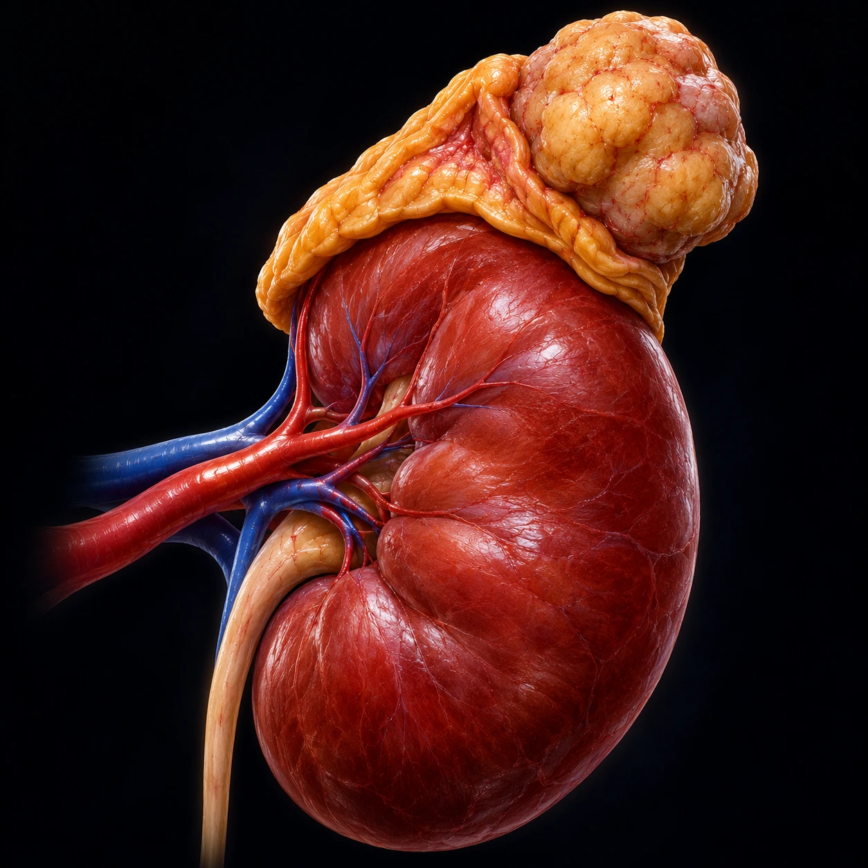
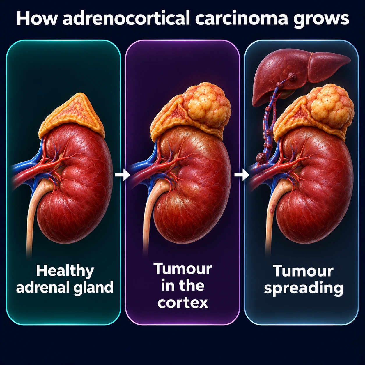

Adrenocortical carcinoma (ACC) is a rare but aggressive malignancy of the adrenal cortex — the outer layer of the adrenal gland that produces cortisol, aldosterone, and androgens. With an incidence of only 1–2 cases per million people per year, ACC is one of the rarest solid tumors in adults, yet it carries a disproportionately high mortality. Approximately half of all ACC tumors are hormonally active, producing excess steroids that cause dramatic clinical syndromes — rapid-onset Cushing's syndrome, virilization in women, or feminization in men — that often precede the cancer diagnosis by months. Even when detected early, ACC has a strong tendency to recur, and the cornerstone of systemic therapy remains mitotane, an adrenal-specific cytotoxin.
Table of Contents
1. Overview and Epidemiology
Adrenocortical carcinoma arises from the cortex of the adrenal gland — a structure that sits atop each kidney and is divided into three functional zones: the zona glomerulosa (aldosterone), zona fasciculata (cortisol), and zona reticularis (androgens and estrogens). ACC is distinct from pheochromocytoma, a tumor of the adrenal medulla (the inner portion), which has an entirely different biology, presentation, and management.
Incidence: ACC is diagnosed in approximately 1–2 people per million per year worldwide, translating to roughly 500–600 new cases in the United States annually. Despite its rarity, ACC is among the most lethal solid tumors: the majority of patients with metastatic disease die within 2 years of diagnosis.
Age distribution: ACC shows a bimodal age distribution that is characteristic and clinically important. The first peak occurs in children under 5 years old, predominantly in Brazil (where TP53 R337H germline mutations are unusually prevalent in the population). The second and larger peak affects adults aged 40–50 years. Unlike most solid tumors, ACC shows a female predominance of approximately 1.5:1 in adults, possibly related to steroidogenic enzyme expression differences or hormonal milieu.
Adrenal incidentaloma context: The widespread use of abdominal computed tomography (CT) and magnetic resonance imaging (MRI) has dramatically increased detection of adrenal masses. Approximately 4–7% of all computed tomography scans detect an adrenal mass incidentally ("incidentaloma"). The vast majority of these (over 80%) are benign cortical adenomas. However, the probability of malignancy rises sharply with lesion size: masses under 4 cm carry less than 2% malignancy risk, those 4–6 cm carry intermediate risk (15–25%), and those over 6 cm are malignant in 25–98% of cases depending on series. The average ACC diameter at diagnosis exceeds 12 cm — reflecting late discovery in many patients.
2. Genetics and Molecular Biology
The majority of ACC is sporadic, but germline genetic predisposition accounts for a meaningful minority of cases, particularly in children and young adults. Molecular characterization of ACC has identified several recurrently altered pathways with diagnostic and potentially therapeutic relevance.
TP53 and Li-Fraumeni syndrome: Germline mutations in TP53, the tumor-suppressor gene on chromosome 17p, are the most important genetic predisposition to ACC. Li-Fraumeni syndrome (LFS), caused by heterozygous germline TP53 mutations, predisposes to ACC, breast cancer, sarcomas, brain tumors, and leukemias. Approximately 50–80% of childhood ACC cases and 3–5% of adult sporadic ACC cases carry germline TP53 mutations. In southern Brazil, a founder mutation (R337H) in TP53 occurs with a carrier frequency of approximately 1 in 300, explaining the extraordinarily high pediatric ACC incidence in that region (15-fold above the global rate). Somatic TP53 mutations are found in approximately 25% of sporadic adult ACC and correlate with higher tumor grade and worse outcome.
Wnt/beta-catenin pathway (CTNNB1): Activating mutations in CTNNB1 (beta-catenin) are found in approximately 16–38% of ACC. Constitutive Wnt signaling drives aberrant adrenocortical cell proliferation. Beta-catenin-mutated ACC may have distinct immunological properties and altered responses to immunotherapy.
Beckwith-Wiedemann syndrome (BWS): BWS, caused by defects at chromosome 11p15 involving IGF2 (insulin-like growth factor 2) overexpression and loss of imprinting, predisposes to childhood overgrowth, Wilms tumor, hepatoblastoma, and ACC. IGF2 overexpression is the most common somatic molecular abnormality in ACC overall (found in 80–90% of tumors), making IGF signaling a heavily studied therapeutic target, though anti-IGF therapies have so far yielded modest clinical results.
Multiple Endocrine Neoplasia type 1 (MEN1): Germline MEN1 mutations predispose to pituitary adenomas, parathyroid adenomas, and pancreatic neuroendocrine tumors; adrenal cortical tumors occur in 20–40% of MEN1 patients, though true ACC (as opposed to adenoma) is uncommon.
Somatic landscape: The Cancer Genome Atlas (TCGA) analysis of ACC (2016) identified three subtypes with distinct clinical outcomes, which could be captured by a DNA-methylation signature of 68 sites; massive DNA loss followed by whole-genome doubling, together with increased TERT expression and shorter telomeres, was associated with an aggressive course. Ki-67 proliferation index above 20% is the single most important pathological predictor of aggressive behavior and early recurrence.
3. Clinical Presentation
ACC presents through two broad routes: hormone excess syndromes (in approximately 50–60% of cases) or symptoms from local tumor mass effect (in 40–50%). The nature of the hormone produced provides important diagnostic clues and has direct implications for quality of life and treatment.
Cortisol excess — Cushing's syndrome: The most common hormone syndrome in ACC. Unlike pituitary-driven Cushing's disease (which has an insidious onset over years), ACC-related Cushing's often develops over weeks to a few months, reflecting the rapid growth rate of the tumor. Classic features include central obesity, moon facies, buffalo hump, purple striae (widened, >1 cm), proximal muscle weakness, easy bruising, hypertension, hyperglycemia, and hypokalemia (from mineralocorticoid-like effect of high cortisol on renal tubules). The rapid pace is a red flag distinguishing ACC from a benign adenoma.
Androgen excess — virilization: Excess dehydroepiandrosterone sulfate (DHEA-S), androstenedione, and testosterone from ACC produces virilization in women: hirsutism, acne, clitoromegaly, deepening of the voice, amenorrhea, and male-pattern baldness. Many ACC tumors co-secrete cortisol and androgens simultaneously, creating a mixed hormonal picture.
Estrogen excess — feminization in males: Rare but highly specific for malignancy. A male patient presenting with gynecomastia, loss of libido, or testicular atrophy in the setting of an adrenal mass is treated as having ACC until proven otherwise. Estrogen-producing ACC is uncommon but carries this distinctive and underrecognized clinical signature.
Aldosterone excess: Primary hyperaldosteronism from ACC is uncommon (more typical of the benign aldosterone-producing adenoma). When it occurs in ACC, it manifests as resistant hypertension, hypokalemia, and suppressed plasma renin activity.
Non-functioning ACC: Approximately 40–50% of ACC produces no clinically apparent hormone excess. These tumors are often discovered incidentally or because of local symptoms — flank pain, abdominal mass, early satiety, nausea, or back discomfort from the large tumor compressing adjacent structures. Non-functioning ACC typically presents at a more advanced stage and may carry a worse prognosis due to delayed detection.
4. Diagnosis and Imaging
Accurate characterization of an adrenal mass requires both structural imaging (computed tomography or magnetic resonance imaging) and comprehensive hormonal evaluation. The critical principle is that pheochromocytoma is excluded biochemically before any surgical intervention on an adrenal mass, as an undiagnosed pheochromocytoma during surgery can cause a fatal hypertensive crisis.
Computed tomography imaging — Hounsfield unit analysis: The unenhanced computed tomography attenuation value (Hounsfield units, HU) is the single most important imaging parameter for characterizing adrenal masses. Lipid-rich adenomas: typically less than 10 HU on unenhanced computed tomography (fat content suppresses attenuation) — a reading under 10 HU is effectively diagnostic of a benign lipid-rich adenoma, requiring no further workup in a patient without a known primary malignancy. Lipid-poor adenomas require washout analysis: an absolute contrast washout above 60% or relative washout above 40% at 15-minute delayed imaging indicates a benign adenoma. ACC imaging characteristics: typically above 10 HU on unenhanced computed tomography, often heterogeneous (necrosis, hemorrhage, calcifications), large (>6 cm), irregular margins, possible invasion of adjacent structures (kidney, inferior vena cava, liver), and incomplete or heterogeneous contrast washout.
Magnetic resonance imaging: Complementary to CT; ACC shows heterogeneous T2 hyperintensity (reflecting necrosis and hemorrhage). Chemical-shift magnetic resonance imaging (in-phase vs. opposed-phase): intracellular lipid droplet loss on opposed-phase (signal drop) is the hallmark of benign lipid-rich adenoma; ACC does not show signal dropout. Magnetic resonance imaging is superior for assessing vascular invasion of the inferior vena cava and adjacent structures.
FDG-PET/computed tomography: ACC is FDG-avid — it strongly takes up fluorodeoxyglucose (FDG), the tracer used in positron emission tomography (PET) (high standard uptake value, SUV), while benign adenomas are typically FDG-negative or show low SUV. FDG-PET is valuable for staging (detecting lymph node, lung, and bone metastases) and for monitoring treatment response.
Biopsy — a critical caveat: Percutaneous biopsy of a suspected ACC is generally contraindicated. Risks include tumor seeding along the biopsy tract, capsule rupture, and hemorrhage into a large vascular tumor. If the mass is resectable, surgical resection provides both diagnosis and therapy simultaneously. Biopsy is reserved for situations where metastatic disease is present and tissue is needed to guide systemic therapy in an unresectable setting — after excluding pheochromocytoma.
Biochemical workup — mandatory for all adrenal masses:
- Pheochromocytoma exclusion (mandatory first): Plasma free metanephrines or 24-hour urine metanephrines and catecholamines. Done before any surgery.
- Cortisol: 1 mg overnight dexamethasone suppression test + morning cortisol; 24-hour urine free cortisol; late-night salivary cortisol (×2).
- Aldosterone/renin ratio: If hypertension or hypokalemia is present.
- Adrenal androgens: DHEA-S, androstenedione, testosterone — if virilization or rapid Cushing's features are present.
- Estrogens: Estradiol and estrone — in males with gynecomastia or feminizing features.
5. Pathology and Staging
Weiss scoring system: The gold standard for distinguishing ACC from adenoma on histopathology. The Weiss system evaluates nine criteria, with a score of 3 or more indicating adrenocortical carcinoma:
- High nuclear grade (Fuhrman grade 3–4)
- Mitotic rate greater than 5 per 50 high-power fields
- Atypical mitotic figures
- Clear cells comprising 25% or less of the tumor
- Diffuse architecture (more than one-third of the tumor is sheet-like)
- Confluent necrosis
- Venous invasion (medium-sized or large veins with smooth muscle wall)
- Sinusoidal invasion (thin-walled vascular channels within adrenal tissue)
- Capsular invasion
Modified versions (Helsinki score, Lin-Weiss-Bisceglia) offer refinements for lipid-rich or oncocytic variants, which can be scored falsely low on classic Weiss criteria.
Ki-67 proliferation index: Ki-67 is the most clinically impactful single pathological marker. Ki-67 above 20% is associated with dramatically shorter recurrence-free and overall survival, even in completely resected Stage I-II tumors. The 2018 European Society of Endocrinology (ESE) guideline suggests adjuvant mitotane after radical surgery when the risk of recurrence is perceived as high: stage III disease, a microscopically positive margin (R1 resection), or Ki-67 above 10%.
ENSAT staging system (European Network for the Study of Adrenal Tumors):
- Stage I: Tumor ≤5 cm, confined to the adrenal gland, no lymph nodes, no metastases.
- Stage II: Tumor >5 cm, confined to the adrenal gland, no lymph nodes, no metastases.
- Stage III: Local invasion into surrounding fat or adjacent organs, or regional lymph node metastases, or tumor thrombus in the renal vein or inferior vena cava.
- Stage IV: Distant metastases (lung, liver, peritoneum, bone, brain).
ENSAT staging is preferred over older TNM systems because it better stratifies survival outcomes and is used in all major ACC clinical trials.
6. Treatment
Surgery — the only potentially curative treatment: Complete surgical resection (R0 resection — microscopically negative margins) is the single most critical prognostic factor in ACC. Even in Stage III disease with local invasion, aggressive en bloc resection including adjacent involved structures can achieve cure or prolonged remission in selected patients.
Open versus laparoscopic adrenalectomy: Surgical approach matters. Open adrenalectomy is strongly preferred for ACC for several important reasons: (1) ACC tumors are typically large (>6 cm), often with local invasion; (2) the adrenal capsule is fragile and easily ruptured — capsule violation upgrades the patient to R1 resection and dramatically worsens prognosis; (3) adequate lymphadenectomy is more easily performed via open approach. Laparoscopic resection is generally limited to small (<6 cm), localized, encapsulated ACC without evidence of local invasion — and to experienced centers. Surgeons convert to an open operation immediately if any concerns arise about capsular integrity or local extension.
Adjuvant therapy: After complete resection, the 2018 ESE guideline suggests adjuvant mitotane for patients with a perceived high risk of recurrence: Stage III, Ki-67 above 10%, or R1 resection (positive margins); Stage II disease with additional risk factors is also sometimes treated. The ADIUVO trial (randomized, 2023) found no significant recurrence-free survival benefit of adjuvant mitotane in low-to-intermediate risk ACC (R0 resection, Stage I-III, Ki-67 ≤10%); its authors wrote that adjuvant mitotane might not be indicated in these patients, but the trial stopped early and cannot rule out a benefit (see section 9). Duration of adjuvant mitotane is typically 2 years or until disease recurrence.
Recurrence management: Even after apparently complete resection, ACC recurs in 50–80% of patients within 2 years. Repeat resection is indicated for isolated resectable recurrence. Local ablative therapies (radiofrequency ablation, stereotactic body radiotherapy) are used for limited metastatic sites. Systemic therapy with EDP-M (see below) is the standard for unresectable or widely metastatic recurrence.
Systemic chemotherapy — EDP-M regimen: For unresectable or metastatic ACC, the FIRM-ACT trial (2012) established etoposide + doxorubicin + cisplatin + mitotane (EDP-M) as the first-line chemotherapy standard. EDP-M was superior to streptozotocin + mitotane for both response rate (23% vs. 9%) and progression-free survival. However, objective response rates remain modest and median overall survival in metastatic ACC is approximately 14–18 months with EDP-M.
FIRM-ACT in numbers: The trial randomly assigned 304 patients with advanced ACC. As first-line therapy, EDP-mitotane gave a response rate of 23.2% versus 9.2% and a median progression-free survival of 5.0 versus 2.1 months, but the difference in overall survival (14.8 versus 12.0 months) was not statistically significant. Patients whose disease progressed switched to the other regimen; among those who never received that second-line switch, median survival was 17.1 months with first-line EDP-mitotane and 4.7 months with streptozocin-mitotane. Rates of serious side effects did not differ significantly between the two regimens.
Immunotherapy: Checkpoint inhibitors have shown limited efficacy in ACC. Pembrolizumab and nivolumab have been tested in Phase 2 trials; in the pembrolizumab trial (39 patients with advanced ACC), 23% had an objective response (95% CI 11–39%), the disease was controlled in 52%, and median overall survival was 24.9 months. Lenvatinib + pembrolizumab combinations are under active investigation. CTNNB1-mutated tumors may be immune-excluded, potentially explaining reduced checkpoint inhibitor benefit in this molecular subset.
7. Mitotane: Pharmacology and Management
Mitotane (o,p'-DDD; 1,1-dichloro-2-(o-chlorophenyl)-2-(p-chlorophenyl)ethane) is the most important drug in ACC and requires specialized management. It is an adrenocorticolytic agent that both inhibits steroidogenesis and directly destroys adrenocortical cells through toxic metabolites.
Mechanism of action: Mitotane undergoes metabolic activation within adrenocortical mitochondria, generating reactive metabolites that bind to adrenocortical proteins and cause cell-specific cytotoxicity. It also inhibits multiple steps in steroid synthesis (CYP11A1, CYP11B1, CYP11B2) and accelerates cortisol catabolism by inducing hepatic cytochrome P450 3A4 (CYP3A4) enzymes. This dual action (cytotoxic + steroidogenesis inhibition) makes it simultaneously a cancer drug and a cause of adrenal insufficiency that requires steroid replacement.
Therapeutic drug monitoring: Mitotane is highly lipophilic and accumulates in adipose tissue. It has a narrow therapeutic window. The target plasma concentration is 14–20 mg/L. Below 14 mg/L, antitumor efficacy is substantially diminished. The 14 mg/L target is supported by a retrospective European multicenter study of 91 patients with advanced ACC: 19% had an objective tumour response, responders were more common among patients reaching 14 mg/L or more, and those levels were linked with longer survival (hazard ratio 0.52). Above 20 mg/L, neurological toxicity (ataxia, dysarthria, cognitive slowing, somnolence) becomes more likely: the U.S. label states that levels above 20 mg/L are associated with a greater incidence of toxicity and that severe neurotoxicity may occur at those levels. The label states that the target level is usually reached within 3 to 5 months of dose escalation, and it describes level checks every 2 weeks after starting treatment and after each dose change, then periodic checks (for example, monthly).
Mandatory adrenal replacement: All patients on mitotane require corticosteroid replacement, because mitotane induces pharmacological adrenal insufficiency and simultaneously accelerates cortisol breakdown (by inducing cytochrome P450 3A4). Hydrocortisone doses needed are often two to three times higher than standard physiological replacement (typically 30–50 mg/day in divided doses). Standard practice includes a medical alert bracelet, injectable hydrocortisone carried for emergencies, and sick-day rules (higher steroid doses during illness), because adrenal crisis is a condition hospitals treat as an emergency. Mineralocorticoid (fludrocortisone) is added if aldosterone deficiency develops.
Drug interactions: Because mitotane is a potent cytochrome P450 3A4 inducer, it dramatically alters the metabolism of many co-administered drugs, including warfarin (higher doses are often needed), oral contraceptives (which can be rendered ineffective, so barrier contraception is the method usually relied on), and other oncological agents metabolized by cytochrome P450 3A4. Any medicine taken alongside mitotane is a candidate for this interaction.
Key toxicities: Gastrointestinal (nausea, vomiting, diarrhea) are the most common early toxicities and limit initial dose escalation. The U.S. label states that the effect of food on absorption of the mitotane tablet is not known, while data from various mitotane formulations suggest that high-fat food enhances absorption; the label's instructions are to take the tablets with food. Taking it with food may also reduce gastrointestinal (GI) effects. Neurological toxicity (ataxia, confusion, tremor) is more frequent at levels above 20 mg/L. Elevated liver enzymes and lipid abnormalities are common. Pregnancy — the U.S. label lists no contraindications, but it states that mitotane can cause fetal harm when given to a pregnant woman; limited postmarketing cases report preterm births and early pregnancy loss, and the label describes effective non-hormonal contraception during treatment and for as long as mitotane remains detectable in the blood.
8. Prognosis and Surveillance
ACC carries a poor overall prognosis, though outcomes vary substantially by stage and molecular features.
Stage-specific survival:
- Stage I: 5-year overall survival approximately 60–80% with complete resection.
- Stage II: 5-year overall survival approximately 50–70%; recurrence risk remains high even after R0 resection.
- Stage III: 5-year overall survival approximately 30–45%; reflects heterogeneous local extension (lymph node-only vs. vascular invasion vs. adjacent organ involvement).
- Stage IV (metastatic): Median overall survival approximately 12–18 months with EDP-M; 5-year survival below 15%.
Prognostic factors: The most powerful predictors of outcome are: (1) ENSAT stage at diagnosis; (2) R0 vs. R1/R2 resection; (3) Ki-67 proliferation index (≤20% vs. >20%); (4) achievement of therapeutic mitotane levels in the adjuvant setting; (5) cortisol-secreting tumors (independently associated with worse outcomes, likely due to immunosuppression from chronic cortisol excess); and (6) molecular features (TP53 mutation, chromosomal instability subgroup).
Post-resection surveillance: After R0 resection, surveillance imaging is critical because 50–80% of patients recur, most within 2 years. Standard surveillance: contrast-enhanced computed tomography of chest/abdomen/pelvis every 3 months for the first 2 years, then every 6 months for years 3–5, then annually thereafter. Adrenal steroid panel (DHEA-S, androstenedione, 24-hour urine cortisol) is obtained at each visit in hormonally active tumors to detect biochemical recurrence that may precede radiographic recurrence.
Germline genetic testing: Germline genetic testing for TP53 is commonly offered to all patients with ACC, regardless of age at diagnosis, with consideration of broader panel testing including MEN1, PRKAR1A (Carney complex), and APC (adrenal tumors linked to familial adenomatous polyposis). First-degree relatives of TP53 germline carriers are candidates for Li-Fraumeni syndrome surveillance protocols.
9. Recent Research and Emerging Therapies
ADIUVO trial (2023): This international randomized phase 3 trial (23 centres in seven countries) enrolled 91 ACC patients with completely resected (R0) low-to-intermediate risk disease (Stage I-III, Ki-67 ≤10%) and randomized them to 2 years of adjuvant mitotane (45 patients) vs. surveillance alone (46 patients). No significant difference in recurrence-free survival was observed: 5-year recurrence-free survival was 79% with mitotane and 75% with surveillance (hazard ratio 0.74, 95% CI 0.30–1.85), and 5-year overall survival was 95% and 86%, also not significantly different. All 42 patients who took mitotane had side effects, and 8 (19%) stopped it. The authors concluded that adjuvant mitotane might not be indicated in low-grade, localised ACC, given these patients' relatively good prognosis and the drug's toxicity — but the trial was stopped early because of slow recruitment and cannot rule out a benefit of treatment.
Targeted therapy — IGF1R pathway: Given that IGF2 overexpression is near-universal in ACC, multiple trials have tested anti-IGF1R antibodies (cixutumumab, ganitumab) and IGF1R/insulin receptor tyrosine kinase inhibitors (linsitinib). The Phase 3 GALACCTIC trial of linsitinib was negative, establishing that broad IGF pathway inhibition does not benefit unselected ACC. Research is ongoing to identify molecular subsets that may respond.
Wnt pathway targeting: Given the prevalence of CTNNB1 mutations in ACC, Wnt inhibitors (porcupine inhibitors, tankyrase inhibitors) are being explored in early-phase trials. Results are preliminary.
Cabozantinib: In a single-arm phase 2 trial at one U.S. cancer center, 18 patients with advanced ACC took cabozantinib, an oral multikinase inhibitor, at 60 mg daily. At 4 months, 13 of 18 (72.2%) were free of progression, and median progression-free survival was 6 months; 11 (61%) had a treatment-related side effect of grade 3 or worse. In a 2025 basket trial that combined cabozantinib with the immunotherapy drug atezolizumab, the ACC group (24 patients) had a response rate of 8.3% and 47.6% survival at 12 months.
Molecular classification: The TCGA comprehensive molecular analysis of ACC (2016) identified three subtypes with distinct clinical outcomes, captured by a 68-site DNA-methylation signature (see section 2). Separately, a 2014 European genomic study split carcinomas by their gene-activity (transcriptome) profile into two groups with opposite outcomes: "C1A", with numerous mutations and DNA-methylation changes and poor outcome, and "C1B", with good prognosis. In a 2022 validation cohort using routine paraffin-embedded tumour samples, 5-year overall survival was 26% in C1A and 100% in C1B, and the grouping predicted survival independently of tumour stage and Ki-67. Such molecular stratification may eventually inform treatment selection.
Steroidogenesis inhibitors: For hormonally active ACC, control of cortisol excess is both a quality-of-life priority and potentially therapeutic. Osilodrostat (a CYP11B1 inhibitor) and levoketoconazole have been approved for Cushing's syndrome from various causes and are increasingly used in ACC. In the context of mitotane treatment, additional steroidogenesis inhibitors may be needed if tumor-driven steroid excess persists despite mitotane.
Expert center care: Given the rarity and complexity of ACC, outcomes are significantly better at high-volume centers with multidisciplinary expertise in adrenal oncology, experienced adrenal surgeons, endocrinologists skilled in mitotane management, and access to clinical trials. The 2018 ESE guideline recommends that all patients with suspected or proven ACC are discussed in a multidisciplinary expert team meeting, and that surgery for suspected ACC is done only by surgeons experienced in adrenal and cancer surgery.
Key Research Papers
- FIRM-ACT Trial: EDP-M vs. Streptozotocin-Mitotane in Advanced ACC
Fassnacht M, Terzolo M, Allolio B, et al. N Engl J Med. 2012;366:2189–2197. — doi:10.1056/NEJMoa1200966 · PubMed PMID: 22551107 - ADIUVO Trial: Adjuvant Mitotane in Low-Risk Resected ACC (RCT)
Terzolo M, Fassnacht M, Perotti P, et al. Lancet Diabetes Endocrinol. 2023;11(10):720–730. — doi:10.1016/S2213-8587(23)00193-6 · PubMed PMID: 37619579 - European Society of Endocrinology Clinical Practice Guidelines for ACC
Fassnacht M, Dekkers OM, Else T, et al. Eur J Endocrinol. 2018;179:G1–G46. — doi:10.1530/EJE-18-0608 · PubMed PMID: 30299884 - TCGA Comprehensive Molecular Characterization of ACC
Zheng S, Cherniack AD, Dewal N, et al. Cancer Cell. 2016;29:723–736. — doi:10.1016/j.ccell.2016.04.002 · PubMed PMID: 27165744 - Weiss Criteria for Histopathological Diagnosis of ACC
Weiss LM, Medeiros LJ, Vickery AL. Am J Surg Pathol. 1989;13(3):202–206. — Search PubMed · PubMed PMID: 2919718 - ENSAT Staging System for Adrenocortical Carcinoma
Fassnacht M, Johanssen S, Quinkler M, et al. Cancer. 2009;115(2):243–250. — doi:10.1002/cncr.24030 · PubMed PMID: 19025987 - Mitotane Serum Level and Outcome in ACC: Multicenter Study
Hermsen IG, Fassnacht M, Terzolo M, et al. J Clin Endocrinol Metab. 2011;96(6):1844–1851. — doi:10.1210/jc.2010-2676 · PubMed PMID: 21470991 - TP53 Germline Mutations and ACC in the Brazilian Population
Ribeiro RC, Sandrini F, Figueiredo B, et al. Proc Natl Acad Sci USA. 2001;98(16):9330–9335. — Search PubMed · PubMed PMID: 11481490 - IGF2 Overexpression and Insulin-like Growth Factor Pathway in ACC
Gicquel C, Bertagna X, Schneid H, Francillard-Leblond M, Luton JP, Girard F, Le Bouc Y. Rearrangements at the 11p15 locus and overexpression of insulin-like growth factor-II gene in sporadic adrenocortical tumors. J Clin Endocrinol Metab. 1994;78(6):1444–1453. — doi:10.1210/jc.78.6.1444 · PubMed PMID: 7911125 - Adrenal Incidentaloma: Management and Imaging Classification
Fassnacht M, Arlt W, Bancos I, et al. Eur J Endocrinol. 2016;175:G1–G34. — doi:10.1530/EJE-16-0467 · PubMed PMID: 27390021 - Ki-67 as Independent Prognostic Marker in Resected ACC
Morimoto R, Satoh F, Murakami O, et al. Endocr J. 2008;55(1):49–55. — Search PubMed · PubMed PMID: 18187873 - Laparoscopic vs. Open Adrenalectomy for ACC: Oncological Outcomes
Miller BS, Ammori JB, Gauger PG, et al. World J Surg. 2010;34(6):1380–1385. — doi:10.1007/s00268-010-0532-2 · PubMed PMID: 20372905 - Integrated Genomic Characterization of ACC: C1A and C1B Groups
Assié G, Letouzé E, Fassnacht M, et al. Nat Genet. 2014;46(6):607–612. — doi:10.1038/ng.2953 · PubMed PMID: 24747642 - Transcriptome in Paraffin Samples for ACC Diagnosis and Prognosis
Jouinot A, Lippert J, Sibony M, et al. Eur J Endocrinol. 2022;186(6):607–617. — doi:10.1530/EJE-21-1228 · PubMed PMID: 35266879 - PD-1 Blockade (Pembrolizumab) in Advanced ACC: Phase II Trial
Raj N, Zheng Y, Kelly V, et al. J Clin Oncol. 2020;38(1):71–80. — doi:10.1200/JCO.19.01586 · PubMed PMID: 31644329 - Cabozantinib Monotherapy for Advanced ACC: Single-Arm Phase 2 Trial
Campbell MT, Balderrama-Brondani V, Jimenez C, et al. Lancet Oncol. 2024;25(5):649–657. — doi:10.1016/S1470-2045(24)00095-0 · PubMed PMID: 38608694 - Cabozantinib plus Atezolizumab in Advanced Endocrine Malignancies (CABATEN/GETNE-T1914)
Capdevila J, Hernando J, Molina-Cerrillo J, et al. Clin Cancer Res. 2025;31(22):4655–4663. — doi:10.1158/1078-0432.CCR-25-2143 · PubMed PMID: 40938918
PubMed Topic Searches
- PubMed: Adrenocortical carcinoma mitotane treatment
- PubMed: Adrenal cortex cancer surgery resection
- PubMed: ACC adrenal carcinoma immunotherapy
- PubMed: Adrenocortical carcinoma genetics TP53
- PubMed: Adrenal Cushing syndrome malignant
- PubMed: Weiss score adrenal pathology
- PubMed: Adrenal incidentaloma management guidelines
- PubMed: Adrenocortical carcinoma prognosis recurrence
- PubMed: Li-Fraumeni syndrome adrenocortical carcinoma
- PubMed: Adrenal cancer EDP chemotherapy