Wilms Tumor
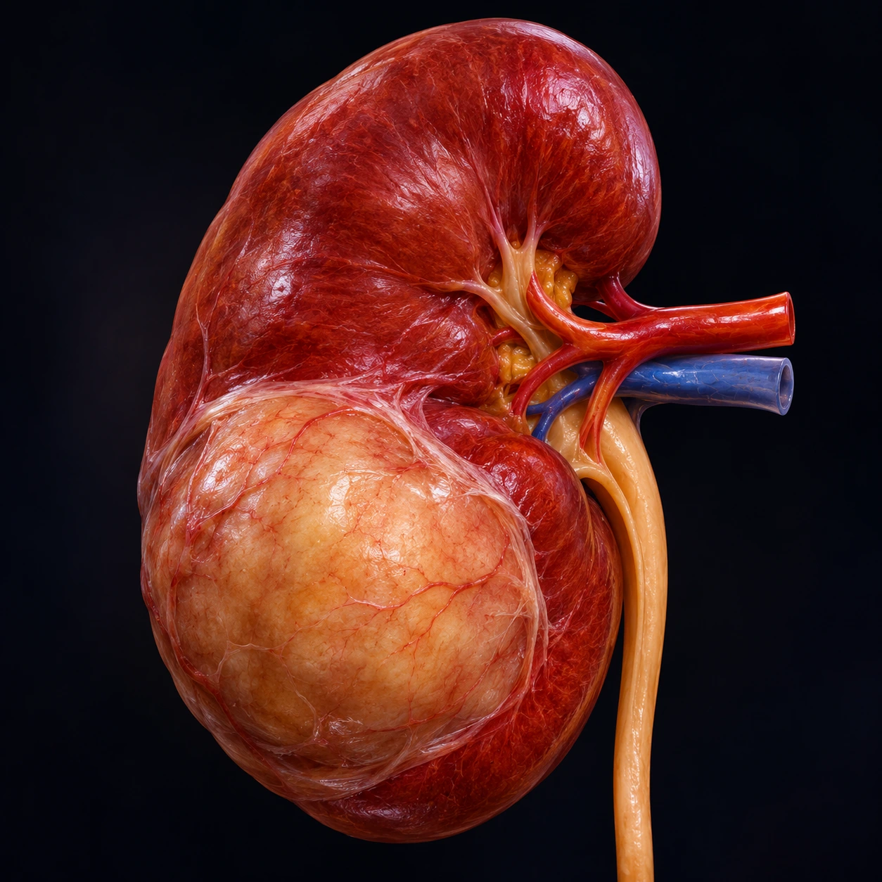
Wilms tumor, or nephroblastoma, is the most common primary renal malignancy in children and one of the great success stories of pediatric oncology — a disease that killed most affected children before the 1950s is now cured in more than 90% of patients through collaborative, protocol-driven treatment. Arising from embryonic kidney precursor cells, Wilms tumor teaches fundamental lessons about pediatric oncogenesis, the genetics of tumor suppressor genes, and the remarkable effectiveness of combining surgery, chemotherapy, and targeted radiotherapy in children. With approximately 500 new cases diagnosed annually in the United States, it remains the fourth most common pediatric malignancy overall.
Table of Contents
1. Overview and Epidemiology
Wilms tumor (nephroblastoma) is the most common primary malignant tumor of the kidney in children, accounting for approximately 85–90% of all pediatric renal tumors and roughly 6% of all childhood cancers in the United States. About 500 new cases are diagnosed annually in the United States, translating to an incidence of approximately 7.6 cases per million children under 15 years of age.
The peak age of diagnosis is 3 to 4 years. The tumor is rare in the first six months of life — when it does occur in infants this young, genetic syndromes are more likely to be involved — and it becomes uncommon after age 15. In the National Wilms Tumor Study registry (children registered 1969–1985), median age at diagnosis was 36.5 months for boys and 42.5 months for girls with a tumor in one kidney, and 23.5 and 30.5 months for boys and girls with tumors in both kidneys. Adult Wilms tumor exists but is rare, comprising less than 1% of adult renal malignancies, and generally carries a worse prognosis than the pediatric form.
Bilateral involvement occurs in approximately 5–10% of patients (Stage V disease). Bilateral tumors are more strongly associated with genetic predisposition syndromes, younger age at diagnosis, and nephrogenic rests. Managing bilateral Wilms tumor requires the additional imperative of preserving enough functional renal parenchyma to avoid dialysis dependence.
Sex distribution is roughly equal, with a slight female predominance in some series. There is a modest excess risk among children of African ancestry compared to Asian-ancestry children, with White and Black American children having similar rates. The disease occurs worldwide with relatively uniform incidence, unlike some adult cancers that show dramatic geographic variation tied to environmental exposures.
Before modern multimodality treatment, Wilms tumor was almost universally fatal. The National Wilms Tumor Study Group (NWTS), established in 1969 and succeeded by the Children's Oncology Group (COG), transformed outcomes through a series of landmark collaborative trials that progressively refined surgery, chemotherapy, and radiotherapy — achieving today's overall five-year survival exceeding 90%. A 2021 review in Nature Reviews Disease Primers states that cure is achievable in 90% of affected children, even those whose disease has spread, yet survival gaps remain within and between countries.
2. Genetics and Predisposing Syndromes
Wilms tumor arises through inactivation of tumor suppressor genes that regulate normal renal development. Approximately 10–15% of cases are associated with recognized genetic syndromes or constitutional mutations; the majority (~85%) are sporadic with no identifiable germline alteration at clinical evaluation, though somatic mutations accumulate during renal development.
WT1 Gene (11p13)
The WT1 gene on chromosome 11p13 encodes a zinc-finger transcription factor essential for normal kidney and gonadal development. Constitutional WT1 mutations or deletions underlie several overgrowth and genitourinary syndromes:
- Denys-Drash syndrome: Caused by specific missense mutations in the zinc-finger DNA-binding domain of WT1. The triad consists of (1) diffuse mesangial sclerosis leading to nephrotic syndrome in infancy, (2) 46,XY male pseudohermaphroditism (ambiguous or female genitalia despite XY karyotype), and (3) Wilms tumor. Children with Denys-Drash syndrome have an approximately 90% lifetime risk of Wilms tumor and typically present at younger ages. Prophylactic nephrectomy is sometimes considered given the near-certainty of tumor development in the dysgenetic kidneys.
- Frasier syndrome: Caused by mutations in the WT1 intron 9 splice-donor site, altering the ratio of WT1 isoforms. Clinical features include 46,XY gonadal dysgenesis (streak gonads), focal segmental glomerulosclerosis progressing to renal failure, and elevated risk of gonadoblastoma. Wilms tumor risk is lower in Frasier syndrome than in Denys-Drash.
- WAGR syndrome: Results from constitutional deletion of chromosome 11p13 encompassing both WT1 and the PAX6 gene. The acronym captures: Wilms tumor, Aniridia (absence of the iris, due to PAX6 deletion), Genitourinary anomalies (in XY individuals: cryptorchidism, hypospadias), and a Range of developmental delays and intellectual disability. Wilms tumor risk in WAGR is approximately 50%. Children with isolated congenital aniridia are routinely tested for WT1 deletion because of this link.
WT2 Locus (11p15) and Beckwith-Wiedemann Syndrome
A second Wilms tumor locus maps to chromosome 11p15.5 — the WT2 imprinting region that also controls the IGF2 (insulin-like growth factor 2) and H19 genes. Disruption of normal genomic imprinting at this locus produces Beckwith-Wiedemann syndrome (BWS), an overgrowth disorder characterized by:
- Macrosomia (large birth weight) and postnatal overgrowth
- Macroglossia (enlarged tongue, often causing feeding difficulty and airway concerns)
- Omphalocele or umbilical hernia
- Hemihypertrophy (asymmetric overgrowth of one body side)
- Neonatal hypoglycemia (from pancreatic islet hyperplasia and elevated IGF2)
- Visceromegaly (enlarged liver, spleen, kidneys, adrenal glands)
In a 2016 meta-analysis of 1,370 people with BWS, 102 (7.4%) developed a tumor, with rates ranging from 2.5% to 22.8% depending on the molecular cause (PubMed PMID: 27372391; see Beckwith-Wiedemann Syndrome), with Wilms tumor being the most common (followed by hepatoblastoma and adrenocortical carcinoma). Children with BWS or isolated hemihypertrophy are enrolled in surveillance protocols with abdominal ultrasound every 3 months; a 2017 surveillance paper describes kidney ultrasound every 3 months through the seventh birthday, plus full abdominal ultrasound and blood alpha-fetoprotein every 3 months through the fourth birthday for hepatoblastoma (PubMed PMID: 28674120). A 2019 pooled analysis of published and unpublished cases found that screening until age 7 detects close to 95% of Wilms tumors in children with Beckwith-Wiedemann spectrum, and that children whose tumor was found by screening were diagnosed at lower stages than children who were not screened. Members of the American Association for Cancer Research Pediatric Cancer Working Group updated these surveillance guidelines, first published in 2017, in 2024.
Other Genetic Alterations
- WTX gene (Xq11.1): Somatically mutated in approximately 30% of sporadic Wilms tumors. WTX encodes a negative regulator of the WNT/beta-catenin pathway; loss of WTX function promotes WNT signaling and tumor growth.
- CTNNB1 (beta-catenin): Activating mutations in approximately 15% of tumors, frequently co-occurring with WT1 mutations; drives WNT pathway activation.
- SIX1/SIX2: Mutations in these developmental transcription factors are found in approximately 10% of Wilms tumors, predominantly in blast-predominant and late-relapse tumors; SIX1/2 normally regulate the transition from renal progenitor cells to differentiated tubular epithelium.
- TP53: Mutations in approximately 75% of anaplastic (unfavorable histology) Wilms tumors; a critical driver of the anaplastic phenotype and chemoresistance.
- Familial Wilms tumor: Bilateral or familial cases without a recognized syndrome account for fewer than 2% of all cases. Germline mutations in WT1, WTX, CTR9, DROSHA, and DGCR8 (miRNA biogenesis genes) have been identified in familial kindreds.
3. Pathology and Histology
Wilms tumor pathology is classified by histology into favorable and unfavorable categories — a distinction that has profound implications for treatment intensity and prognosis.
Favorable Histology (FH): The Classic Triphasic Pattern
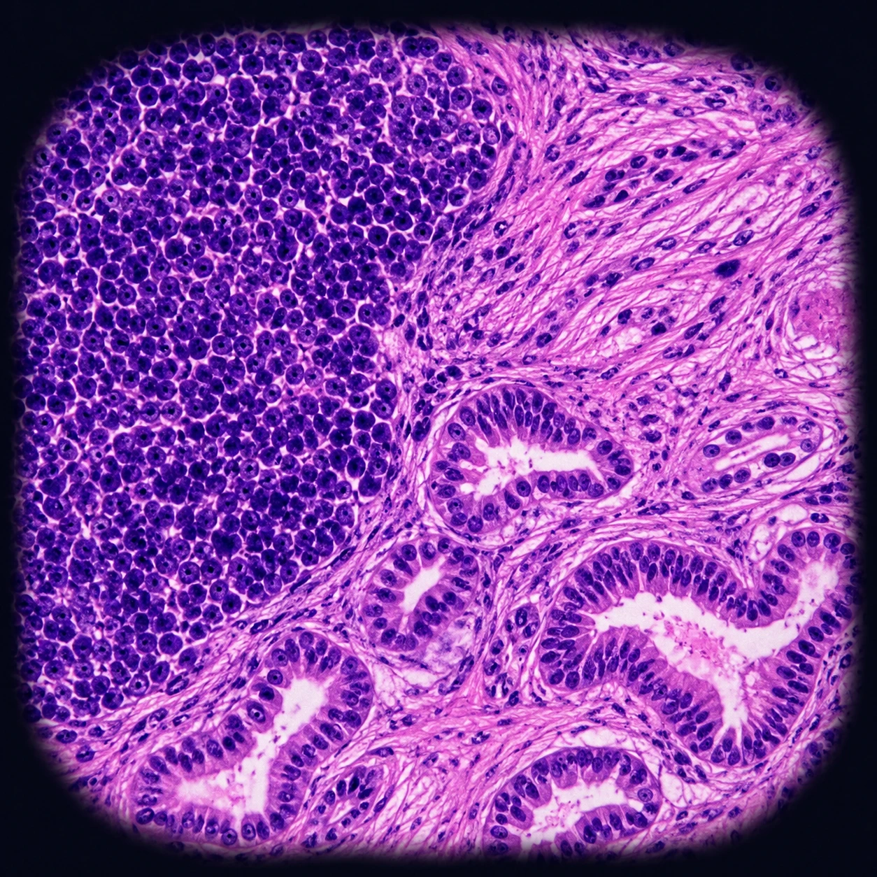
The characteristic Wilms tumor is a triphasic tumor containing three tissue types that recapitulate normal metanephric development:
- Blastemal component: Densely packed, small, round blue cells with scanty cytoplasm resembling primitive metanephric blastema; highly mitotically active; drives tumor growth and is the element most responsive to actinomycin D and vincristine.
- Stromal component: Loose mesenchymal-type tissue that may differentiate toward skeletal muscle, smooth muscle, fat, or cartilage; anaplasia can arise within the stromal compartment.
- Epithelial component: Attempts to form tubular or glomeruloid structures; the most differentiated element; least proliferative.
Not all tumors are equally triphasic; some are predominantly blastemal, others predominantly stromal or epithelial. Predominantly blastemal tumors in the SIOP (European) protocol after preoperative chemotherapy carry higher relapse risk and are treated as intermediate-risk.
Unfavorable Histology (UH): Anaplasia
Anaplasia is defined by three strict histologic criteria: (1) multipolar polyploid mitotic figures, (2) marked nuclear enlargement (nuclei at least 3 times the diameter of adjacent cells), and (3) nuclear hyperchromasia. In the fifth National Wilms Tumor Study (NWTS-5), anaplasia was found in 281 of 2,596 enrolled children (10.8%), but accounts for approximately 50% of Wilms tumor deaths — a dramatic overrepresentation driven by chemoresistance linked to TP53 mutations.
- Focal anaplasia: Confined to one or a few discrete nodules within the primary tumor; favorable prognosis approaching FH when completely resected; treated with intensified but not the most aggressive regimens.
- Diffuse anaplasia: Anaplasia present throughout the tumor or in more than one region, or found in any extra-renal site (lymph node, surgical margin, metastasis); the highest-risk Wilms tumor subgroup. In NWTS-5, 4-year event-free survival was 64.7% for stage III and 33.3% for stage IV diffuse anaplasia; in the later Children's Oncology Group AREN0321 study, children with stage II–IV diffuse anaplasia given a carboplatin-containing regimen had 4-year event-free survival of 67.7% and overall survival of 73.7%.
Nephrogenic Rests: The Precursor Lesions
Nephrogenic rests are foci of abnormally persistent embryonic kidney cells that normally involute after birth. They are found in approximately 1% of infant kidneys at autopsy but in approximately 35% of kidneys harboring unilateral Wilms tumor and in nearly all bilateral Wilms tumors. They are the direct precursors to Wilms tumor:
- Perilobar nephrogenic rests (PLNRs): Located at the periphery of renal lobes; associated with BWS, hemihypertrophy, and WT2/11p15 alterations; tend to be more numerous and are associated with bilateral Wilms tumor risk.
- Intralobar nephrogenic rests (ILNRs): Scattered within the renal parenchyma; associated with WAGR syndrome and WT1 mutations; carry higher individual transformation risk.
When nephrogenic rests are diffuse and bilateral, the condition is termed nephroblastomatosis — a finding that mandates surveillance and often preoperative chemotherapy to reduce bilateral tumor burden before nephron-sparing surgery.
4. Clinical Presentation
The overwhelming majority of Wilms tumor cases present as a smooth, firm, asymptomatic abdominal mass discovered incidentally — most commonly by a parent bathing or dressing the child, or by a clinician during a routine well-child visit. This presentation pattern reflects the tumor's typical encapsulation and the relatively large abdominal cavity in toddlers, which allows the tumor to grow to substantial size (often exceeding 500 grams) without causing symptoms.
Common Symptoms and Signs
- Abdominal mass: Present in approximately 90% of patients at diagnosis; smooth, nontender, does not cross the midline (unlike neuroblastoma, which often does); confined to one side of the abdomen in unilateral disease.
- Hematuria: Gross or microscopic hematuria in approximately 20–25% of patients; results from tumor invasion of the collecting system; not a reliable indicator of Stage or prognosis.
- Hypertension: Present in approximately 25% of patients; caused by renin secretion from compressed or ischemic renal parenchyma adjacent to the tumor, or from direct invasion of the renal vasculature; usually resolves after nephrectomy.
- Abdominal pain: Less common than with neuroblastoma; when present, may result from rapid tumor expansion, intratumoral hemorrhage, or capsule distension.
- Fever: Low-grade fever in 20–30% of patients; cause uncertain — possibly related to tumor necrosis or cytokine release.
- Anemia: Subcutaneous hemorrhage into the tumor flank can produce anemia; intravascular extension to the inferior vena cava (IVC; see below) can cause varicocele or hepatomegaly.
- Polycythemia: Rare; due to erythropoietin secretion by tumor cells.
Acquired von Willebrand Disease
An important and under-recognized association is acquired von Willebrand disease (aVWD), which occurs in approximately 8% of Wilms tumor patients. The tumor absorbs and degrades von Willebrand factor multimers, producing a coagulopathy that can cause significant intraoperative bleeding. Treatment teams check clotting before surgery (including a von Willebrand factor activity assay) and treat aVWD before the operation with desmopressin (DDAVP) or von Willebrand factor-containing concentrates.
Why Forceful Abdominal Examination Is a Known Hazard
Once Wilms tumor is suspected, repeated or forceful abdominal palpation is a recognized hazard. The tumor capsule, while usually intact at presentation, can be ruptured by aggressive examination — an event that upstages the patient to Stage III (requiring abdominal radiation) and significantly worsens prognosis. Pediatric teams examine such a child gently and teach staff caring for the child about this risk. Abdominal ultrasound, not repeated physical examination, is the usual next step for a child with an abdominal mass.
5. Diagnosis and Imaging
The diagnostic workup for a child with a suspected renal mass follows a logical sequence from rapid bedside assessment to detailed cross-sectional staging imaging. Tissue biopsy is generally deferred until after definitive nephrectomy in the North American (COG) approach, both to avoid upstaging from tumor spillage and because Wilms tumor diagnosis is usually confirmable by imaging alone.
Abdominal Ultrasound: First-Line Assessment
Abdominal ultrasound is the usual first test, done promptly, in any child with a palpable abdominal mass. Ultrasound provides rapid, radiation-free assessment of:
- Intrinsic renal origin of the tumor versus adrenal/retroperitoneal origin (critical for distinguishing from neuroblastoma)
- Contralateral kidney appearance and the presence of bilateral tumors
- Intravascular extension: Wilms tumor extends into the renal vein or IVC in approximately 4–10% of cases; Doppler ultrasound can identify IVC thrombus, which critically alters surgical planning
- Tumor vascularity and internal architecture (solid versus cystic components)
Computed Tomography Scan of Chest, Abdomen, and Pelvis
Contrast-enhanced computed tomography (CT) of the chest, abdomen, and pelvis is the standard staging modality in North America. Key findings include:
- The "claw sign": A rim of normal renal parenchyma displaced around the tumor mass, demonstrating the intrinsic renal origin — a critical imaging feature distinguishing Wilms tumor from neuroblastoma, which displaces the kidney downward or laterally rather than arising from within it.
- Contralateral kidney: Examined for synchronous Wilms tumor, nephrogenic rests, or cysts.
- Lymph node assessment: Hilar, para-aortic, and pericaval lymphadenopathy.
- IVC and cardiac extension: computed tomography angiography or magnetic resonance imaging (MRI) is preferable for detailed delineation of intravascular thrombus extending into the right atrium, which requires cardiopulmonary bypass during surgical resection.
- Pulmonary metastases: The lungs are the most common site of distant metastasis, occurring in approximately 20% of patients at diagnosis; computed tomography chest identifies lesions missed on plain radiographs. Even small pulmonary nodules (<1 cm) that are computed tomography-detected-only affect staging and treatment decisions.
- Liver metastases: Second most common distant site; enhancing hepatic lesions on contrast computed tomography require biopsy if liver-directed therapy is planned.
Magnetic Resonance Imaging
Magnetic resonance imaging of the abdomen is preferred over computed tomography in several circumstances: bilateral Wilms tumor (superior delineation of residual normal parenchyma for nephron-sparing planning), horseshoe kidney Wilms tumor (complex anatomy), intraspinal extension (rare), and in centers seeking to minimize radiation dose. Magnetic resonance imaging angiography delineates vascular anatomy and IVC thrombus with excellent detail.
Distinguishing Wilms Tumor from Neuroblastoma
This distinction is the most critical in pediatric abdominal oncology and can usually be made by imaging before pathology:
- Wilms tumor: intrarenal origin (claw sign), well-encapsulated, calcification rare (~10%), crosses midline less commonly, does not encase the aorta or celiac axis
- Neuroblastoma: adrenal or paraspinal origin, often crosses midline, calcification common (~90%), encases rather than displaces great vessels, associated with elevated urine catecholamine breakdown products (vanillylmandelic acid and homovanillic acid, VMA/HVA), bone marrow involvement, and skeletal metastases
Urine VMA and HVA are commonly measured in children with a suspected renal or retroperitoneal mass to help exclude neuroblastoma before surgery.
6. Staging
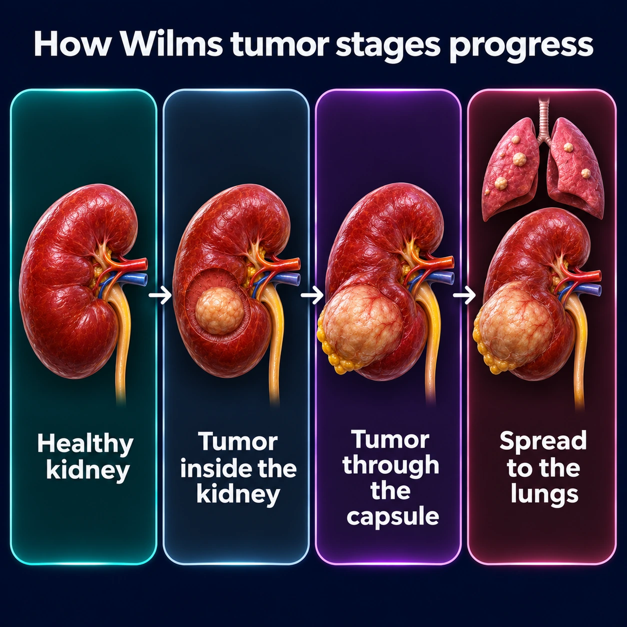
The COG/NWTS staging system, used in North America, is a surgical-pathological staging system — meaning final stage is assigned after nephrectomy based on both intraoperative findings and pathologic assessment. This is distinct from the SIOP (European) system, which stages after preoperative chemotherapy. The COG stages are:
- Stage I: Tumor is limited to the kidney and was completely resected. The renal capsule is intact, with no rupture or biopsy prior to surgery. No residual tumor. Renal sinus vasculature is not involved. Approximately 40–45% of cases.
- Stage II: Tumor extends beyond the kidney (through the renal capsule into perirenal fat, or involves the renal sinus vessels) but is completely excised with negative surgical margins. No residual disease at or beyond the surgical margins. Regional lymph nodes are negative. Approximately 20–25% of cases.
- Stage III: Residual non-hematogenous tumor is confined to the abdomen. This includes: positive lymph nodes (regional or para-aortic), surgical margin positivity, tumor spillage before or during surgery (including pre-surgical biopsy), peritoneal implants, or tumor removal in multiple pieces. Approximately 20–25% of cases.
- Stage IV: Hematogenous metastases to lung, liver, bone, brain, or other distant sites. Approximately 10–15% of cases at diagnosis. Pulmonary metastases are by far the most common site.
- Stage V: Bilateral renal involvement at diagnosis. Occurs in approximately 5–8% of cases. Each kidney is substaged independently (I–III) based on its own resectability and margins, but Stage V designation captures the bilateral nature and the overriding priority of renal preservation.
Intraoperative Staging Notes
Intraoperative findings critically determine final stage. Standard surgical protocol for Wilms nephrectomy includes:
- Careful exploration of all abdominal compartments for peritoneal implants or spread
- Sampling of ipsilateral hilar and para-aortic lymph nodes — even if they appear grossly normal — because microscopic nodal positivity upstages from II to III and changes the treatment plan
- Documentation of whether the tumor capsule was violated during resection
- Identification and documentation of intraoperative spillage, which carries significant staging and prognostic consequences (Stage III, abdominal radiation required)
7. Treatment
Treatment of Wilms tumor is governed by large collaborative group protocols — principally the Children's Oncology Group (COG) in North America and the Société Internationale d'Oncologie Pédiatrique (SIOP) in Europe and most of the rest of the world. The two approaches differ fundamentally in their sequencing of surgery and chemotherapy, but both achieve excellent results.
COG (North American) Approach: Surgery First
The standard COG approach for most patients is immediate nephrectomy followed by risk-stratified chemotherapy. This allows precise surgical-pathological staging (histology drives treatment), avoids the possibility of chemotherapy-induced histologic changes masking anaplasia, and provides tumor tissue for biologic studies.
Chemotherapy Regimens by Stage and Histology
- Stage I and II, Favorable Histology: Two-drug regimen of actinomycin D (dactinomycin) + vincristine for 18 weeks. This achieves greater than 95% four-year survival with minimal toxicity. Neither radiation nor doxorubicin is required. The landmark NWTS-4 trial demonstrated that pulse-intensive scheduling of these agents was equally effective with fewer hospitalizations compared to standard scheduling.
- Stage III, Favorable Histology: Three-drug regimen adding doxorubicin to actinomycin D + vincristine; plus flank radiation (10.8 Gy whole-abdominal for diffuse spillage; flank-only for localized Stage III). Duration approximately 24 weeks.
- Stage IV, Favorable Histology: Three-drug regimen (actinomycin D + vincristine + doxorubicin) plus flank radiation; whole-lung radiation (12 Gy) for patients with persistent pulmonary nodules after 6 weeks of chemotherapy. Patients achieving complete pulmonary response with chemotherapy alone may avoid whole-lung radiation in current COG protocols, reducing the risk of late cardiac and pulmonary toxicity.
- Stage I–II, Focal Anaplasia: Same three-drug regimen as Stage III FH (actinomycin D + vincristine + doxorubicin, regimen DD4A), plus radiation: in the COG AREN0321 and AREN03B2 studies, stage I–III focal anaplasia was treated with DD4A plus radiation. In the older NWTS-5 study, stage I anaplasia was treated with vincristine and dactinomycin alone, without radiation.
- Stage III–IV, Focal Anaplasia; any Stage Diffuse Anaplasia: Highly intensified regimens adding cyclophosphamide and etoposide plus radiation (regimen I in NWTS-5; regimen UH-1, which adds carboplatin to the regimen I drugs, in the COG AREN0321 study of stage II–IV diffuse anaplasia and stage IV focal anaplasia). In AREN0321, 4-year event-free survival for stage II–IV diffuse anaplasia was 67.7% and overall survival 73.7%, compared with 57.5% and 59.2% in NWTS-5; drug doses were reduced partway through the study because of side effects, and 4 of 66 children (6%) died of treatment toxicity.
In the Children's Oncology Group AREN0533 trial, 292 children with favorable-histology Wilms tumor that had spread only to the lungs were treated according to how their lung spots responded. In the 133 children whose spots disappeared after 6 weeks of three-drug chemotherapy, lung radiation was left out: their 4-year overall survival was 96.1%, though relapses and other events were more common than the trial expected (20.2% against 15%; 4-year event-free survival 79.5%). The 159 children whose spots remained (or whose tumor had lost parts of chromosomes 1p and 16q) received lung radiation and four cycles of cyclophosphamide and etoposide on top of the three drugs; their 4-year event-free survival was 88.5% and overall survival 95.4%. Across the whole trial, 4-year overall survival was 95.6%, up from 84.0% in NWTS-5.
Radiation Therapy
Radiation is reserved for Stage III+ favorable histology, Stage II+ anaplasia, and pulmonary metastases failing to resolve with chemotherapy. Radiation fields and doses are carefully tailored to minimize late effects — particularly vertebral growth asymmetry, hepatotoxicity (right lobe radiation), gonadal injury, and second malignancy risk — while maintaining locoregional control.
SIOP (European) Approach: Preoperative Chemotherapy
In the SIOP protocol, children aged 6 months and older with imaging-typical Wilms tumor receive 4 weeks of preoperative chemotherapy (actinomycin D + vincristine) before nephrectomy. Advantages of this approach include:
- Tumor shrinkage in approximately 80% of cases, facilitating complete resection
- Reduction in the rate of intraoperative tumor rupture and spillage
- Downstaging (from Stage III to Stage II in some series), potentially sparing abdominal radiation
Disadvantages include: treatment of a small proportion of patients who ultimately have a non-Wilms renal tumor without a tissue diagnosis, and modification of tumor histology making anaplasia identification more challenging. Long-term survival outcomes are equivalent between COG and SIOP protocols.
Bilateral Wilms Tumor (Stage V): Nephron-Sparing Surgery
The management of Stage V disease prioritizes renal function preservation above all else. Standard approach:
- Initial biopsy of both tumors (percutaneous or open, with careful technique to minimize spillage) to confirm Wilms tumor histology and anaplastic status
- Preoperative chemotherapy — in the COG AREN0534 trial, three drugs (vincristine, dactinomycin and doxorubicin) for 6 or 12 weeks depending on the response on imaging, with definitive surgery completed by 12 weeks from diagnosis — to shrink the tumors as much as possible
- Nephron-sparing surgery: Partial nephrectomy of one or both kidneys, preserving as much functional renal parenchyma as possible while achieving clear surgical margins. The goal is to avoid bilateral nephrectomy and lifelong dialysis.
- If one kidney can be safely removed (dominant tumor, small/non-functional remnant) and the contralateral tumor fully resected with nephron-sparing, this may be the preferred approach.
- Post-surgical chemotherapy is determined by final histology and stage of each kidney independently.
AREN0534, the first prospective trial for children with tumors in both kidneys, enrolled 208 children, of whom 189 could be evaluated. After the three-drug chemotherapy, 84% had definitive surgery on at least one kidney by 12 weeks, and 39% kept parts of both kidneys. Four-year event-free survival was 82.1% and overall survival 94.9%; in the earlier NWTS-5 study, 4-year event-free survival for children with tumors in both kidneys had been 56%.
8. Prognosis and Survival
Wilms tumor is among the most curable of all pediatric solid tumors. The dramatic improvement in outcomes over the NWTS trial era — from approximately 30% survival in the 1960s to greater than 90% overall today — represents one of the landmark achievements of pediatric oncology. However, significant variation by histology and stage persists, and late treatment effects are an increasingly recognized challenge as survivors reach adulthood.
Survival by Stage and Histology
- Stage I, Favorable Histology: >98%; excellent prognosis with minimal-intensity treatment
- Stage II, Favorable Histology: ~95%; similarly excellent with standard two-drug regimen
- Stage III, Favorable Histology: ~85–90%; three-drug regimen + flank radiation
- Stage IV, Favorable Histology: 4-year overall survival 95.6% and event-free survival 85.4% for children with lung metastases in the COG AREN0533 trial (292 children), up from 84.0% and 72.5% in NWTS-5; treatment is adjusted to the lung metastases' response to chemotherapy
- Stage I–II, Focal Anaplasia: 100% 4-year event-free and overall survival (21 children) with the three-drug regimen DD4A plus radiation in the pooled COG AREN0321 and AREN03B2 data; in NWTS-5, stage I focal anaplasia treated with two drugs alone had 4-year overall survival of 88.9%
- Stage III–IV, Diffuse Anaplasia: in NWTS-5, 4-year event-free survival 64.7% for stage III and 33.3% for stage IV (overall survival similar); 4-year overall survival 73.7% for stage II–IV in COG AREN0321; still the dominant unmet need in Wilms tumor therapy
- Bilateral (Stage V): 4-year event-free survival 82.1% and overall survival 94.9% in the COG AREN0534 trial (189 children), up from 4-year event-free survival of 56% in NWTS-5; varies with the histology in each kidney and whether nephron-sparing surgery is achievable
Newer Children's Oncology Group trials changed the outlook for anaplastic Wilms tumor. In AREN0321, 66 children with stage II–IV diffuse anaplasia received a regimen that added carboplatin to vincristine, doxorubicin, cyclophosphamide and etoposide plus radiation: 4-year overall survival was 73.7%, compared with 59.2% in NWTS-5, but doses had to be reduced partway through because of side effects, and 6% of the children died of treatment toxicity. In the pooled AREN0321 and AREN03B2 data on focal anaplasia, 4-year overall survival was 100% for stage I–II (21 children), 87.8% for stage III (17 children) and 85.7% for stage IV (7 children).
Relapse: Salvage and Prognosis
Approximately 15–20% of patients relapse after first-line therapy. Prognosis at relapse depends critically on initial treatment intensity:
- Patients initially treated with actinomycin D + vincristine only (Stage I–II FH) can be salvaged with doxorubicin-containing regimens plus radiation; approximately 80% second complete remission rate
- Patients relapsing after three-drug chemotherapy have lower salvage rates; high-dose chemotherapy with autologous stem cell rescue is used in some centers for selected patients
- Diffuse anaplasia relapse carries a very poor prognosis; clinical trials with novel agents (including cabozantinib, checkpoint immunotherapy) are under evaluation
Late Effects of Treatment
As more than 90% of children with Wilms tumor become long-term survivors, late effects of therapy have become a major focus of survivorship research:
- Renal insufficiency: Children with a solitary kidney after nephrectomy have a lifetime risk of hypertension and progressive chronic kidney disease (CKD); approximately 1% develop end-stage renal disease (ESRD) from their solitary kidney over 20 years, rising with bilateral disease, nephron-sparing surgery complications, or radiation nephrotoxicity. Survivor follow-up typically includes yearly blood pressure checks and kidney function tests for life.
- Radiation-related musculoskeletal effects: Vertebral irradiation in growing children causes scoliosis and vertebral body hypoplasia; soft-tissue hypoplasia of the irradiated flank; leg-length discrepancy (if pelvic radiation given). These effects are mitigated by symmetric whole-abdomen radiation when indicated (rather than asymmetric flank radiation).
- Hepatotoxicity: Right flank radiation and actinomycin D together cause hepatic sinusoidal obstruction syndrome (SOS, also called veno-occlusive disease, VOD) in a small fraction of patients; clinically significant hepatic fibrosis is rare with current doses.
- Cardiac toxicity: Doxorubicin (used in Stage III–IV FH and all anaplastic disease) carries a dose-dependent risk of cardiomyopathy; echocardiographic surveillance and lifetime cardiology follow-up are standard for doxorubicin-exposed survivors.
- Second malignancies: Risk is approximately 1–2% at 15 years; breast cancer, leukemia, sarcoma, and thyroid cancer are among the most common second neoplasms; radiation field substantially increases sarcoma risk within the treatment field.
- Reproductive outcomes: Female survivors with uterine radiation have elevated rates of premature labor, low birth weight infants, and uterine structural abnormalities; gonadal shielding and ovarian transposition reduce but do not eliminate ovarian radiation exposure.
Wilms tumor survivors are usually followed long term in a dedicated pediatric oncology survivorship clinic, with the frequency and content of monitoring set by the treatment received.
Key Research Papers
- Breslow N, Beckwith JB, Ciol M, Sharples K. Age distribution of Wilms' tumor: report from the National Wilms' Tumor Study. Cancer Res. 1988;48(6):1653–1657. Foundational epidemiologic dataset establishing peak age distribution and bilateral incidence. — PubMed PMID: 2830967
- Dome JS, Graf N, Geller JI, et al. Advances in Wilms tumor treatment and biology: progress through international collaboration. J Clin Oncol. 2015;33(27):2999–3007. Comprehensive review of NWTS/COG and SIOP protocol convergence and emerging biologic insights. — PubMed PMID: 26304882
- Spreafico F, Fernandez CV, Brok J, et al. Wilms tumour. Nat Rev Dis Primers. 2021;7(1):75. Primer covering epidemiology, genetics, staging, and global treatment approaches. — PubMed PMID: 34650095
- Treger TD, Chowdhury T, Pritchard-Jones K, Behjati S. The genetic changes of Wilms tumour. Nat Rev Nephrol. 2019;15(4):240–251. Detailed review of WT1, WTX, CTNNB1, SIX1/2, and miRNA pathway mutations with therapeutic implications. — PubMed PMID: 30705419
- Dix DB, Seibel NL, Chi YY, et al. Treatment of Stage IV Favorable Histology Wilms Tumor With Lung Metastases: A Report From the Children's Oncology Group AREN0533 Study. J Clin Oncol. 2018;36(16):1564–1570. Practice-changing trial supporting omission of whole-lung radiation in Stage IV FH patients achieving complete pulmonary response. — PubMed PMID: 29659330
- Fukuzawa R, Heathcott RW, More HE, Reeve AE. Sequential WT1 and CTNNB1 mutations and alterations of beta-catenin localisation in intralobar nephrogenic rests and associated Wilms tumours: two case studies. J Clin Pathol. 2007;60(9):1013–1016. Demonstrates stepwise molecular progression from nephrogenic rest to Wilms tumor, validating the precursor model. — PubMed PMID: 17172473
- Grundy PE, Breslow NE, Li S, et al. Loss of heterozygosity for chromosomes 1p and 16q is an adverse prognostic factor in favorable-histology Wilms tumor: a report from the National Wilms Tumor Study Group. J Clin Oncol. 2005;23(29):7312–7321. Identifies 1p and 16q LOH as independent adverse prognostic markers in Stage I–II FH, leading to current COG risk-stratification incorporating these molecular markers. — PubMed PMID: 16129848
- Dome JS, Cotton CA, Perlman EJ, et al. Treatment of anaplastic histology Wilms' tumor: results from the fifth National Wilms' Tumor Study. J Clin Oncol. 2006;24(15):2352–2358. Defines the differential outcomes of focal vs diffuse anaplasia and established the rationale for intensified therapy in diffuse anaplasia. — PubMed PMID: 16710034
- Pritchard-Jones K, Bergeron C, de Camargo B, et al. Omission of doxorubicin from the treatment of stage II–III, intermediate-risk Wilms' tumour (SIOP WT 2001): an open-label, non-inferiority, randomised controlled trial. Lancet. 2015;386(9999):1156–1164. Demonstrates non-inferiority of actinomycin D + vincristine without doxorubicin for intermediate-risk SIOP Stage II–III, reducing cardiotoxicity exposure. — PubMed PMID: 26164096
- Tournade MF, Com-Nougue C, de Kraker J, et al. Optimal duration of preoperative therapy in unilateral and nonmetastatic Wilms' tumor in children older than 6 months: results of the Ninth International Society of Paediatric Oncology Wilms' Tumor Trial and Study. J Clin Oncol. 2001;19(2):488–500. Established the 4-week preoperative chemotherapy standard in the SIOP system and demonstrated equivalent outcomes with shorter preoperative duration. — PubMed PMID: 11208843
- Daw NC, Chi YY, Kalapurakal JA, et al. Activity of Vincristine and Irinotecan in Diffuse Anaplastic Wilms Tumor and Therapy Outcomes of Stage II to IV Disease: Results of the Children's Oncology Group AREN0321 Study. J Clin Oncol. 2020;38(14):1558–1568. — PubMed PMID: 32134700
- Armstrong AE, Daw NC, Renfro LA, et al. Treatment of focal anaplastic Wilms tumor: A report from the Children's Oncology Group AREN0321 and AREN03B2 studies. Cancer. 2025;131(2):e35713. — PubMed PMID: 39803937
- Ehrlich P, Chi YY, Chintagumpala MM, et al. Results of the First Prospective Multi-institutional Treatment Study in Children With Bilateral Wilms Tumor (AREN0534): A Report From the Children's Oncology Group. Ann Surg. 2017;266(3):470–478. — PubMed PMID: 28795993
- Mussa A, Duffy KA, Carli D, et al. The effectiveness of Wilms tumor screening in Beckwith-Wiedemann spectrum. J Cancer Res Clin Oncol. 2019;145(12):3115–3123. — PubMed PMID: 31583434
- Kalish JM, Becktell KD, Bougeard G, et al. Update on Surveillance for Wilms Tumor and Hepatoblastoma in Beckwith-Wiedemann Syndrome and Other Predisposition Syndromes. Clin Cancer Res. 2024;30(23):5260–5269. — PubMed PMID: 39320341