Autoimmune Hepatitis
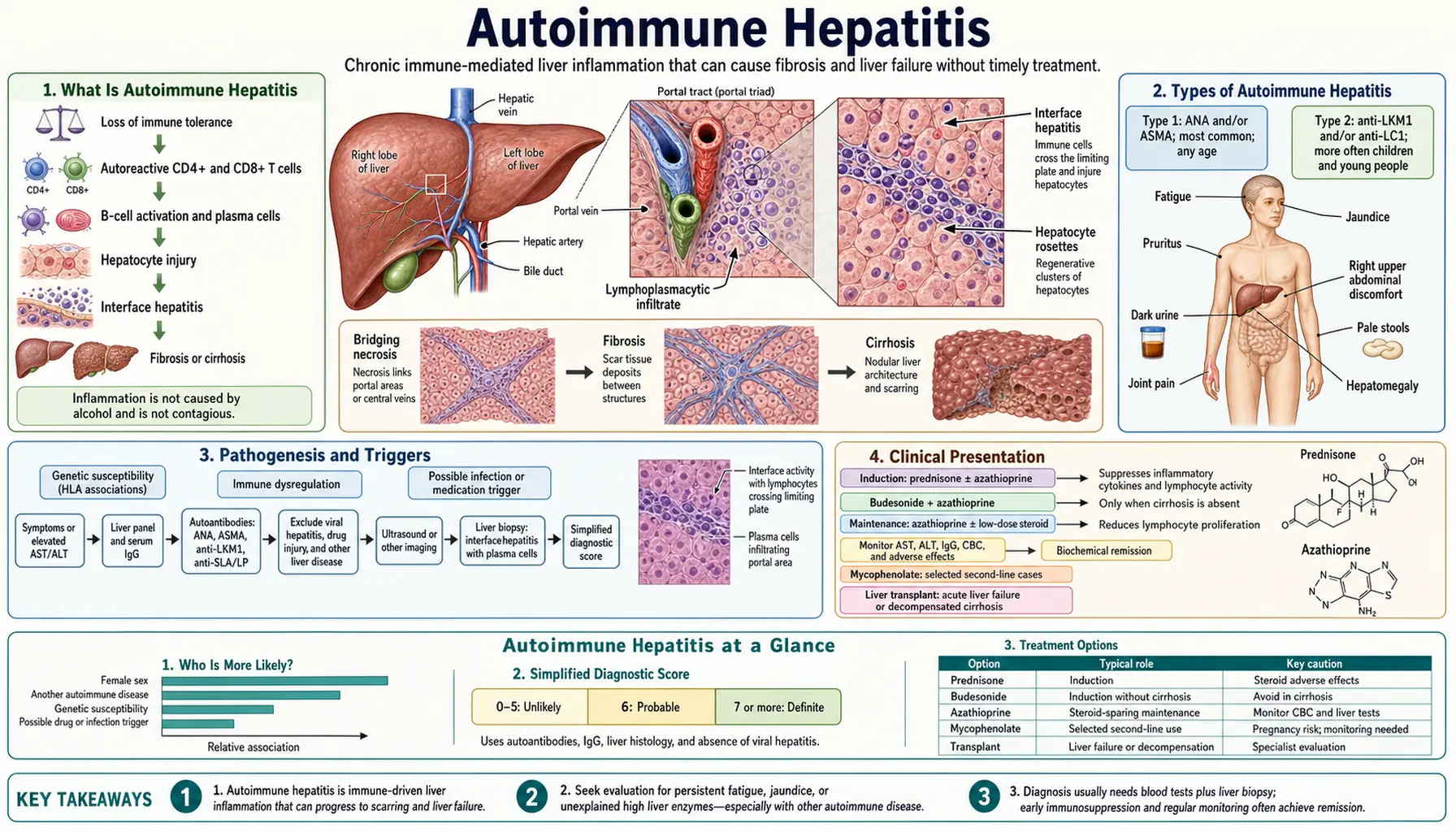
Autoimmune hepatitis (AIH) is a chronic inflammatory liver disease caused by the immune system attacking the body's own hepatocytes. It affects predominantly women (70%; the 2025 British Society of Gastroenterology (BSG) guideline gives about 3 women to 1 man in the UK) and follows a bimodal age distribution — a first peak in adolescence and young adulthood, and a second peak around perimenopause. AIH can masquerade as virtually every other liver disease: it may present as acute hepatitis indistinguishable from viral infection, as chronic hepatitis discovered incidentally on blood tests, or as cirrhosis presenting with its complications. The treacherous part is that without treatment, AIH progresses silently to cirrhosis in the majority of patients — yet with the right treatment, most patients achieve complete remission and a normal life expectancy.
- Overview
- Type 1 vs Type 2 AIH
- Pathophysiology
- Clinical Presentation
- Diagnosis and the Simplified IAIHG Criteria
- Liver Biopsy and Histology
- Treatment
- Overlap Syndromes: AIH-PSC and AIH-PBC
- Pregnancy With Autoimmune Hepatitis
- Key Research Papers
- Connections
Overview
Autoimmune hepatitis is defined by three features: (1) elevated serum transaminases (aspartate aminotransferase (AST)/alanine aminotransferase (ALT)), typically 5–50 times the upper limit of normal; (2) characteristic autoantibodies (antinuclear antibody (ANA), anti-smooth muscle antibody (ASMA), anti-liver-kidney microsomal antibody type 1 (anti-LKM1)); and (3) interface hepatitis on liver biopsy — a histological pattern of lymphocytic and plasma cell infiltration eroding the boundary between the portal tract and the liver parenchyma (the "limiting plate"). A fourth feature, elevated immunoglobulin G (IgG), is present in 85% of patients and is an important diagnostic clue.
AIH is rare — prevalence approximately 10–20 per 100,000 in European and North American populations — but its importance lies in its treatability. A 2023 meta-analysis of 37 studies gives a global prevalence of 15.65 per 100,000, rising from 9.95 per 100,000 in 1970–1999 to 27.91 per 100,000 in 2015–2022, with about 1.28 new cases per 100,000 people per year. Untreated, approximately 40% of patients with severe AIH die within 6 months. With standard immunosuppressive treatment, 80–90% achieve biochemical remission and 5-year survival exceeds 90%. This dramatic treatment benefit makes early recognition critical.
Key clinical associations:
- Other autoimmune diseases occur in 30–50% of AIH patients: autoimmune thyroiditis (Hashimoto's), rheumatoid arthritis, type 1 diabetes, celiac disease, vitiligo, Sjogren's syndrome
- First-degree relatives have increased rates of autoimmune diseases and elevated aminotransferases
- HLA-DR3 (DRB1*0301) and HLA-DR4 (DRB1*0401) are the two major susceptibility alleles in European-descent populations; HLA-DR3 is associated with younger age of onset and more aggressive disease
Type 1 vs Type 2 AIH
AIH is divided into two subtypes based on autoantibody profile:
Type 1 AIH (most common, ~80% of AIH)
Autoantibody markers:
- antinuclear antibody (ANA): positive at titer ≥1:40 in ~70% of Type 1 AIH patients; homogeneous or speckled pattern on immunofluorescence; non-specific (also positive in many healthy individuals, systemic lupus erythematosus (SLE), drug reactions)
- ASMA (anti-smooth muscle antibody): positive in ~70% of Type 1; specificity increases substantially at titers ≥1:80; directed against actin (F-actin specificity is most specific for AIH)
- Anti-SLA/LP (anti-soluble liver antigen/liver-pancreas): present in 10–30% of Type 1; highly specific (>99%) for AIH when positive; associated with more severe disease and higher relapse rate
- Type 1 can occur at any age; accounts for the majority of adult and elderly AIH
Type 2 AIH (less common, ~20%, predominantly children and adolescents)
Autoantibody markers:
- Anti-LKM1 (anti-liver-kidney microsomal antibody type 1): directed against cytochrome P450 2D6 (CYP2D6); positive in ~95% of Type 2; negative in Type 1; can be induced by hepatitis C infection (hepatitis C virus (HCV) can trigger anti-LKM1, which is why anti-LKM1-positive patients are tested for hepatitis C virus)
- Anti-LC1 (anti-liver cytosol antibody type 1): directed against formiminotransferase cyclodeaminase; often co-positive with anti-LKM1; can be the sole marker in Type 2
- Type 2 AIH tends to present more acutely, at younger ages (2–14 years most typical), with higher severity at presentation
- Type 2 has higher rate of progression to cirrhosis if untreated
Practical note: Type 1 and Type 2 are not distinguished by treatment — both receive the same prednisolone ± azathioprine protocol. The distinction matters for diagnostic workup and for understanding disease behavior.
Pathophysiology
AIH results from breakdown of immune tolerance to hepatocyte antigens, with proposed roles for both regulatory T cells (Tregs; disputed, see below) and aberrant effector immune responses.
Molecular Mimicry Hypothesis
Environmental triggers (viruses, drugs, environmental antigens) present peptide sequences that share structural similarity with hepatocyte proteins. Cytochrome P450 2D6 in Type 2 AIH is a clear example: a peptide from the hepatitis C virus (HCV) mimics the cytochrome P450 2D6 epitope targeted by anti-LKM1, potentially explaining how hepatitis C virus infection can trigger Type 2 AIH. Measles virus and herpes simplex virus have also been proposed as molecular mimics for other AIH autoantigens.
Defective Regulatory T Cell (Treg) Suppression
Tregs normally suppress autoreactive effector T cells. Whether they are defective in AIH is disputed. Earlier studies described Tregs as quantitatively reduced in peripheral blood and liver, and functionally impaired — failing to suppress CD4+ and CD8+ T cells that recognize hepatocyte antigens. A 2012 study by Peiseler and colleagues, using staining that separates Tregs from activated T cells, reported that "the frequency and function of circulating Treg cells is not impaired in AIH"; it noted that the earlier studies "had used only CD25" as a marker, and found Treg frequency in the liver higher than in fatty-liver inflammation (non-alcoholic steatohepatitis), rising with inflammation. A 2023 meta-analysis of 29 studies found Treg proportions in blood generally decreased, with results depending on the markers used, ethnicity and disease activity. A 2026 study of 57 patients found more Tregs in the liver but markedly reduced suppressive capacity in the laboratory. It has also been proposed that mutations in FoxP3 (the master transcription factor of Tregs) and IL-2 signaling pathways (shared with Type 1 diabetes) account for part of any defect; this is not established.
Th1/Th17 Effector Response
Autoreactive CD4+ T helper cells in AIH are skewed toward Th1 (producing IFN-γ and tumor necrosis factor alpha (TNF-α)) and Th17 (producing IL-17) phenotypes. These cytokines activate:
- Macrophage and Kupffer cell cytotoxicity against hepatocytes
- CD8+ cytotoxic T lymphocyte killing of hepatocytes expressing the autoantigens on MHC class I
- B cell production of autoantibodies (ANA, ASMA, anti-LKM1) — the antibodies themselves are largely epiphenomenal (markers, not primary drivers) but serve as diagnostic surrogates
Plasma Cell Infiltration
A distinctive feature of AIH histology is hepatic plasma cell infiltration. Activated B cells mature into plasma cells within the liver, producing local immunoglobulin G that is reflected in the elevated serum immunoglobulin G. This polyclonal immunoglobulin G elevation (immunoglobulin G typically 1.5–3× upper limit of normal in active AIH) is a key diagnostic feature and a biomarker of treatment response (immunoglobulin G normalizes with effective immunosuppression).
Genetic Architecture
HLA-DR3 and HLA-DR4 present autoantigenic peptides on CD4+ T cells in the context of hepatic injury. HLA-DR3 (DRB1*0301) is associated with more severe disease, younger onset, and higher relapse rates; HLA-DR4 (DRB1*0401) is associated with milder disease, older onset, and better response to azathioprine. Non-HLA loci include PTPN22 (lymphocyte phosphatase, shared with rheumatoid arthritis (RA) and type 1 diabetes) and SH2B3.
Clinical Presentation
AIH has three main presentations:
1. Acute Hepatitis Presentation (30–40% of Cases)
- Sudden onset jaundice, fatigue, right upper quadrant pain, sometimes nausea/vomiting
- Transaminases strikingly elevated: aspartate aminotransferase/alanine aminotransferase typically 10–100× upper limit of normal
- May be mistaken for acute viral hepatitis (hepatitis A virus (HAV), hepatitis B virus (HBV), Epstein-Barr virus (EBV)) without careful antibody workup
- "Acute severe" AIH: subset presenting with international normalized ratio (INR) >1.5 and encephalopathy — liver failure risk is high; biopsy urgently needed to confirm diagnosis before initiating high-dose steroids (or determine whether transplant is needed)
- ~5–10% of patients develop acute liver failure requiring emergency transplantation
2. Chronic Asymptomatic Presentation (30–50% of Cases)
- Discovered incidentally during routine blood tests or workup for fatigue
- Alanine aminotransferase/aspartate aminotransferase elevated 3–10× upper limit of normal
- No jaundice, no abdominal symptoms
- Many of these patients are found to have advanced fibrosis or cirrhosis on biopsy — the disease was silently progressing for years
3. Presentation with Established Cirrhosis
- First presentation as cirrhosis complications: ascites, variceal hemorrhage, hepatic encephalopathy
- Transaminases may be normal or only mildly elevated if hepatocyte mass is significantly reduced
- AIH is a recognized possibility in any patient with "cryptogenic" cirrhosis — unrecognized burned-out AIH is a common finding in transplant explants
Physical Examination
- Jaundice and scleral icterus (in acute or decompensated presentations)
- Hepatomegaly (tender in acute presentations)
- Spider angiomata, palmar erythema (chronic disease / cirrhosis)
- Other autoimmune disease findings: thyroid enlargement, joint swelling/tenderness, skin depigmentation (vitiligo)
Laboratory Pattern
- Alanine aminotransferase > aspartate aminotransferase (hepatocellular pattern, not cholestatic)
- Alkaline phosphatase (ALP) and gamma-glutamyl transferase (GGT): normal or mildly elevated unless overlap with primary sclerosing cholangitis (PSC) or primary biliary cholangitis (PBC)
- Elevated immunoglobulin G: often 1.5–3× upper limit of normal; can be dramatically elevated (>30 g/L) in acute severe disease
- Low platelets and albumin if cirrhosis present
- Normal or mildly elevated bilirubin unless acute or decompensated
Diagnosis and the Simplified IAIHG Criteria
AIH diagnosis requires integrating clinical, serological, and histological data. The International Autoimmune Hepatitis Group (IAIHG) has published two scoring systems: the original complex scoring system and the simplified 2008 criteria.
IAIHG Simplified Criteria (2008)
| Variable | Finding | Points |
|---|---|---|
| Antinuclear antibody or ASMA | ≥1:40 | +1 |
| Antinuclear antibody or ASMA | ≥1:80 | +2 |
| Anti-LKM1 | ≥1:40 | +2 |
| Anti-SLA/LP | Positive | +2 |
| Immunoglobulin G | >upper limit of normal | +1 |
| Immunoglobulin G | >1.10 × upper limit of normal | +2 |
| Liver histology | Compatible with AIH | +1 |
| Liver histology | Typical of AIH | +2 |
| Absence of viral hepatitis | Yes | +2 |
Score Interpretation
- ≥7 points: definite AIH (81% sensitivity, 99% specificity in the 2008 validation set)
- 6 points: probable AIH (88% sensitivity, 97% specificity at a cutoff of ≥6 in the 2008 validation set)
- <6 points: AIH unlikely
Practical Workup Sequence
- Viral hepatitis excluded: hepatitis A virus immunoglobulin M (IgM), hepatitis B surface antigen (HBsAg), hepatitis B core antibody IgM, hepatitis C virus antibody (with reflex RNA if positive), hepatitis E virus (HEV) IgM
- Autoantibodies measured: antinuclear antibody (immunofluorescence on Hep-2 cells), ASMA (immunofluorescence), anti-LKM1 (immunofluorescence), anti-SLA/LP (enzyme-linked immunosorbent assay, ELISA)
- Immunoglobulin G measured (and immunoglobulin A (IgA), immunoglobulin M to assess pattern)
- Liver biopsy (virtually always required for definitive diagnosis and fibrosis staging)
- Abdominal ultrasound (baseline; assess liver echogenicity, portal flow)
- If cholestatic features (elevated alkaline phosphatase/GGT): magnetic resonance cholangiopancreatography (MRCP) to evaluate for overlap with primary sclerosing cholangitis
Drug-Induced AIH
Many drugs cause AIH-like reactions with identical serological and histological findings: minocycline, nitrofurantoin, statins, methyldopa (alpha-methyldopa), hydralazine, isoniazid. Drug-induced AIH may resolve with drug withdrawal alone. A detailed drug history is part of the workup, including herbal supplements: kava, comfrey and germander have caused AIH-like drug reactions.
Liver Biopsy and Histology
Liver biopsy is essential in AIH for three reasons: (1) to confirm the diagnosis, (2) to stage fibrosis, and (3) to establish a histological baseline for monitoring treatment response.
Characteristic Histological Features
Interface Hepatitis (the Cardinal Lesion)
Lymphocytes and plasma cells spill from portal tracts across the limiting plate into the periportal hepatocyte parenchyma, destroying periportal hepatocytes in clusters. This periportal hepatitis eroding the limiting plate is the defining histological feature and is responsible for the "rosette" formation described below.
Plasma Cell Infiltrate
Dense plasma cell infiltration — often described as the "most striking" feature by experienced pathologists — is highly characteristic of AIH. Plasma cells are rare or absent in viral hepatitis and metabolic dysfunction-associated steatotic liver disease (MASLD), making their presence a strong diagnostic pointer. In acute severe AIH, plasma cells may be sparse ("plasma-cell-poor AIH"), which can mislead the pathologist.
Hepatocyte Rosette Formation
Remaining periportal hepatocytes, hemmed in by the inflammatory infiltrate, arrange themselves in a gland-like ring (rosette) around a dilated bile canaliculus or central hepatocyte. Rosettes are not specific to AIH but are characteristic.
Emperipolesis
Lymphocytes penetrating into hepatocyte cytoplasm (lymphocyte emperipolesis) — a more specific feature of AIH when prominent; distinct from hepatocyte apoptosis which produces Councilman bodies.
Fibrosis Staging
Staged F0–F4 by the Metavir or Ishak system. Approximately 30% of newly-diagnosed AIH patients already have F3–F4 fibrosis at first biopsy, emphasizing the silent progression of untreated disease.
Treatment
AIH treatment has two phases: induction of remission and long-term maintenance.
Indications for Treatment
- Active AIH with aspartate aminotransferase ≥10× upper limit of normal, or aspartate aminotransferase ≥5× upper limit of normal + IgG ≥2× upper limit of normal, or bridging/multilobular necrosis on biopsy: treated immediately (approximately 40% of patients with severe AIH die within 6 months untreated, see Overview)
- Asymptomatic AIH with interface hepatitis and ≥F2 fibrosis: treated
- Asymptomatic AIH with interface hepatitis and F0–F1: treated (prevents progression)
- AIH with F4 (cirrhosis) and active inflammation: treated cautiously
Standard Induction — Regimens in Liver-Society Guidance
Option 1 (preferred for women, young patients, type 2 diabetes risk):
- Prednisolone 30–40 mg/day × 2 weeks → taper by 5 mg every 2 weeks to 10 mg/day
- Azathioprine 50 mg/day is added from week 2 → increased to 1–2 mg/kg/day once transaminases are improving (azathioprine monotherapy in active hepatitis risks worsening via immunosuppression of the regulatory arm)
- Reach maintenance: prednisolone 5–10 mg/day + azathioprine 1–2 mg/kg/day
Option 2 (monotherapy with prednisolone alone):
- Prednisolone 40–60 mg/day → taper more slowly
- Reserved in older guidance for cytopenias making azathioprine risky, pregnancy (azathioprine U.S. Food and Drug Administration (FDA) Category D, as a 2016 review notes), malignancy history. The position on pregnancy has changed: the 2025 BSG guideline states "there is no apparent relationship between the use of azathioprine in pregnancy and adverse outcomes" and that "continuation of therapy during pregnancy is justified" (see Pregnancy)
Budesonide (for Non-Cirrhotic AIH)
- Budesonide 3 mg three times daily (9 mg/day total) + azathioprine
- 90% first-pass liver metabolism in a normal liver (2025 BSG guideline) = fewer systemic steroid side effects (less Cushingoid appearance, osteoporosis, type 2 diabetes risk)
- Important contraindication: cirrhosis — extensive portosystemic shunting bypasses first-pass extraction, eliminating budesonide's safety advantage; the 2025 BSG guideline states budesonide "should not be used" in cirrhosis, where prednisolone is used
Monitoring Remission
- Biochemical remission: normalization of aspartate aminotransferase, alanine aminotransferase, immunoglobulin G, bilirubin — target within 6–12 months; immunoglobulin G normalization is the most reliable marker
- Histological remission: older practice was a biopsy at 3 years of biochemical remission to confirm; histological remission lags behind biochemical by 3–6 months. The 2025 BSG guideline states that liver biopsy to confirm histological remission in those with complete biochemical remission "is not routinely required, although it might sometimes be useful"
- Histological remission (≤minimal activity on biopsy) required in older guidance before considering withdrawal of therapy; the 2025 BSG guideline does not recommend routine liver biopsy in adults before withdrawal
Treatment Withdrawal
- Older guidance: treatment withdrawal considered only after ≥2 years of biochemical + histological remission. The 2025 BSG guideline describes considering withdrawal of azathioprine after sustained complete biochemical response over 3–4 years
- Relapse rate after withdrawal: approximately 60–80% — most patients require lifelong treatment. The 2025 BSG guideline gives 74% (range 25%–100%) overall and 62% (36%–67%) over 12 months, with most relapses in the first year; a 2016 review gives 50% to 87% depending on length of follow-up
- Relapse is defined as aspartate aminotransferase/alanine aminotransferase >3× upper limit of normal after withdrawal; prednisolone + azathioprine is restarted at initial doses
- After a second relapse, guidance describes long-term maintenance therapy indefinitely
Second-Line Therapies (azathioprine Intolerance, Refractory Disease)
- Mycophenolate mofetil (MMF) 1–2 g/day: first-choice azathioprine alternative; achieves remission in ~60–75% of azathioprine-intolerant patients. A pooled review of 14 studies and almost 400 patients in the 2025 BSG guideline gives a biochemical response of 82% in azathioprine-intolerant patients but only 32% in those who responded inadequately to azathioprine. Safety fact: the same guideline calls mycophenolate mofetil "highly teratogenic" (it causes birth defects) and states it should be stopped 12 weeks before conception, with two contraception methods for women during treatment and for 6 weeks after stopping, and contraception for men during treatment and for 90 days after
- Tacrolimus: effective for refractory AIH; monitored by trough levels; nephrotoxicity risk
- Cyclosporine: less preferred than tacrolimus; similar efficacy; more drug interactions
- Rituximab (anti-CD20): emerging option for refractory AIH; small case series; targets B cell lineage and plasma cells
- Infliximab: described as used with caution, because tumor necrosis factor-alpha inhibitors can paradoxically cause drug-induced AIH in some patients
Newer Trials and Guidelines
- Mycophenolate mofetil compared with azathioprine as first treatment (CAMARO trial): a randomized open-label trial of 70 people with newly found autoimmune hepatitis, both drugs given with prednisolone. Remission at 24 weeks occurred in 56.4% on mycophenolate mofetil and 29.0% on azathioprine; 5.1% and 25.8% stopped because of side effects. At 3 years, 76% and 57% were still on the assigned drug with normal liver enzymes (p = 0.099, not statistically significant).
- Zetomipzomib (under study): a small placebo-controlled phase 2a trial in 23 people with hard-to-control disease. Complete remission occurred in 50% on the drug and 42.9% on placebo, a difference whose confidence interval (−38.5 to 48.3) includes zero; injection site reactions occurred in 93.8% and 57.1%.
- 2025 guidelines: the European (EASL) and British (BSG) liver societies both published new autoimmune hepatitis guidelines in 2025.
Liver Transplantation
- Indicated for AIH presenting as acute liver failure (not responding to steroids within 7 days), or decompensated AIH-cirrhosis not manageable medically
- Post-transplant outcomes: 5-year survival 80–85%
- Recurrent AIH in transplanted liver: 20–35% at 10 years; usually responds to augmented immunosuppression
Overlap Syndromes: AIH-PSC and AIH-PBC
AIH overlaps with the two other autoimmune liver diseases, primary sclerosing cholangitis and primary biliary cholangitis, in approximately 7–10% of cases. These overlap syndromes require modified treatment.
AIH-PSC Overlap
- More common in children and adolescents
- Typical: elevated aspartate aminotransferase/alanine aminotransferase and positive ANA/ASMA (AIH features) PLUS ALP/gamma-glutamyl transferase elevation and biliary strictures on MRCP (primary sclerosing cholangitis features)
- Histology: both interface hepatitis AND onion-skin periductal fibrosis (rare to find both on one biopsy)
- Treatment: prednisolone + azathioprine (as for AIH) — but ursodeoxycholic acid may also be added; response to immunosuppression is better than pure primary sclerosing cholangitis but worse than pure AIH; transplant rates higher than pure AIH
- Pediatric AIH-PSC ("autoimmune sclerosing cholangitis"): originally described as a childhood variant; now recognized as the juvenile form of AIH-PSC overlap
AIH-PBC Overlap (Paris Criteria Diagnosis)
Paris Criteria require at least 2 of 3 features from each disease:
- AIH features: alanine aminotransferase ≥5× upper limit of normal, IgG ≥2× upper limit of normal or positive ASMA, interface hepatitis on biopsy
- primary biliary cholangitis features: alkaline phosphatase ≥2× upper limit of normal or gamma-glutamyl transferase ≥5× upper limit of normal, positive antimitochondrial antibody (AMA) at titer ≥1:40, florid duct lesion on biopsy
- Treatment: both ursodeoxycholic acid (13–15 mg/kg/day) AND prednisolone ± azathioprine; ursodeoxycholic acid alone inadequate; prednisolone alone does not address the biliary component
The 2025 BSG guideline recommends that the revised and simplified IAIHG scores not be used to diagnose these variant syndromes, and states that the Paris criteria can be used "but do not identify all patients".
Pregnancy With Autoimmune Hepatitis
In a two-center study of 197 pregnancies in 112 women with autoimmune hepatitis, 88% ended in a live birth and 24% in preterm delivery. Preterm birth was more common with cirrhosis (33% vs 16%), although live birth rates were similar. Loss of disease control on blood tests was more common after delivery than during pregnancy (25% vs 6%). Intrahepatic cholestasis of pregnancy occurred in 9% and preeclampsia in 8%.
The 2025 BSG guideline states that azathioprine, tacrolimus, prednisolone and budesonide "appear safe in pregnancy and also, during breastfeeding", and recommends continuing corticosteroids with or without thiopurines throughout pregnancy. Mycophenolate mofetil is the exception: it causes birth defects (see Second-Line Therapies above).
Key Research Papers
- Manns MP et al. "Diagnosis and Management of Autoimmune Hepatitis." Hepatology 2010 (AASLD practice guidelines) — PubMed PMID: 20513004
- Hennes EM et al. "Simplified criteria for the diagnosis of autoimmune hepatitis." Hepatology 2008 (simplified IAIHG scoring) — PubMed PMID: 18537184
- European Association for the Study of the Liver. "EASL Clinical Practice Guidelines: Autoimmune hepatitis." Journal of Hepatology 2015. — PubMed PMID: 26341719
- Johnson PJ, McFarlane IG. "Meeting report: International Autoimmune Hepatitis Group." Hepatology 1993 (original IAIHG scoring system) — PubMed PMID: 8406375
- Vergani D et al. "Liver autoimmune serology: a consensus statement from the committee for autoimmune serology of the International Autoimmune Hepatitis Group." Journal of Hepatology 2004. — PubMed PMID: 15464251
- Lohse AW, Mieli-Vergani G. "Autoimmune hepatitis." Journal of Hepatology 2011. — PubMed PMID: 21167232
- Czaja AJ. "Diagnosis and Management of Autoimmune Hepatitis: Current Status and Future Directions." Gut and Liver 2016. — PubMed PMID: 26934884
- Heneghan MA et al. "Autoimmune hepatitis." Lancet 2013. — PubMed PMID: 23768844
- Gregorio GV et al. "Autoimmune hepatitis in childhood: a 20-year experience." Hepatology 1997 (Type 2 AIH in children) — PubMed PMID: 9049195
- Chazouillères O et al. "Primary biliary cirrhosis–autoimmune hepatitis overlap syndrome: clinical features and response to therapy." Hepatology 1998 (Paris criteria AIH-PBC) — PubMed PMID: 9695990
- Zachou K et al. "Review article: autoimmune hepatitis -- current management and challenges." Alimentary Pharmacology & Therapeutics 2013. — PubMed PMID: 24010812
- Mack CL et al. "Diagnosis and Management of Autoimmune Hepatitis in Adults and Children." Hepatology 2020. — PubMed PMID: 31863477
- Gleeson D et al. "British Society of Gastroenterology guidelines for diagnosis and management of autoimmune hepatitis." Gut 2025. — PubMed PMID: 40169244
- European Association for the Study of the Liver. "EASL Clinical Practice Guidelines on the management of autoimmune hepatitis." Journal of Hepatology 2025. — PubMed PMID: 40348684
- Snijders RJALM et al. "An open-label randomised-controlled trial of azathioprine vs. mycophenolate mofetil for the induction of remission in treatment-naive autoimmune hepatitis." Journal of Hepatology 2024. — PubMed PMID: 38101756
- Slooter CD et al. "Azathioprine versus mycophenolate mofetil in treatment-naive autoimmune hepatitis: 3-year follow-up of the CAMARO-trial." JHEP Reports 2026. — PubMed PMID: 42880409
- Lammert CS et al. "Safety and efficacy of zetomipzomib in relapsed or insufficiently responding autoimmune hepatitis: Results from the randomized, double-blind, placebo-controlled, phase 2a PORTOLA study and open-label extension." Hepatology 2026. — PubMed PMID: 42770396
- Chung Y et al. "Pregnancy outcomes in an international cohort of autoimmune hepatitis." JHEP Reports 2026. — PubMed PMID: 42217578
- Hahn JW et al. "Global incidence and prevalence of autoimmune hepatitis, 1970-2022: a systematic review and meta-analysis." EClinicalMedicine 2023. — PubMed PMID: 37876996
- Peiseler M et al. "FOXP3+ regulatory T cells in autoimmune hepatitis are fully functional and not reduced in frequency." Journal of Hepatology 2012. — PubMed PMID: 22425700
- Huang Z et al. "The proportion of regulatory T cells in peripheral blood of patients with autoimmune hepatitis: A systematic review and meta-analysis." International Immunopharmacology 2023. — PubMed PMID: 37390643
- Kwon MH et al. "Expanded but dysfunctional regulatory T cells in treatment-naive autoimmune hepatitis." Hepatology International 2026. — PubMed PMID: 41364306
Connections
- Nephrology & Hepatology
- Cirrhosis
- Primary Biliary Cholangitis
- Primary Sclerosing Cholangitis
- Liver Disease
- Hepatic Encephalopathy
- Metabolic Dysfunction-Associated Steatotic Liver Disease
- Alcoholic Hepatitis
- Hepatitis C
- Hepatitis — the umbrella overview of liver inflammation; viral hepatitis must be excluded before AIH can be diagnosed.
- Systemic Lupus Erythematosus
- Hashimoto's Thyroiditis
- Aspartate Aminotransferase (AST)
- Alanine Aminotransferase (ALT)
- Bilirubin
- Herbs covered on this site that discuss this condition: Noni (Morinda citrifolia) · Tinospora / Guduchi (Tinospora cordifolia) · Skullcap