Alcoholic Hepatitis
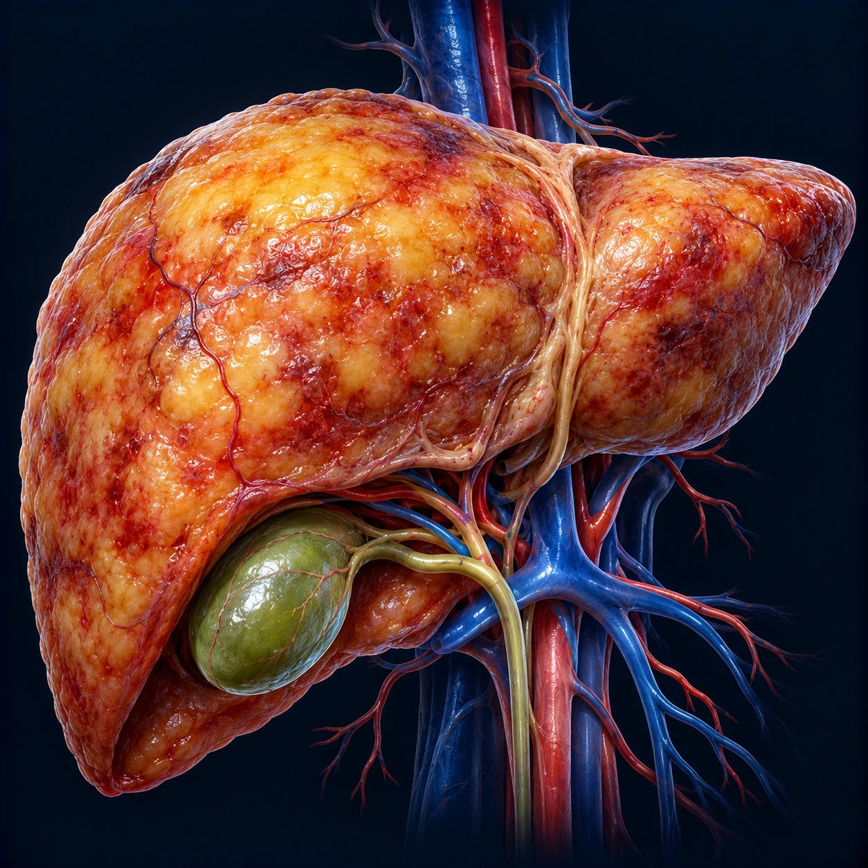
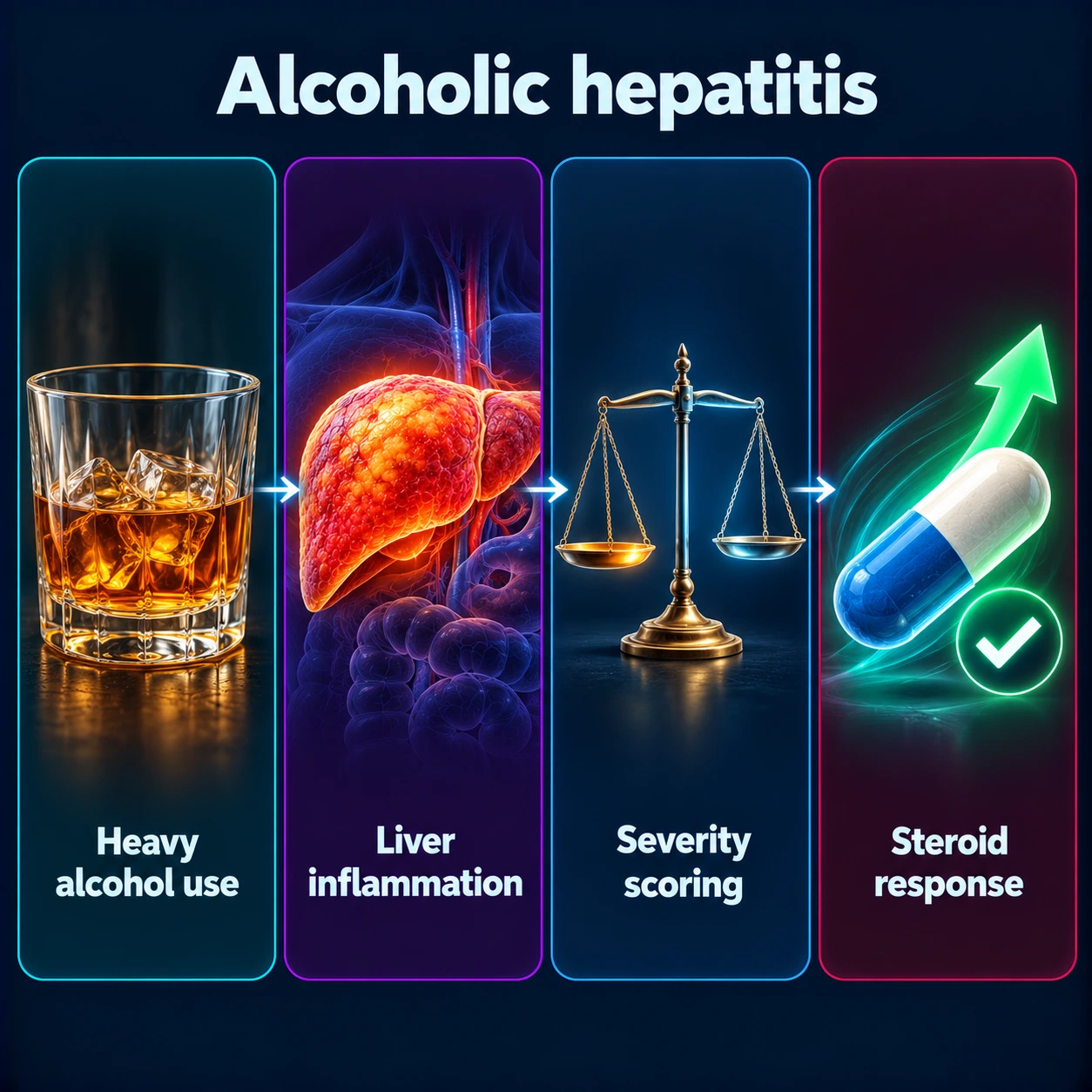
Alcoholic hepatitis is a severe, life-threatening inflammatory liver disease that develops suddenly in people with sustained heavy alcohol use — usually after decades of daily drinking exceeding 80–100 grams of alcohol per day. The paradox of alcoholic hepatitis is its timing: it can strike without warning in someone who has been drinking heavily for years without apparent liver trouble, and it can also develop even after a person reduces or stops drinking. Severe alcoholic hepatitis carries a 30-day mortality of 25–35% and a 90-day mortality up to 50% without treatment, making it one of the most immediately dangerous liver conditions in clinical medicine.
- Overview
- Epidemiology and Risk Factors
- Pathophysiology
- Clinical Presentation and Diagnosis
- Severity Scoring: Maddrey DF and MELD
- Liver Biopsy and Histology
- Treatment
- The Lille Score: Monitoring Steroid Response
- Key Research Papers
- Connections
Interactive Visualization What Alcohol Actually Does Inside Your Liver Ethanol becomes acetaldehyde becomes acetate — and the reduced nicotinamide adenine dinucleotide (NADH) that piles up shuts down fat burning, stalls your blood sugar and drives uric acid up. Nothing speeds it up. Launch →
Overview
Alcoholic hepatitis (AH) sits at the severe end of the alcohol-associated liver disease (ALD) spectrum, which ranges from alcoholic fatty liver (steatosis) through alcoholic hepatitis to alcoholic cirrhosis. The term "alcoholic hepatitis" specifically refers to the acute-on-chronic inflammatory syndrome — not to mildly elevated liver enzymes in a drinker. The key features are:
- Onset of jaundice (bilirubin rising rapidly, often to 5–20 mg/dL) in a person with known heavy alcohol use
- Hepatomegaly (liver enlargement, often painful)
- Fever (38–39°C) in the absence of infection — infection is ruled out first, because it can cause the same fever
- Leukocytosis (elevated WBC, typically 12,000–20,000/μL)
- Characteristic aspartate aminotransferase (AST):alanine aminotransferase (ALT) ratio >2:1 — both enzymes are elevated, but aspartate aminotransferase is disproportionately high due to alcohol's specific mitochondrial toxicity (alcohol depletes mitochondrial pyridoxal-5-phosphate, which is an alanine aminotransferase cofactor)
- Both aspartate aminotransferase and alanine aminotransferase typically <300 international units (IU)/L even in severe disease — values >500 suggest a different diagnosis (viral hepatitis, ischemic hepatitis)
AH can develop in:
- People still actively drinking at the time of admission (most common)
- People who have recently reduced or stopped alcohol — alcohol reduction can paradoxically trigger AH within weeks
- People who develop AH on a background of previously unrecognized cirrhosis (acute-on-chronic liver failure)
Epidemiology and Risk Factors
AH accounts for approximately 50,000–70,000 hospitalizations annually in the United States. In-hospital mortality from severe AH is 25–40%; 90-day mortality approaches 30–50% in MELD ≥21 cases.
Risk factors for developing AH from chronic alcohol use:
- Quantity and duration: typically >80 g/day (>6 standard drinks/day) for >5–10 years; women develop AH at lower amounts and shorter durations due to reduced first-pass alcohol metabolism, lower body water, and possibly hormonal effects on hepatic stellate cells
- Pattern of drinking: binge drinking superimposed on daily drinking appears to confer greater risk than sustained steady consumption
- Obesity: body mass index (BMI) >30 kg/m² synergistically worsens alcohol-induced hepatic injury
- Female sex: women develop AH with less alcohol and shorter drinking history
- Genetic factors: variants in PNPLA3 (I148M) and TM6SF2 increase susceptibility to AH
- Poor nutrition: protein-calorie malnutrition is near-universal in severe AH and worsens outcomes independently
- Concurrent hepatitis C: hepatitis C virus (HCV) coinfection accelerates progression from AH to cirrhosis
Pathophysiology
The central injury in AH flows from acetaldehyde toxicity and oxidative stress:
Step 1 — Alcohol metabolism and acetaldehyde: Hepatocytes metabolize >90% of ingested alcohol. Ethanol is oxidized by ADH (alcohol dehydrogenase) to acetaldehyde, and then by ALDH2 (aldehyde dehydrogenase) to acetate. Acetaldehyde is highly reactive — it forms protein adducts (Mallory-Denk bodies) and mitochondrial adducts that impair oxidative phosphorylation.
Step 2 — NADH-to-NAD+ imbalance: Alcohol oxidation by ADH shifts the hepatic redox state dramatically toward reduced nicotinamide adenine dinucleotide (NADH), raising the ratio of NADH to its oxidized form, nicotinamide adenine dinucleotide (NAD+). This inhibits gluconeogenesis (causing hypoglycemia) and fatty acid beta-oxidation (causing fatty acid accumulation/steatosis). Excess NADH also drives lactic acidosis.
Step 3 — Cytochrome P450 2E1 (CYP2E1) induction: Chronic alcohol use induces cytochrome P450 2E1, an alternative oxidative pathway that generates reactive oxygen species (ROS) — superoxide, hydrogen peroxide, hydroxyl radicals. Reactive oxygen species attack hepatocyte membranes, mitochondria, and nuclear DNA.
Step 4 — Gut barrier disruption and endotoxin translocation: Alcohol disrupts tight junctions between intestinal epithelial cells, increasing gut permeability. Bacterial endotoxin (lipopolysaccharide, LPS) floods the portal circulation, activating hepatic Kupffer cells via TLR4 signaling. Kupffer cells release massive amounts of tumor necrosis factor alpha (TNF-α), interleukin-1 beta (IL-1β), interleukin-6 (IL-6), and IL-8, creating the inflammatory cytokine storm that drives hepatocyte necrosis.
Step 5 — Neutrophilic infiltration: Unlike most forms of hepatitis where lymphocytes predominate, AH is characterized by neutrophilic infiltration of the liver parenchyma — a histological hallmark that reflects the acute-phase cytokine response.
Step 6 — Stellate cell activation and acute fibrosis: Simultaneously, activated hepatic stellate cells produce collagen at an accelerated rate, sometimes causing fibrosis to progress weeks to months in AH that would have taken years in metabolic dysfunction-associated steatotic liver disease (MASLD).
Clinical Presentation and Diagnosis
AH is diagnosed by a combination of clinical presentation, laboratory findings, and exclusion of competing diagnoses. Liver biopsy is confirmatory but not always required.
Clinical features:
- Recent or current heavy alcohol use (the amount and duration are part of the diagnosis)
- Jaundice developing over days to weeks (may be preceded by pruritis)
- Hepatomegaly — often tender
- Fever (38–39°C) — infection is ruled out before fever is put down to AH itself
- Spider angiomata, palmar erythema, Dupuytren's contracture (signs of chronic liver disease often co-present)
- Malnutrition stigmata: muscle wasting, parotid enlargement, peripheral neuropathy
Laboratory findings:
- Bilirubin: elevated, often markedly (5–30 mg/dL); predominantly conjugated
- Aspartate aminotransferase:alanine aminotransferase ratio >2:1: virtually diagnostic when combined with alcohol history; rarely exceeds 8:1; both typically <300 international units/L
- Gamma-glutamyl transferase (GGT): markedly elevated (a sensitive marker of alcohol use)
- Alkaline phosphatase: mildly elevated
- International normalized ratio (INR): elevated from impaired hepatic synthetic function; NOT from vitamin K deficiency — giving vitamin K does not fully correct international normalized ratio in AH
- Albumin: often low (malnutrition + impaired synthesis)
- WBC: elevated (12,000–30,000/μL) even without infection
- Creatinine: elevated in hepatorenal syndrome (a serious complication of severe AH)
- Platelet count: low if concurrent portal hypertension/hypersplenism from cirrhosis
Key differential diagnoses:
- Viral hepatitis (HAV, HBV, HCV, HEV): serologies required to exclude
- Ischemic hepatitis ("shock liver"): aspartate aminotransferase/alanine aminotransferase typically >1000 international units/L
- Drug-induced liver injury (DILI): medication review critical
- Autoimmune hepatitis: ANA, ASMA, IgG; may coexist or mimic AH
Severity Scoring: Maddrey DF and MELD
Severity stratification guides treatment decisions in AH. Two scores are widely used:
Maddrey Discriminant Function (DF):
Formula: DF = 4.6 × (PTpatient − PTcontrol) + serum bilirubin (mg/dL)
- DF <32: mild to moderate AH — 30-day mortality ~9%; managed with supportive care, without steroids
- DF ≥32: severe AH — 30-day mortality 35–45% without treatment; prednisolone is considered
- The DF was developed from data where patients in the placebo arm of early steroid trials had 30-day mortality ~35%; DF ≥32 defined the group where treatment benefit exceeded harm
MELD Score (Model for End-Stage Liver Disease):
Formula: 9.57 × log(creatinine) + 3.78 × log(bilirubin) + 11.2 × log(international normalized ratio) + 6.43
- MELD ≥21: severe AH; similar threshold as Maddrey DF ≥32 in predicting 30-day mortality
- MELD ≥25: very high 90-day mortality (>50%)
- MELD is increasingly preferred over Maddrey DF because it incorporates renal function (creatinine), which is an independent prognostic variable in AH
- The 2024 American College of Gastroenterology (ACG) guideline names the MELD score as the most accurate score for grading the severity of alcohol-associated hepatitis, with a score above 20 defining severe disease; it puts the 1-month risk of death in severe forms at 20%–50%
ABIC Score (Age-Bilirubin-INR-Creatinine):
- Stratifies into low, intermediate, high mortality risk
- Less widely used but validated in European cohorts
- In the 2008 derivation study (103 patients with biopsy-proven AH, confirmed in a second cohort of 80), cutoffs of 6.71 and 9.0 split patients into low, intermediate and high risk, with 90-day survival of 100%, 70% and 25% respectively
Liver Biopsy and Histology
Liver biopsy in AH is confirmatory when diagnosis is clinically uncertain. Histological findings:
Characteristic features (Desmet-Scheuer criteria):
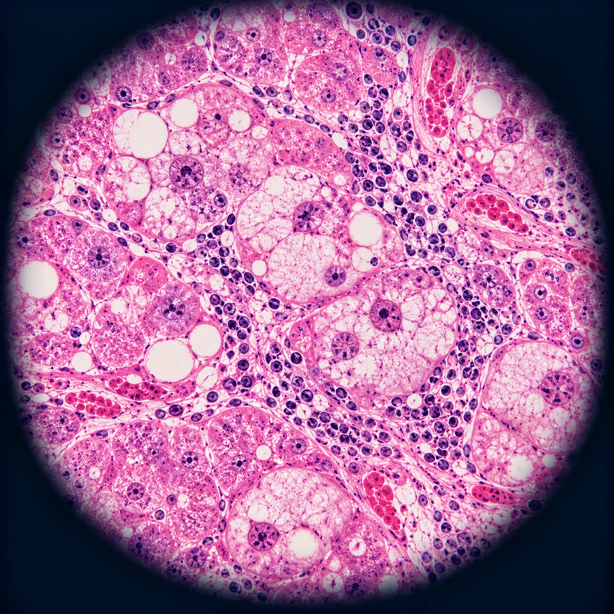
- Hepatocyte ballooning: swollen hepatocytes with pale, wispy cytoplasm — the cardinal lesion; represents ER stress and cytoskeletal disruption
- Mallory-Denk bodies: eosinophilic, irregularly-shaped cytoplasmic inclusions composed of cross-linked cytokeratin 8/18 and p62/ubiquitin; highly characteristic of AH (also seen in Wilson's disease, metabolic dysfunction-associated steatotic liver disease)
- Lobular neutrophilic infiltration: neutrophils — not lymphocytes — predominate; reflect acute-phase cytokine response; absent in most other hepatitides
- Megamitochondria: giant eosinophilic mitochondria visible on H&E; a specific marker of alcohol-related injury
- Zone 3 necrosis and inflammation: injury concentrated around hepatic venules (zone 3 / centrilobular region) — the zone with highest cytochrome P450 2E1 expression and lowest oxygen tension
- Variable steatosis: macrovesicular fat is common; may be absent if patient has been abstinent
Fibrosis patterns:
- Pericellular/sinusoidal fibrosis (chicken-wire fibrosis) in zone 3
- Central hyaline sclerosis (perivenular fibrosis)
- May show bridging fibrosis or established cirrhosis in long-standing disease
Biopsy can be performed transjugularly in coagulopathic patients (international normalized ratio >1.5 is the usual threshold for percutaneous biopsy; transjugular route avoids bleeding risk).
Treatment
Treatment of severe AH (Maddrey DF ≥32 or MELD ≥21) has four pillars:
1. Absolute abstinence
Abstinence is the single most important intervention across all AH severity levels. Even in severe AH where corticosteroids are used, abstinence is the cornerstone — it reduces 6-month and 1-year mortality, prevents relapse, and may allow reversal of fibrosis. Addiction medicine consultation and social support during hospitalization improve long-term abstinence rates.
2. Nutritional support
Protein-calorie malnutrition is present in nearly all patients with AH and independently worsens outcomes:
- Feeding through the gut (enteral nutrition) is preferred over intravenous (parenteral) feeding when the gut is working
- Target: 35–40 kcal/kg/day; 1.2–1.5 g protein/kg/day
- Protein is not restricted in these regimens: restricting it out of fear of hepatic encephalopathy is not evidence-based in AH, and hypocaloric feeding worsens outcomes
- Thiamine (vitamin B1) 100 mg intravenous (IV) × 3 days before any glucose administration — prevents Wernicke's encephalopathy
- Zinc, folate, multivitamin supplementation
3. Corticosteroids (prednisolone) for severe AH
The STOPAH trial (Thursz et al., NEJM 2015) is the landmark trial:
- 1103 patients with severe AH (Maddrey DF ≥32)
- Prednisolone 40 mg/day × 28 days: 28-day mortality was lower but the difference did not reach statistical significance (odds ratio (OR) 0.72, 95% confidence interval 0.52–1.01, P=0.06); no significant difference at 90 days or 1 year
- Serious infections occurred in 13% of patients given prednisolone versus 7% of those not given it (P=0.002)
- Pentoxifylline: no benefit at any time point (overturning two decades of prior belief based on smaller trials)
- Absolute contraindications to prednisolone: active infection (checked with blood cultures, CXR, UA, and ascitic fluid paracentesis before starting), uncontrolled upper gastrointestinal (GI) bleeding, renal failure
A 2018 meta-analysis of individual patient data from 11 randomized trials (2,111 patients) found that corticosteroids lowered the risk of death within 28 days compared with placebo or control (hazard ratio 0.64, 95% confidence interval 0.48–0.86), but there was no significant difference in 6-month mortality; adding pentoxifylline to corticosteroids made no difference to 28-day mortality. The 2024 American College of Gastroenterology guideline describes corticosteroids as the only drug treatment with proven efficacy in severe alcohol-associated hepatitis, giving a 1-month survival benefit in 50%–60% of patients.
Prednisolone is initiated at 40 mg/day orally × 28 days, followed by taper over 2 weeks. Response is assessed at day 7 using the Lille score (see below).
4. Liver transplantation for non-responders
Early liver transplantation for carefully selected severe AH non-responders was tested in a 2011 cohort from seven centres (Mathurin and colleagues): 26 patients were placed on the transplant list within a median of 13 days after failing to respond to medical therapy, and fewer than 2% of patients admitted with severe alcoholic hepatitis were selected:
- Stringent selection in that study: no prior episodes of alcoholic hepatitis, a Lille score of 0.45 or higher (or rapid worsening despite medical therapy), supportive family members, no severe coexisting conditions, and a commitment to alcohol abstinence
- 6-month survival in transplanted patients: 77% vs 23% in matched non-transplanted controls; the benefit was maintained through 2 years of follow-up
- Post-transplant alcohol relapse in that study: three patients resumed drinking, at 720, 740 and 1,140 days after transplantation
- Not yet universally available; requires center expertise in addiction psychiatry and ethics committee review
The Lille Score: Monitoring Steroid Response
The Lille score, calculated on day 7 of prednisolone, predicts the likelihood of benefit from continuing corticosteroids.
How it is built: the Lille model (Louvet and colleagues, 2007) is a logistic-regression model that combines six measurements — age, kidney failure (renal insufficiency), albumin, prothrombin time, bilirubin, and the change in bilirubin by day 7 — into a score between 0 and 1. It was developed in 320 patients with severe AH treated with corticosteroids and checked in a further 118. It predicted death at 6 months better than the Child-Pugh and Maddrey scores, and in the validation group better than the MELD and Glasgow scores. Patients above the cutoff of 0.45 — about 40% of cases — had 25% 6-month survival, against 85% for the others, and this cutoff picked out about 75% of the deaths.
Interpretation:
- Lille score ≤0.45: good response to prednisolone; continue to complete 28-day course; 6-month survival approximately 85%
- Lille score >0.45: non-response; stopping prednisolone at day 7 does not worsen outcomes and avoids the infection risk of continued immunosuppression; 6-month survival approximately 25–30%
- Lille score >0.56 (complete non-response): 6-month mortality exceeds 75% without transplant
The Lille score is now the standard of care in managing steroid therapy for AH and is incorporated into EASL, AASLD, and ACG guidelines. Continuing prednisolone in a Lille non-responder offers no survival benefit and increases infection (including aspergillosis) risk.
Key Research Papers
- — Thursz MR et al. "Prednisolone or Pentoxifylline for Alcoholic Hepatitis." New England Journal of Medicine 2015. — PubMed PMID: 25901427 (STOPAH trial)
- — Louvet A et al. "The Lille model: a new tool for therapeutic strategy in patients with severe alcoholic hepatitis treated with steroids." Hepatology 2007;45(6):1348-1354. (Lille score derivation) — PubMed PMID: 17518367
- — Mathurin P et al. "Early liver transplantation for severe alcoholic hepatitis." New England Journal of Medicine 2011. — PubMed PMID: 22070476
- — European Association for the Study of the Liver. "EASL Clinical Practice Guidelines: Management of alcohol-related liver disease." Journal of Hepatology 2018;69(1):154-181. — PubMed PMID: 29628280
- — Dunn W et al. "MELD accurately predicts mortality in patients with alcoholic hepatitis." Hepatology 2005. — PubMed PMID: 15660383
- — Dominguez M et al. "A new scoring system for prognostic stratification of patients with alcoholic hepatitis." American Journal of Gastroenterology 2008;103(11):2747-2756. (ABIC score) — PubMed PMID: 18721242
- — Singal AK et al. "ACG clinical guideline: alcoholic liver disease." American Journal of Gastroenterology 2018. — PubMed PMID: 29336434
- — Lucey MR et al. "Minimal criteria for placement of adults on the liver transplant waiting list: a report of a national conference organized by the American Society of Transplant Physicians and the American Association for the Study of Liver Diseases." Transplantation 1998;66(7):956-962. — PubMed PMID: 9798717
- — Maddrey WC et al. "Corticosteroid therapy of alcoholic hepatitis." Gastroenterology 1978. — PubMed PMID: 352788 (Maddrey DF original)
- — Crabb DW et al. "Standard Definitions and Common Data Elements for Clinical Trials in Patients With Alcoholic Hepatitis." Gastroenterology 2016. — PubMed PMID: 26921783
- — Lazarus JV et al. "Advancing the global public health agenda for NAFLD." Nature Reviews Gastroenterology & Hepatology 2022. — Search PubMed
- — O'Shea RS et al. "Alcoholic liver disease." Hepatology 2010;51(1):307-328. (AASLD practice guidelines) — PubMed PMID: 20034030
- — Louvet A et al. "Corticosteroids Reduce Risk of Death Within 28 Days for Patients With Severe Alcoholic Hepatitis, Compared With Pentoxifylline or Placebo-a Meta-analysis of Individual Data From Controlled Trials." Gastroenterology 2018;155(2):458-468.e8. — PubMed PMID: 29738698
- — Jophlin LL et al. "ACG Clinical Guideline: Alcohol-Associated Liver Disease." American Journal of Gastroenterology 2024;119(1):30-54. — PubMed PMID: 38174913
Connections
- Nephrology & Hepatology
- What Alcohol Does Inside Your Liver — interactive animation
- Cirrhosis
- Liver Disease
- Hepatic Encephalopathy
- Metabolic Dysfunction-Associated Steatotic Liver Disease (Metabolic-Associated Steatotic Liver Disease)
- Autoimmune Hepatitis
- Hepatitis C
- Non-Alcoholic Fatty Liver Disease
- Pancreatitis
- Vitamin B1 (Thiamine)
- Folate (Vitamin B9)
- Aspartate Aminotransferase (AST)
- Alanine Aminotransferase (ALT)
- Bilirubin
- Viral Hepatitis — a distinct, infectious cause of liver inflammation that must be excluded by serology, and a common co-factor: hepatitis C virus coinfection accelerates progression to cirrhosis.
- Herbs covered on this site that discuss this condition: Wormwood · Elephant's Foot (Elephantopus scaber) · Noni (Morinda citrifolia)