Nutritional Deficiencies and Bone Health in Celiac Disease
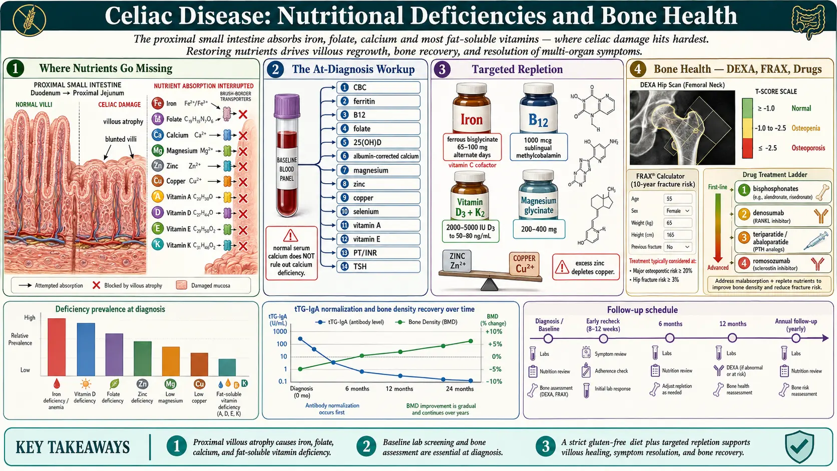
Table of Contents
- Why Nutrients Go Missing in Celiac
- The At-Diagnosis Nutritional Workup
- Iron — The Signature Celiac Deficiency
- Vitamin B12 and Folate
- Vitamin D and Calcium
- Magnesium
- Zinc and Copper — The Balancing Act
- Fat-Soluble Vitamins: A, E, K
- Protein, Albumin, and Selenium
- Bone Health: DEXA, Osteopenia, Osteoporosis
- When Bone Drugs Are Used
- Lactose Intolerance, Dermatitis Herpetiformis, and Pediatric Growth
- The Recheck Schedule — 3, 6, 12 Months and Beyond
- Realistic Healing Timeline
- Key Research Papers
- Connections
- Featured Videos
Why Nutrients Go Missing in Celiac
Celiac disease damages the proximal small intestine — the duodenum and upper jejunum — exactly where your body absorbs iron, folate, calcium, and most fat-soluble vitamins. The flattened villi and inflamed epithelium cannot grip and transport nutrients across the gut wall, so a person eating a seemingly adequate diet can still run low on everything. The longer undiagnosed celiac has been active, the deeper and more numerous the deficiencies tend to be.
This is not a minor footnote to your diagnosis. Nutrients drive the repair of the villi themselves, the rebuilding of bone that has been quietly thinning for years, the regeneration of red blood cells, and the normal function of every enzyme your body runs. Getting the numbers right matters as much as the gluten-free diet itself, and the first year after diagnosis is the window where it all gets sorted out.
The good news: most deficiencies correct on a strict gluten-free diet within 6 to 12 months as the villi regrow, with judicious supplementation to speed the process. Bone density takes longer — typically two years or more — but it does recover in the majority of adult patients. The work is front-loaded: one thorough workup at diagnosis, careful repletion, and two or three rechecks in the first year.
The At-Diagnosis Nutritional Workup
The 2013 American College of Gastroenterology (ACG) guideline states that people with newly diagnosed celiac disease should undergo testing and treatment for micronutrient deficiencies, and that the deficiencies considered for testing should include, but not be limited to, iron, folic acid, vitamin D, and vitamin B12. The 2023 ACG update lists further tests that may be used: complete blood count, liver enzymes, vitamins A, D, E and B12, copper, zinc, folic acid, ferritin, and iron. The panel below is a fuller version of that baseline. Tests taken at diagnosis, before any supplements are started, give a clean baseline that shows which nutrients are low and, equally important, which are not, so they are not over-supplemented.
- Complete blood count (CBC) with indices. The key figure is the MCV (mean corpuscular volume). Microcytic anemia (MCV under 80) points to iron deficiency; macrocytic anemia (MCV over 100) points to B12 or folate deficiency. A mixed picture is common in celiac because both patterns coexist — the MCV may look "normal" while hiding both.
- Ferritin, serum iron, total iron-binding capacity (TIBC), transferrin saturation. Ferritin is the best single marker of body iron stores and is almost always the first abnormality in celiac — iron is absorbed in the duodenum, ground zero for celiac damage.
- Vitamin B12 (serum) and ideally methylmalonic acid if B12 is low-normal.
- Folate — red blood cell (RBC) folate is more reliable than serum folate, which fluctuates with recent meals.
- 25-hydroxyvitamin D (25(OH)D). Some practitioners aim for 50–80 ng/mL during celiac recovery; most newly diagnosed patients sit well below 30.
- Serum calcium with albumin correction. Important caveat: blood calcium often looks normal even when total-body calcium is severely depleted, because the body will rob bone to keep serum levels in range. A normal serum calcium does not rule out calcium deficiency.
- Magnesium — RBC magnesium is often preferred over serum magnesium, which misses intracellular depletion.
- Zinc (plasma or serum).
- Copper (serum) and ceruloplasmin. Copper can be depleted either by celiac itself or iatrogenically later if zinc supplementation is pushed too high.
- Selenium.
- Vitamin A (retinol) and vitamin E (alpha-tocopherol).
- Vitamin K — measured functionally via prothrombin time / international normalized ratio (PT/INR). A prolonged PT in an unanticoagulated patient points to vitamin K deficiency.
- Albumin and total protein — markers of chronic malabsorption and protein-losing enteropathy.
- Thyroid-stimulating hormone (TSH) — autoimmune thyroid disease is the single most common overlap with celiac.
This panel sounds long, but it is one blood draw and almost every line item is covered by insurance when the indication is a new celiac diagnosis. A personal copy of every result is useful for tracking recovery.
Iron — The Signature Celiac Deficiency
Iron deficiency is so typical of undiagnosed celiac that unexplained iron-deficiency anemia in an adult is widely treated as a reason to test for celiac disease, with or without gastrointestinal (GI) symptoms; the 2023 ACG guideline lists chronic iron deficiency and unexplained anemia among the conditions in which testing for celiac disease is considered. Duodenal villi are the main site of iron absorption; when they are blunted, iron never enters the body, no matter how much is in the diet.
Oral repletion. For mild to moderate deficiency, ferrous bisglycinate 65–100 mg elemental iron is a form practitioners describe as well tolerated and, for many patients, better absorbed than ferrous sulfate. The dosing interval matters as much as the dose. In two randomized trials in iron-depleted women without celiac disease, Stoffel and colleagues (2017) gave 40 women 60 mg of iron either every day for 14 days or every other day (alternate-day dosing) for 28 days, 14 doses in each group. Cumulative fractional iron absorption was 16.3% with daily dosing and 21.8% with alternate-day dosing, and total iron absorbed was 131.0 mg against 175.3 mg; serum hepcidin (the hormone that blocks iron uptake from the gut) was higher in the daily group. In the second trial, splitting the dose into two doses a day raised hepcidin with no significant gain in absorption. A 2020 study by the same group in women with iron-deficiency anemia found that the rise in hepcidin after a dose persists for 24 hours, lowering absorption from iron taken later the same or the next day. A commonly described practice is to take iron on an empty stomach with vitamin C (250–500 mg), at least two hours apart from coffee, tea, calcium, or dairy.
When intravenous (IV) iron is used. Oral iron fails roughly 30% of the time, either because the gut cannot tolerate it (nausea, constipation, cramping) or because absorption is still too damaged. A threshold practitioners describe for switching to intravenous iron is ferritin below 20 ng/mL with oral-iron intolerance or failure after 8–12 weeks. Two modern formulations dominate the U.S. market: ferric carboxymaltose, typically given as two 750 mg doses a week apart, and ferric derisomaltose, which can deliver a full 1000 mg in a single 20-minute infusion. They differ in how often they lower blood phosphate. Two randomized trials published in 2020 (245 adults with iron-deficiency anemia who could not tolerate, or did not respond to, oral iron) found low blood phosphate (hypophosphatemia, serum phosphate below 2.0 mg/dL) within 35 days in 75.0% and 73.7% of patients after ferric carboxymaltose, against 7.9% and 8.1% after ferric derisomaltose. The authors state that further research is needed to determine the clinical importance of this difference.
Iron deficiency that persists after a year of strict gluten-free eating is one of the first clues that gluten exposure is ongoing or that refractory celiac disease is developing — see the refractory celiac article.
Vitamin B12 and Folate
These two B vitamins both cause macrocytic anemia, and celiac can drop either one. Folate is absorbed in the proximal small intestine (where celiac damage is worst), so folate deficiency is common. B12 is absorbed in the terminal ileum, which is usually spared, but concurrent autoimmune gastritis, H. pylori, or pernicious anemia (all over-represented in celiac patients) frequently takes out B12 anyway.
Folate repletion. A commonly used regimen is 1 mg of folic acid (or methylfolate for patients with methylenetetrahydrofolate reductase (MTHFR) variants who feel better on the methylated form) daily for 1–3 months, followed by a maintenance multivitamin dose.
B12 repletion. For mild deficiency, a commonly used dose is 1000 mcg sublingual methylcobalamin daily, which practitioners report works for most people. For severe deficiency, neurological symptoms, or documented pernicious anemia, a common regimen starts with 1000 mcg intramuscular cyanocobalamin weekly for 4–8 weeks, then monthly for maintenance. Neurological symptoms are time-sensitive — peripheral neuropathy and cognitive changes from B12 deficiency can become permanent if repletion is delayed more than a year.
Vitamin D and Calcium
Roughly two-thirds of newly diagnosed celiac adults are vitamin D deficient, and bone loss follows directly. Correcting vitamin D is the single most important intervention for long-term bone health in celiac disease.
Vitamin D dosing. Commonly used regimens start at vitamin D3 2000–5000 international units (IU) daily, adjusted to a 25(OH)D blood level of 50–80 ng/mL. When the starting level is under 20 ng/mL, another regimen in use is a loading dose of 50,000 international units weekly for 8 weeks followed by daily maintenance. Some practitioners pair it with vitamin K2 (menaquinone-7, MK-7) 100–200 mcg daily, on the proposed idea that vitamin K2 helps direct the extra calcium into bone rather than arteries.
Calcium. Adults need roughly 1000–1200 mg of elemental calcium per day from all sources combined (food plus supplement). Food sources include dairy if tolerated, fortified plant milks, sardines with bones, kale, and almonds. Among supplements, calcium citrate (500–600 mg elemental once or twice daily is a common amount) absorbs better than carbonate, especially in the low-acid stomach environment many celiac patients have. Calcium and iron compete for absorption when taken together.
As noted in the workup, serum calcium is a poor screening test because the body holds it constant at the expense of the bones. 25(OH)D, urinary calcium, and parathyroid hormone (PTH) give a truer picture. An elevated parathyroid hormone with a "normal" serum calcium is a classic sign of calcium and vitamin D insufficiency.
Magnesium
Magnesium deficiency is common in celiac and chronically under-diagnosed because standard serum magnesium is insensitive — only about 1% of body magnesium lives in the blood. RBC magnesium is the better test. Symptoms of deficiency are vague but suggestive: muscle cramps (especially at night or in the calves), eyelid twitches, migraines, anxiety, poor sleep, constipation, and palpitations.
Repletion. A commonly used form is magnesium glycinate 200–400 mg elemental daily, taken in the evening; glycinate is described as gentle on the gut and well absorbed. Magnesium citrate works too but tends to loosen stools, which may or may not be welcome depending on a person’s gastrointestinal state. Magnesium oxide is cheap but poorly absorbed. Kidney disease reduces magnesium clearance, which makes extra magnesium a concern for people with kidney disease.
Zinc and Copper — The Balancing Act
Zinc is directly required for villous regeneration, wound healing, immune function, and taste. Deficiency is common in active celiac and can actually slow gut healing. Zinc picolinate 15–30 mg elemental daily for 2–3 months is a commonly used repletion dose.
Here is the trap: zinc and copper compete for absorption in the gut. Supplementing zinc at doses above 30–40 mg daily for more than a few months reliably induces copper deficiency, which then causes its own anemia and neurological problems. A common practice for people taking 30 mg or more of zinc beyond the first few months is to add copper 1–2 mg daily at a separate time of day, or to check serum copper and ceruloplasmin before and after zinc repletion and stop supplementing once levels normalize.
Copper can also be depleted by celiac disease itself (again, duodenal absorption), so a normal copper level cannot be assumed in someone who has not been taking zinc; only a blood test shows it.
Fat-Soluble Vitamins: A, E, K
All three fat-soluble vitamins depend on intact fat absorption, which celiac disrupts. Deficiencies are less often symptomatic than iron or B12, but they show up on lab panels and matter for long-term health.
- Vitamin A. Low retinol slows villous regeneration and impairs dim-light vision. Diet sources include eggs, liver, and dairy; a modest supplement (2500–5000 international units daily) is a common addition if labs remain low. Vitamin A is stored in the liver and can become toxic with sustained intakes above 10,000 international units/day.
- Vitamin E. Low alpha-tocopherol can cause peripheral neuropathy and ataxia in severe cases. Mixed-tocopherol supplements (200–400 international units daily) correct mild deficiency.
- Vitamin K. Functional deficiency shows up as a prolonged PT/INR. Green leafy vegetables plus K2 (menaquinone-7) cover most cases; persistent deficiency points to ongoing fat malabsorption.
Protein, Albumin, and Selenium
Chronic malabsorption pulls down albumin and total protein. An albumin below 3.5 g/dL in newly diagnosed celiac is not rare and usually corrects within 6 months of a strict gluten-free diet plus adequate protein intake. Intakes of 1.0–1.2 g of protein per kilogram of body weight per day are commonly described during recovery — roughly 70–100 g/day for most adults. This is higher than standard recommendations because tissue repair is running at an accelerated rate.
Selenium deficiency is under-recognized in celiac and contributes to thyroid autoimmunity. Two Brazil nuts a day, or a 100–200 mcg supplement for 2–3 months, are the amounts commonly described for correcting it.
Bone Health: DEXA, Osteopenia, Osteoporosis
The 2023 ACG guideline update lists bone density testing by DEXA (dual-energy X-ray absorptiometry), along with vaccines, among the preventive care that is a goal of follow-up after a celiac diagnosis. Bone loss is one of the most common silent consequences of untreated celiac, driven by years of calcium malabsorption, vitamin D deficiency, and low-grade inflammation.
Bone loss shows up as broken bones. A 2015 meta-analysis found that in case-control and cross-sectional studies, fractures were almost twice as common in people with clinically diagnosed celiac disease as in people without it. In prospective studies, celiac disease at the start was linked to a 30% higher risk of any fracture (95% confidence interval 1.14 to 1.50) and a 69% higher risk of hip fracture (95% confidence interval 1.10 to 2.59).
A DEXA scan reports two numbers:
- T-score compares your bone density to a healthy young adult of the same sex. Used for adults over 50 or postmenopausal women.
- Z-score compares your bone density to someone your own age and sex. Used for adults under 50, premenopausal women, and children.
T-score cutoffs:
- Normal: T-score above −1.0
- Osteopenia (low bone mass): T-score between −1.0 and −2.5
- Osteoporosis: T-score at or below −2.5
- Severe osteoporosis: T-score at or below −2.5 plus one or more fragility fractures
The FRAX calculator (an online tool from the University of Sheffield) uses T-score, age, sex, weight, height, prior fracture history, parental hip fracture, smoking, alcohol, glucocorticoid use, and rheumatoid arthritis to estimate your 10-year probability of a major osteoporotic fracture. It is widely used in deciding whether a borderline DEXA result leads to treatment.
The recovery curve. On a strict gluten-free diet with adequate calcium and vitamin D, bone mineral density (BMD) improves measurably within the first year and continues to improve for 2–5 years. A 2012 review by Larussa and colleagues states that adherence to a strict gluten-free diet reverses the intestinal damage and the biochemical signs of calcium malabsorption, “resulting in rapid increase of BMD”, but that the diet “improves BMD but does not normalize it in all patients, even after the recovery of intestinal mucosa.” The same review notes that the question of who to refer for a DEXA scan in celiac disease, and when, is still open. A repeat DEXA at about 2 years is a commonly described follow-up for adult celiac patients who were osteopenic or osteoporotic at diagnosis.
When bone density fails to improve after 2 years on a strict diet, the causes usually looked for widen to: persistent gluten exposure, refractory celiac, coexisting hyperparathyroidism, vitamin D malabsorption, or an endocrine cause unrelated to celiac.
When Bone Drugs Are Used
Most adults with celiac-associated osteopenia are not treated with bone drugs; the gluten-free diet plus vitamin D, calcium, protein, and weight-bearing exercise rebuilds bone in many patients. Drug therapy is generally considered in a narrower set of situations:
- T-score at or below −2.5 (osteoporosis by density criteria)
- History of a fragility fracture (a fracture from a fall from standing height or less) regardless of T-score
- FRAX 10-year risk of major osteoporotic fracture above 20%, or hip fracture above 3%
The drug options, in rough order of use:
- Bisphosphonates — alendronate or risedronate. Oral weekly dosing; the usual first choice for most patients. The tablets are taken on an empty stomach with a full glass of water, sitting upright for 30 minutes, to protect the esophagus. Intravenous zoledronic acid once a year is an alternative for patients who cannot tolerate oral dosing.
- Denosumab. A subcutaneous injection every 6 months. Used for patients with kidney impairment who cannot take bisphosphonates, but has a rebound bone-loss problem if stopped abruptly — treatment is effectively lifelong or is followed by a bisphosphonate when it stops.
- Teriparatide or abaloparatide. Daily subcutaneous injections for up to 2 years. Anabolic — they build new bone rather than just preventing loss. Used mainly for severe osteoporosis or patients who have already fractured on antiresorptive therapy.
- Romosozumab. Monthly subcutaneous injection for 12 months; both builds bone and reduces resorption. Used for severe osteoporosis, with cardiovascular cautions.
Vitamin D repletion (at least 30 ng/mL; some practitioners aim for 50 or more) and adequate calcium intake are usually confirmed before a bone drug is started, because bone drugs do not work well in a vitamin-D-deficient patient.
Lactose Intolerance, Dermatitis Herpetiformis, and Pediatric Growth
Temporary lactose intolerance. The enzyme that digests milk sugar — lactase — lives on the tips of intestinal villi, which are exactly what celiac destroys. Many newly diagnosed patients are secondarily lactose intolerant and feel worse when they eat dairy. This is usually temporary, resolving within 6–12 months as the villi regrow. A short dairy-free period at diagnosis often improves symptoms markedly, and reintroducing dairy after about 6 months shows whether the intolerance was only temporary.
Dermatitis herpetiformis (DH). DH is celiac disease expressed in the skin — an intensely itchy, blistering rash on the elbows, knees, buttocks, and scalp, driven by the same gluten-triggered immunoglobulin A (IgA) immunology. Patients with DH almost always have some degree of small-intestinal villous damage whether or not they have gastrointestinal symptoms, and the nutritional workup above applies identically. The rash responds to strict gluten-free eating, though it can take months, and oral dapsone speeds relief while the diet works.
Pediatric growth failure. Short stature and delayed puberty are classic presenting signs of celiac disease in children. The encouraging news is that catch-up growth on a gluten-free diet is the rule, not the exception, when diagnosis happens before puberty closes the growth plates. Height often gains 5–10 cm over baseline projections in the first two years, and most children reach their genetic height potential. Celiac disease is among the conditions looked for in a child with unexplained short stature, and an earlier diagnosis leaves more time for catch-up growth.
The Recheck Schedule — 3, 6, 12 Months and Beyond
The 2023 ACG guideline update states that clinical follow-up after diagnosis may require multiple visits during the first year (for example at 3, 6, and 12 months) and regular visits twice a year or yearly after that. It lists tissue transglutaminase (tTG) and deamidated gliadin peptide (DGP) antibodies for monitoring, and other tests that may include a complete blood count, liver enzymes, vitamins A, D, E and B12, copper, zinc, folic acid, ferritin, and iron, with follow-up blood tests individualized to confirm that abnormal baseline results have corrected. The 2013 guideline judged annual follow-up reasonable until more evidence is available. A typical schedule along those lines:
- At 3 months: complete blood count, ferritin, 25(OH)D, tissue transglutaminase immunoglobulin A antibody (tTG-IgA), basic metabolic panel; a check of symptom response.
- At 6 months: a repeat of the items that were low at baseline; tTG-IgA, which is expected to be trending down; a review of diet adherence.
- At 12 months: a full recheck of every baseline abnormality; tTG-IgA is expected to be normal or near-normal with strict adherence. The 2023 guideline states that upper endoscopy with intestinal biopsies is helpful when symptoms do not respond or come back despite a gluten-free diet.
- At 24 months: a repeat DEXA if the baseline scan showed osteopenia or osteoporosis. The 2023 guideline states that a follow-up biopsy could be considered to check mucosal healing in adults without symptoms after 2 years on a gluten-free diet, after shared decision-making between patient and provider; it does not recommend follow-up biopsy in children without symptoms.
- Annually thereafter: tTG-IgA, complete blood count, ferritin, 25(OH)D, thyroid-stimulating hormone (thyroid overlap is common), and comprehensive metabolic panel.
Realistic Healing Timeline
A typical course, roughly:
- Weeks 1–4: Most gastrointestinal symptoms begin to settle. Energy starts rising as inflammation falls.
- Months 1–3: Folate, magnesium, and zinc typically normalize quickly with repletion. Antibodies begin dropping.
- Months 3–6: Ferritin, B12, and vitamin D climb into normal range with consistent supplementation. Lactose tolerance often returns.
- Months 6–12: Antibodies are usually negative by 12 months; the 2013 ACG guideline reports that 80% of people test negative after 6–12 months on a gluten-free diet. The gut lining itself heals more slowly in adults: the 2023 guideline cites a US study in which the median time to mucosal healing in adults was 3 years, and states that in adults the intestine will often fail to heal despite negative antibodies and no symptoms. In children, mucosal healing was observed in 95% within 2 years.
- Years 1–3: Bone mineral density climbs measurably. Weight and muscle mass return to normal. Energy and cognition continue to improve.
- Years 2–5: Peak bone recovery. Many osteopenic patients return to normal DEXA ranges without drugs. What is not known is why some do not: the 2012 Larussa review states that the gluten-free diet does not normalize BMD in all patients, even after the gut lining recovers, and that other mechanisms besides calcium and vitamin D malabsorption are thought to be involved — inflammatory signalling molecules (proinflammatory cytokines), parathyroid function abnormalities, and an imbalance in the bone-remodelling signals of the RANK/RANKL/osteoprotegerin system.
When someone is not on this curve — especially when ferritin stays low, antibodies stay high, or symptoms persist after 12 months of what the person believes is strict gluten-free eating — the 2023 ACG guideline describes a systematic workup, especially for unintentional gluten contamination; the usual first step is a hard look at hidden gluten exposure (see the gluten-free diet practical guide), and if exposure is truly excluded, evaluation for refractory celiac disease, which the guideline describes as a rare cause (see the refractory celiac article).
Key Research Papers
- Rubio-Tapia A, Hill ID, Kelly CP, Calderwood AH, Murray JA; American College of Gastroenterology. ACG clinical guidelines: diagnosis and management of celiac disease. Am J Gastroenterol. 2013;108(5):656-76. — PubMed PMID: 23609613
- Rubio-Tapia A, Hill ID, Semrad C, Kelly CP, Greer KB, Limketkai BN, Lebwohl B. American College of Gastroenterology Guidelines Update: Diagnosis and Management of Celiac Disease. Am J Gastroenterol. 2023;118(1):59-76. — PubMed PMID: 36602836
- Stoffel NU, Cercamondi CI, Brittenham G, Zeder C, Geurts-Moespot AJ, Swinkels DW, Moretti D, Zimmermann MB. Iron absorption from oral iron supplements given on consecutive versus alternate days and as single morning doses versus twice-daily split dosing in iron-depleted women: two open-label, randomised controlled trials. Lancet Haematol. 2017;4(11):e524-e533. — PubMed PMID: 29032957
- Stoffel NU, Zeder C, Brittenham GM, Moretti D, Zimmermann MB. Iron absorption from supplements is greater with alternate day than with consecutive day dosing in iron-deficient anemic women. Haematologica. 2020;105(5):1232-1239. — PubMed PMID: 31413088
- Wolf M, Rubin J, Achebe M, Econs MJ, Peacock M, Imel EA, Thomsen LL, Carpenter TO, Weber T, Brandenburg V, Zoller H. Effects of Iron Isomaltoside vs Ferric Carboxymaltose on Hypophosphatemia in Iron-Deficiency Anemia: Two Randomized Clinical Trials. JAMA. 2020;323(5):432-443. — PubMed PMID: 32016310
- Larussa T, Suraci E, Nazionale I, Abenavoli L, Imeneo M, Luzza F. Bone mineralization in celiac disease. Gastroenterol Res Pract. 2012;2012:198025. — doi:10.1155/2012/198025
- Heikkilä K, Pearce J, Mäki M, Kaukinen K. Celiac disease and bone fractures: a systematic review and meta-analysis. J Clin Endocrinol Metab. 2015;100(1):25-34. — PubMed PMID: 25279497
PubMed Topic Searches
For further reading, the following PubMed topic searches return current peer-reviewed work on nutrient deficiencies and bone health in celiac disease:
- PubMed: Celiac disease and iron deficiency anemia
- PubMed: Celiac disease and vitamin D deficiency
- PubMed: Celiac disease, bone mineral density, and DEXA
- PubMed: Celiac disease, B12 and folate deficiency
- PubMed: Celiac disease, zinc, and copper
- PubMed: Celiac disease, osteoporosis, and bisphosphonates
- PubMed: Alternate-day oral iron dosing and hepcidin
- PubMed: Celiac disease, gluten-free diet, and mucosal healing
- PubMed: Dermatitis herpetiformis and celiac disease
- PubMed: Pediatric celiac disease and catch-up growth
Connections
- Gastroenterology
- Celiac Disease Overview
- Gluten-Free Diet: A Practical Guide
- Dermatitis Herpetiformis
- Endoscopy, Biopsy, and Marsh Classification
- Osteoporosis
- Calcium
- Vitamin D3
- Iron
- Vitamin B12
- Magnesium
- Anemia
- Homocysteine Lab Test
- Inflammatory Bowel Disease Fatigue, Anemia, and Nutrient Deficiencies
- Associated Autoimmune Conditions
- Genetic Testing HLA DQ2 and DQ8
- Celiac Serology Testing tTG DGP EMA
- Non Celiac Gluten Sensitivity
- Refractory Celiac and EATL
- Lab Tests