Metabolic Syndrome and Cardiovascular Risk
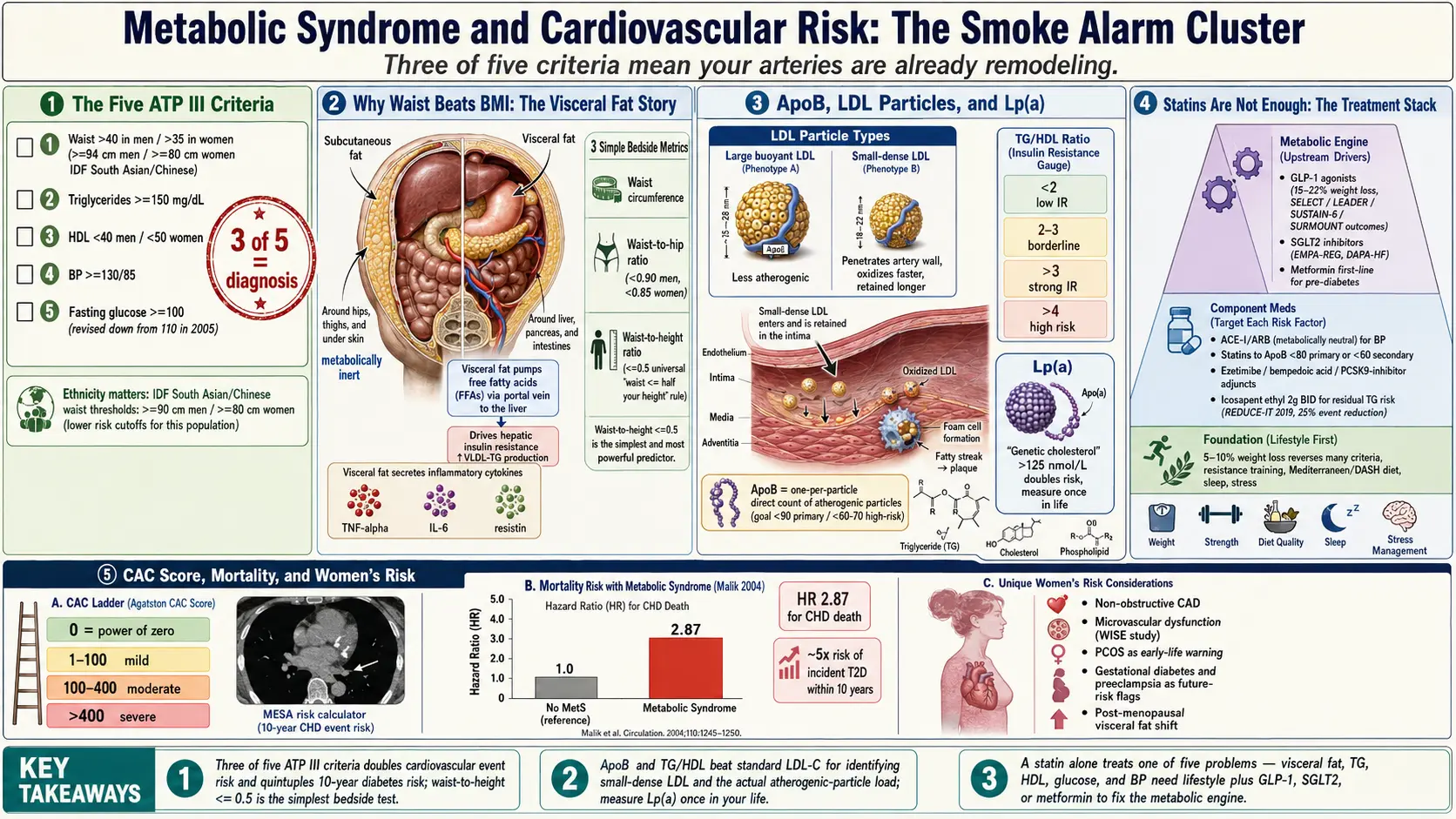
Table of Contents
- What Metabolic Syndrome Actually Is
- The Five Criteria — ATP III and IDF
- Cardiovascular and All-Cause Mortality
- Why Waist Circumference Beats Body Mass Index
- The Triglyceride-to-HDL Ratio
- Low-Density Lipoprotein Particle Count, ApoB, and Small-Dense Low-Density Lipoprotein
- Lp(a) — The Genetic Cholesterol
- Why Statins Alone Do Not Fix the Metabolic Engine
- Coronary Artery Calcium Score, CCTA, and Refining Risk
- Inflammatory and Ancillary Markers
- The Treatment Hierarchy and the Order of Steps
- Women's Risk — Under-Recognized
- ASCVD, PREVENT, and Family History
- Key Research Papers
- Connections
- Featured Videos
What Metabolic Syndrome Actually Is
Metabolic syndrome is not a disease in the way pneumonia or appendicitis is a disease. It is a cluster of findings that travel together because they share a common upstream driver: insulin resistance plus visceral adiposity. When enough of the findings pile up in one person, the ten-year risk of heart attack, stroke, and progression to type 2 diabetes rises steeply. The label exists because clinicians kept noticing that the same patients tended to have central fat, borderline blood pressure, borderline sugar, low high-density lipoprotein (HDL), and high triglycerides all at once — and that each of those findings, even at pre-disease levels, added to cardiovascular risk.
Think of it as a smoke alarm for your blood vessels. None of the five individual findings, on its own, is catastrophic. A triglyceride of 160 mg/dL is not going to kill you next week. Neither is a blood pressure of 135/88. But three of those findings together mean your metabolism is actively remodeling your arteries — laying down visceral fat, generating small-dense low-density lipoprotein (LDL) particles, lowering high-density lipoprotein's protective capacity, raising blood pressure, and nudging fasting glucose upward. Each year you carry that profile is a year of accelerated atherosclerosis.
The Five Criteria — ATP III and IDF
The most widely used definition in the United States is the NCEP ATP III criteria, originally published in 2001 and revised by the American Heart Association and National Heart, Lung, and Blood Institute in 2005 (Grundy et al). You have metabolic syndrome if you meet three of the following five:
- Waist circumference >40 inches (102 cm) in men, >35 inches (88 cm) in women.
- Triglycerides ≥150 mg/dL, or currently on triglyceride-lowering therapy.
- High-density lipoprotein cholesterol <40 mg/dL in men, <50 mg/dL in women, or on therapy.
- Blood pressure ≥130/85 mm Hg, or on antihypertensive therapy.
- Fasting glucose ≥100 mg/dL, or on glucose-lowering therapy.
The 2005 revision is important because it dropped the fasting glucose threshold from 110 to 100 mg/dL — meaning a lot of people who had been "metabolically normal" under the 2001 version woke up one morning meeting criteria. That was intentional. The evidence by then was clear that glucose in the 100–110 range was already doing measurable vascular damage.
The International Diabetes Federation (IDF) 2005 criteria are stricter, especially on waist. The IDF treats central obesity as a required criterion rather than one of five optional ones, and adjusts the waist threshold by ethnicity:
- Europid, Middle Eastern, Sub-Saharan African: ≥94 cm (37 in) men, ≥80 cm (31.5 in) women.
- South Asian, Chinese, South and Central American: ≥90 cm (35.5 in) men, ≥80 cm (31.5 in) women.
- Japanese: ≥90 cm men, ≥80 cm women.
The ethnicity adjustment matters. A South Asian man with a 36-inch waist has more visceral fat and more cardiovascular risk than a Northern European man at the same waist. The IDF criteria recognize that. The ATP III criteria, using a single 40-inch cutoff, systematically undercount metabolic syndrome in South Asian, East Asian, and Hispanic patients. For people in those groups, the IDF waist thresholds above are the ones written for their ethnicity, even where U.S. practice uses the ATP III cutoff.
Cardiovascular and All-Cause Mortality
The reason metabolic syndrome gets its own label, rather than being filed under "pre-diabetes plus a few extras," is the hard-endpoint data. Malik et al. 2004, following 6,255 U.S. men and women aged 30 to 75 from the NHANES II survey for a mean of 13.3 years, found that people with metabolic syndrome (and no prior cardiovascular disease) had a hazard ratio of 2.02 for coronary heart disease death, 1.82 for cardiovascular death and 1.40 for all-cause mortality compared with people who had neither. Meta-analyses have found similar or larger effects: Gami 2007 (37 studies, 172,573 people) found a relative risk of 1.78 for cardiovascular events and death, and Mottillo 2010 (87 studies, 951,083 patients) found a relative risk of 2.35 for cardiovascular disease and 1.58 for all-cause mortality.
Diabetes risk is even sharper. People with metabolic syndrome are roughly 5 times more likely to develop type 2 diabetes within ten years than people without. The risk is not linear — each additional component multiplies it. Meeting all five criteria is not twice as bad as meeting three; it is closer to four or five times as bad.
In practice, meeting three of the ATP III criteria is not "borderline": it places a person in a high-risk vascular category, which guidelines address with lifestyle change and, in many cases, medication for the specific components.
Why Waist Circumference Beats Body Mass Index
Body mass index (BMI) is a population metric that works poorly for individuals. It cannot distinguish a muscular 200-pound man with a 32-inch waist from a deconditioned 200-pound man with a 42-inch waist. Their cardiovascular risks are wildly different. The muscular man may be in the best quintile; the deconditioned man is in the worst.
What actually drives metabolic risk is visceral adipose tissue — the fat packed around the liver, pancreas, and intestines. Visceral fat is metabolically active. It pumps free fatty acids directly into the portal vein, driving hepatic insulin resistance, and it secretes inflammatory cytokines (tumor necrosis factor-alpha, interleukin-6 (IL-6), resistin) that make muscle and liver cells ignore insulin. Subcutaneous fat on the hips and thighs does none of this and is, if anything, mildly protective.
Waist circumference is a rough but cheap surrogate for visceral fat. The standard method measures at the top of the hip bone (iliac crest), parallel to the floor, at the end of a normal breath out, on bare skin and without pulling the stomach in. Two ratios add further signal:
- Waist-to-hip ratio (WHR). Target <0.90 for men, <0.85 for women. Higher ratios indicate central adiposity.
- Waist-to-height ratio (WHtR). Target ≤0.5 for all adults. This is the simplest rule in the lot: a waist less than half of height. For a 5'10" adult (70 inches), that is a waist under 35 inches. WHtR outperforms body mass index in every major study that has compared them for cardiovascular prediction.
Of the numbers a person can track at home, waist circumference is the most useful. It moves faster than weight when you fix insulin resistance through diet and exercise, and it is a better predictor of whether you are actually improving the metabolic engine versus just losing water or muscle.
The Triglyceride-to-HDL Ratio
Of all the ratios you can compute from a standard lipid panel, triglycerides divided by high-density lipoprotein is the single best marker of insulin resistance and small-dense low-density lipoprotein — both of which matter more for heart attack risk than total or low-density lipoprotein cholesterol on their own.
- TG/HDL <2: low insulin resistance, predominantly large buoyant low-density lipoprotein (less atherogenic).
- TG/HDL 2–3: borderline.
- TG/HDL >3: strongly suggestive of insulin resistance and a small-dense low-density lipoprotein phenotype.
- TG/HDL >4: high risk; the metabolic engine is failing.
The threshold is ethnicity-dependent. In Black patients, triglycerides tend to run lower for the same level of insulin resistance, so a cutoff of >2 rather than >3 has been proposed. Using the generic cutoff undercounts metabolic syndrome in African American patients.
At a TG/HDL of 1.2, the standard lipid panel tells most of the story about low-density lipoprotein particle number. At a TG/HDL of 5, the standard lipid panel can hide the problem, and advanced testing shows what is actually happening.
Low-Density Lipoprotein Particle Count, ApoB, and Small-Dense Low-Density Lipoprotein
Standard lipid panels report low-density lipoprotein cholesterol (LDL-C), which is an estimate of the mass of cholesterol inside all low-density lipoprotein particles combined. The problem is that particles come in different sizes. You can have 1,000 big fluffy low-density lipoprotein particles carrying a lot of cholesterol, or 2,500 small-dense low-density lipoprotein particles carrying the same total cholesterol. The low-density lipoprotein cholesterol number looks the same. The vascular risk does not.
Small-dense low-density lipoprotein particles penetrate the arterial wall more easily, oxidize faster, and are retained longer. They are the atherogenic workhorse. Insulin resistance and high triglycerides drive the liver to produce them preferentially.
Two better markers than low-density lipoprotein cholesterol:
- Low-density lipoprotein particle number (low-density lipoprotein-P). Measured by nuclear magnetic resonance (NMR) or ion-mobility advanced lipid panels offered by commercial laboratories. Goal under 1,000 nmol/L for most patients, under 700 for high-risk.
- Apolipoprotein B (ApoB). Every atherogenic particle — low-density lipoprotein, VLDL, IDL, Lp(a) — carries exactly one ApoB molecule. So ApoB in mg/dL is essentially a direct count of the particles that cause atherosclerosis. Sniderman et al. 2019 and every major review since have argued ApoB should replace low-density lipoprotein cholesterol as the primary lipid target. Goal under 90 mg/dL for primary prevention, under 60–70 for high-risk. Cost is about $15–$30; most insurers cover it if coded properly (CPT 82172).
When low-density lipoprotein cholesterol looks fine but TG/HDL is high, an ApoB test is the one that shows the particle count. A low-density lipoprotein cholesterol of 110 mg/dL can hide an ApoB of 120 mg/dL — a particle count that puts that person in a much higher risk stratum than the standard panel suggested.
Lp(a) — The Genetic Cholesterol
Lipoprotein(a), pronounced "L-P-little-a," is a low-density lipoprotein particle with an extra protein (apolipoprotein(a)) bolted on. It is strongly genetic, set largely at birth, and unaffected by diet, exercise, or statins. About 20–25% of the general population has elevated Lp(a), and most have no idea.
High Lp(a) independently raises cardiovascular risk by 2–3×, drives calcific aortic valve disease, and contributes to early heart attacks in people with otherwise normal lipids. The threshold of concern is approximately:
- <75 nmol/L (<30 mg/dL): normal.
- 75–125 nmol/L (30–50 mg/dL): borderline.
- >125 nmol/L (>50 mg/dL): high; doubles or triples risk.
- >250 nmol/L: very high; aggressive management of every other risk factor is warranted.
Lp(a) needs to be measured only once in a lifetime, because it is set genetically and does not meaningfully change. A single test in adulthood is enough, and it is especially informative for anyone with a family history of early coronary disease, aortic stenosis, or unexplained heart attack before age 55 (men) or 65 (women).
No current drug reliably lowers Lp(a). Niacin nudges it down modestly. PCSK9 inhibitors lower it by 20–30%. The real story is two antisense/siRNA drugs in late-stage trials: pelacarsen (Phase 3 HORIZON trial) and olpasiran (Phase 3 OCEAN(a)-Outcomes). In its dose-ranging trial, pelacarsen lowered Lp(a) by 35% to 80% depending on dose (80% at 20 mg weekly; Tsimikas 2020); olpasiran's dose-ranging trial found placebo-adjusted reductions of about 70% to 101% (O'Donoghue 2022). If the outcome trials read positive, they will be the first true Lp(a)-specific therapy approved. Until then, the approach described for people with high Lp(a) is aggressive control of every other modifiable risk factor — ApoB, blood pressure, visceral adiposity, and smoking — to compensate.
Why Statins Alone Do Not Fix the Metabolic Engine
Statins work. They lower low-density lipoprotein cholesterol and ApoB by blocking HMG-CoA reductase in the liver, and they have additional pleiotropic effects — reducing vascular inflammation, stabilizing existing plaque, and improving endothelial function. A pooled analysis of 26 statin trials with about 170,000 participants (Cholesterol Treatment Trialists' Collaboration 2010) found major vascular events fell by about 22% (rate ratio 0.78) for each 1.0 mmol/L (about 39 mg/dL) of low-density lipoprotein cholesterol lowered. The benefit is real.
But statins do not fix insulin resistance. They do not shrink visceral fat. They do not raise high-density lipoprotein meaningfully. They do not lower triglycerides by much. They do not touch Lp(a). And at higher doses, some statins mildly worsen fasting glucose and modestly increase the incidence of new-onset type 2 diabetes — a trade-off that is worth it in higher-risk patients but still a real effect.
The point is not that statins are bad. The point is that if you have metabolic syndrome, a statin alone treats one out of five problems. You still have the visceral fat, the high triglycerides, the low high-density lipoprotein, the rising glucose, and the blood pressure creep. Those require metabolic interventions — diet, exercise, sleep, and in many cases medications that target the metabolic engine directly (glucagon-like peptide-1 (GLP-1) agonists, sodium-glucose cotransporter-2 (SGLT2) inhibitors, metformin). A patient on a statin whose ApoB is now 75 mg/dL but whose waist is 44 inches and hemoglobin A1c (HbA1c) is 6.2% is not "fixed." They are partially treated.
Coronary Artery Calcium Score, CCTA, and Refining Risk
When you are stuck in the middle of a risk calculator — ten-year ASCVD risk of, say, 8% — imaging can tell you whether your arteries are actually behaving like an 8% person or like a 25% person. Two tools:
Coronary Artery Calcium (CAC) score. A low-radiation non-contrast computed tomography (CT) of the chest that counts and quantifies calcified plaque in the coronary arteries. Cost $75–$200 out of pocket in the United States (most insurers do not cover it yet, though that is changing). Results are reported as an Agatston score:
- 0: no detectable calcified plaque. Ten-year event risk very low — often below 1%. The "power of zero" is real.
- 1–100: mild plaque. Intermediate risk.
- 100–400: moderate plaque. High risk; aggressive prevention warranted.
- >400: severe plaque. Very high risk; statin indicated regardless of low-density lipoprotein cholesterol.
The MESA Risk Calculator (mesa-nhlbi.org) lets you plug in your numbers and compare predicted versus actual plaque. For a 55-year-old with metabolic syndrome, a coronary artery calcium of 0 is commonly taken as a reason aggressive drug therapy can wait; a coronary artery calcium of 300 in the same person falls in the 100–400 high-risk band above, where statins are commonly started.
Coronary computed tomography Angiography (CCTA) goes further, using contrast to visualize the artery lumen and characterize plaque as calcified, non-calcified, or mixed. In stable chest pain, the SCOT-HEART trial found that adding CCTA to standard care lowered the 5-year rate of coronary death or heart attack (2.3% vs. 3.9%; Newby 2018), while the PROMISE trial found that starting with CCTA instead of functional testing did not improve clinical outcomes over a median of 2 years (Douglas 2015). Cost is higher ($500–$2,000) and it is typically reserved for patients with symptoms or an elevated coronary artery calcium.
Inflammatory and Ancillary Markers
A few additional labs sharpen the picture:
- high-sensitivity C-reactive protein (hsCRP): a nonspecific inflammation marker. Under 1 mg/L is low risk, 1–3 intermediate, over 3 high. Elevated high-sensitivity C-reactive protein in the setting of metabolic syndrome amplifies cardiovascular risk and identifies patients most likely to benefit from anti-inflammatory therapy (colchicine, as tested in LoDoCo2 and COLCOT).
- Ferritin: elevated in inflammatory states (and in hereditary hemochromatosis). Mildly elevated ferritin in metabolic syndrome is a surrogate for hepatic inflammation and fatty liver. Dramatically elevated ferritin warrants iron-studies workup.
- Homocysteine: elevated levels are mildly associated with cardiovascular risk; B-vitamin repletion lowers the number but has not consistently lowered events in trials. Worth checking once; not worth chasing aggressively.
- Uric acid: insulin resistance reduces renal clearance of urate, so high-normal or high uric acid often travels with metabolic syndrome, predisposes to gout, and independently correlates with hypertension and kidney disease. Fructose consumption in particular drives both insulin resistance and uric acid.
The Treatment Hierarchy and the Order of Steps
Metabolic syndrome is reversible. The hierarchy matters because the interventions stack, and skipping the foundation makes every drug on top of it less effective.
1. Lifestyle first, always. Nothing substitutes for losing visceral fat through diet change and exercise. Even a 5–10% weight loss reverses many of the criteria. See the sibling Diet Strategy and Exercise Protocol articles for the details. Sleep and stress also matter: see Sleep & Stress.
2. Treat the specific components that remain.
- Blood pressure: angiotensin-converting enzyme inhibitor (ACE inhibitor) or angiotensin receptor blocker (ARB) as first-line, especially in diabetic or pre-diabetic patients, because they are renoprotective and metabolically neutral. Thiazides and beta-blockers can mildly worsen glucose, which is why they are used more cautiously in metabolic syndrome.
- Glucose and weight: glucagon-like peptide-1 receptor agonists (GLP-1s) such as semaglutide and liraglutide, and the dual glucose-dependent insulinotropic polypeptide (GIP)/GLP-1 receptor agonist tirzepatide, produce large weight loss (tirzepatide 15–21% at 72 weeks in the SURMOUNT-1 weight trial). Semaglutide reduced major cardiovascular events in SUSTAIN-6 and SELECT, and liraglutide did so in LEADER. Sodium-glucose cotransporter-2 inhibitors (empagliflozin, dapagliflozin) reduce cardiovascular death and heart-failure hospitalization independent of glucose lowering: in type 2 diabetes (EMPA-REG OUTCOME) and in heart failure with or without diabetes (DAPA-HF, EMPEROR). Metformin remains useful as first-line in pre-diabetes and type 2.
- Low-density lipoprotein/ApoB: statin at the dose needed to reach ApoB <80 (primary prevention) or <60 (secondary prevention or high risk). Add ezetimibe, bempedoic acid, or a PCSK9 inhibitor if the statin alone is insufficient.
- Residual triglyceride risk: icosapent ethyl (eicosapentaenoic acid, EPA) 2 g twice daily reduced cardiovascular events by 25% in patients with high triglycerides already on statins in the REDUCE-IT trial (Bhatt et al. 2019). This is a specific eicosapentaenoic acid-only formulation, not fish oil from the supplement aisle — combination eicosapentaenoic acid/docosahexaenoic acid (DHA) products failed in STRENGTH.
See Medications and glucagon-like peptide-1 for the prescribing detail on each drug class.
Women's Risk — Under-Recognized
Historically, metabolic syndrome and coronary disease were studied primarily in men, and women's cardiovascular risk has been under-recognized as a result. A few specifics:
- Non-obstructive coronary artery disease (CAD). Women are more likely than men to have diffuse, non-obstructive coronary disease — plaque that narrows arteries modestly without producing the focal 70% stenosis that triggers an abnormal stress test. They get told their angiogram was "clean" and sent home, then present with a heart attack months later.
- Microvascular dysfunction. The WISE study (Women's Ischemia Syndrome Evaluation) documented that a large fraction of women with chest pain and no obstructive disease on angiography have coronary microvascular dysfunction — impaired dilation of the small arterioles that actually feed heart muscle. This is a real cause of angina and of adverse outcomes, and it tracks with metabolic syndrome.
- Polycystic ovary syndrome (PCOS) as an early warning. Polycystic ovary syndrome is essentially metabolic syndrome presenting in the reproductive years. For this reason, early and repeated screening for the full metabolic-syndrome panel is described for women with polycystic ovary syndrome. See the sibling polycystic ovary syndrome Connection article.
- Pregnancy history. Gestational diabetes, preeclampsia, and pregnancy-induced hypertension all flag future cardiovascular risk. They belong in a cardiovascular history but are routinely skipped.
- Post-menopausal shift. Visceral fat accumulation accelerates in the years around menopause as estrogen falls. Waist, triglycerides, and blood pressure can all drift into metabolic-syndrome territory within five years of the last period in a woman who was previously metabolically healthy.
ASCVD, PREVENT, and Family History
Two calculators estimate your ten-year cardiovascular risk:
- ACC/AHA ASCVD Risk Estimator Plus (tools.acc.org) — the 2013 Pooled Cohort Equations. Widely used; systematically overestimates risk in some contemporary populations.
- AHA PREVENT 2023 equations (professional.heart.org/en/guidelines-and-statements/prevent-calculator) — the newer replacement, incorporating kidney function, metabolic factors, and social determinants; predicts ten- and thirty-year risk for ASCVD, heart failure, and their composite. PREVENT estimates are generally lower than ASCVD for the same patient and are probably more accurate for current populations.
Neither calculator captures Lp(a) or family history adequately. A first-degree relative (parent or sibling) with early coronary disease — heart attack, bypass, or stent before age 55 in men or 65 in women — is commonly estimated to multiply the calculator's figure by approximately 1.5–2×. Early family history is one of the strongest independent risk factors and is not fully modeled.
The honest summary: calculators estimate probability; coronary artery calcium imaging shows what is actually happening in your arteries; ApoB and Lp(a) show the particles driving it; and waist, TG/HDL, and fasting glucose show whether the metabolic engine is still running hot. Together they give a fuller picture than any one in isolation.
Key Research Papers
Foundational publications defining metabolic syndrome diagnostic criteria, quantifying its cardiovascular risk burden, and demonstrating that ApoB — not low-density lipoprotein cholesterol — is the most accurate atherogenic-particle marker. Each citation links to the full text via DOI.
- Expert Panel on Detection, Evaluation, and Treatment of High Blood Cholesterol in Adults. Executive Summary of the Third Report of the National Cholesterol Education Program (NCEP) Adult Treatment Panel III (ATP III). JAMA. 2001;285(19):2486–2497. — doi:10.1001/jama.285.19.2486
- Grundy SM, Cleeman JI, Daniels SR, et al. Diagnosis and Management of the Metabolic Syndrome: An American Heart Association/National Heart, Lung, and Blood Institute Scientific Statement. Circulation. 2005;112(17):2735–2752. — doi:10.1161/CIRCULATIONAHA.105.169404
- Alberti KGMM, Eckel RH, Grundy SM, et al. Harmonizing the Metabolic Syndrome: A Joint Interim Statement of the IDF, NHLBI, AHA, WHF, IAS, and IASO. Circulation. 2009;120(16):1640–1645. — doi:10.1161/CIRCULATIONAHA.109.192644
- Mottillo S, Filion KB, Genest J, et al. The Metabolic Syndrome and Cardiovascular Risk: A Systematic Review and Meta-Analysis. Journal of the American College of Cardiology. 2010;56(14):1113–1132. — doi:10.1016/j.jacc.2010.05.034
- Malik S, Wong ND, Franklin SS, et al. Impact of the Metabolic Syndrome on Mortality from Coronary Heart Disease, Cardiovascular Disease, and All Causes in United States Adults. Circulation. 2004;110(10):1245–1250. — doi:10.1161/01.CIR.0000140677.20606.0E
- Sniderman AD, Thanassoulis G, Glavinovic T, et al. Apolipoprotein B Particles and Cardiovascular Disease: A Narrative Review. JAMA Cardiology. 2019;4(12):1287–1295. — doi:10.1001/jamacardio.2019.3780
- Bhatt DL, Steg PG, Miller M, et al. Cardiovascular Risk Reduction with Icosapent Ethyl for Hypertriglyceridemia (REDUCE-IT). New England Journal of Medicine. 2019;380(1):11–22. — doi:10.1056/NEJMoa1812792
- Reaven GM. Banting Lecture 1988. Role of Insulin Resistance in Human Disease. Diabetes. 1988;37(12):1595–1607. — doi:10.2337/diab.37.12.1595
- Petersen MC, Shulman GI. Mechanisms of Insulin Action and Insulin Resistance. Physiological Reviews. 2018;98(4):2133–2223. — doi:10.1152/physrev.00063.2017
- Saltiel AR, Olefsky JM. Inflammatory Mechanisms Linking Obesity and Metabolic Disease. Journal of Clinical Investigation. 2017;127(1):1–4. — doi:10.1172/JCI92035
- Marso SP, Daniels GH, Brown-Frandsen K, et al. Liraglutide and Cardiovascular Outcomes in Type 2 Diabetes (LEADER). New England Journal of Medicine. 2016;375(4):311–322. — doi:10.1056/NEJMoa1603827
- Zinman B, Wanner C, Lachin JM, et al. Empagliflozin, Cardiovascular Outcomes, and Mortality in Type 2 Diabetes (EMPA-REG OUTCOME). New England Journal of Medicine. 2015;373(22):2117–2128. — doi:10.1056/NEJMoa1504720
- Gami AS, Witt BJ, Howard DE, et al. Metabolic Syndrome and Risk of Incident Cardiovascular Events and Death: A Systematic Review and Meta-Analysis of Longitudinal Studies. Journal of the American College of Cardiology. 2007;49(4):403–414. — PubMed PMID: 17258085
- Cholesterol Treatment Trialists’ (CTT) Collaboration; Baigent C, Blackwell L, Emberson J, et al. Efficacy and Safety of More Intensive Lowering of LDL Cholesterol: A Meta-Analysis of Data from 170,000 Participants in 26 Randomised Trials. Lancet. 2010;376(9753):1670–1681. — PubMed PMID: 21067804
- Tsimikas S, Karwatowska-Prokopczuk E, Gouni-Berthold I, et al. Lipoprotein(a) Reduction in Persons with Cardiovascular Disease. New England Journal of Medicine. 2020;382(3):244–255. — PubMed PMID: 31893580
- O’Donoghue ML, Rosenson RS, Gencer B, et al. Small Interfering RNA to Reduce Lipoprotein(a) in Cardiovascular Disease. New England Journal of Medicine. 2022;387(20):1855–1864. — PubMed PMID: 36342163
- SCOT-HEART Investigators; Newby DE, Adamson PD, Berry C, et al. Coronary CT Angiography and 5-Year Risk of Myocardial Infarction. New England Journal of Medicine. 2018;379(10):924–933. — PubMed PMID: 30145934
- Douglas PS, Hoffmann U, Patel MR, et al. Outcomes of Anatomical versus Functional Testing for Coronary Artery Disease (PROMISE). New England Journal of Medicine. 2015;372(14):1291–1300. — PubMed PMID: 25773919
PubMed Topic Searches
Live PubMed queries that update as new papers are indexed.
- PubMed: metabolic syndrome and CV mortality
- PubMed: waist circumference and visceral adiposity
- PubMed: triglyceride-to-HDL ratio
- PubMed: apolipoprotein B and CV risk
- PubMed: lipoprotein(a) and CVD
- PubMed: coronary artery calcium and MESA
- PubMed: GLP-1 agonists and CV outcomes
- PubMed: SGLT2 inhibitors and heart failure
- PubMed: women and microvascular coronary disease
- PubMed: AHA PREVENT equations
Connections
- Endocrinology
- Blood Sugar & Insulin — interactive animation
- Reversal Protocol: Diet and Fasting
- CGM for Non-Diabetics
- Insulin Resistance and Polycystic Ovary Syndrome
- Metformin, Berberine and Glucagon-Like Peptide-1 Receptor Agonists
- Exercise and Muscle Insulin Sensitivity
- Insulin Resistance
- Metabolic Syndrome
- Diabetes
- Hypertension
- Fasting Insulin and Homeostatic Model Assessment of Insulin Resistance Testing
- Coronary Artery Disease
- ApoB
- Cardiovascular Disease
- Atherosclerosis
- Lipid Panel
- Visceral Fat
- Uric Acid
- Polycystic Ovary Syndrome
- Blood Sugar Support for Insulin Resistance — the food-and-supplement side of lowering the insulin resistance that drives every metabolic syndrome criterion on this page.
- Cardiology