Raynaud's Phenomenon
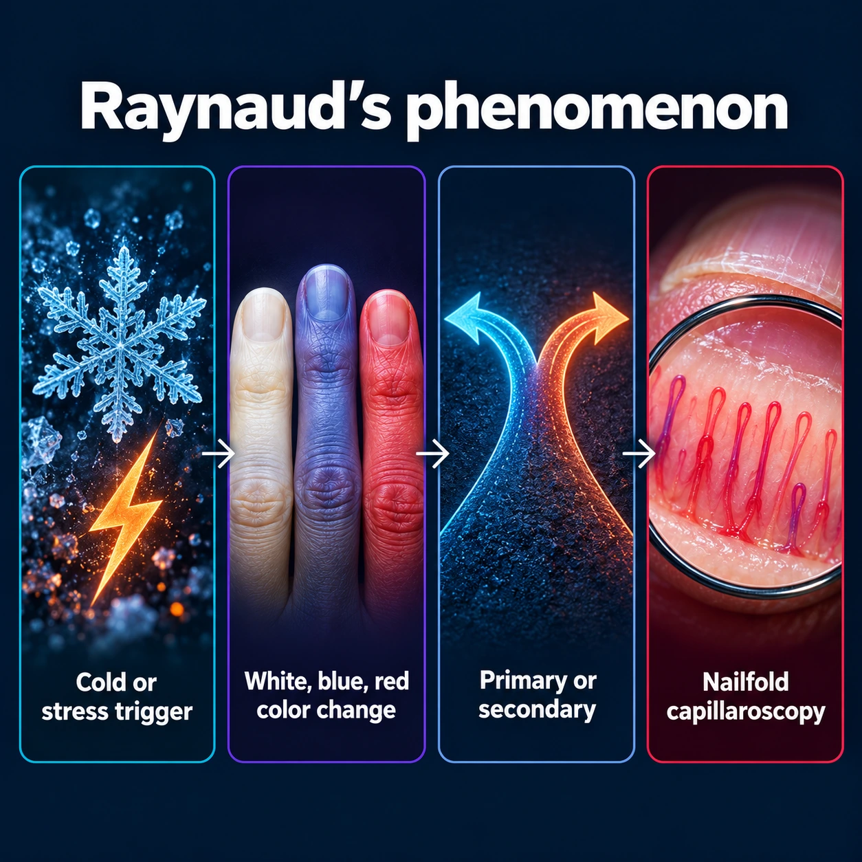
Raynaud's phenomenon is an episodic, reversible constriction of the small blood vessels supplying the fingers and toes — and sometimes the ears, nose, and nipples — triggered by cold exposure or emotional stress. The result is a dramatic color change in the affected digits that can be alarming the first time you see it. The condition ranges from a harmless nuisance in otherwise healthy young women to an early warning sign of serious autoimmune disease — and knowing which type you have changes everything about treatment. Maurice Raynaud, a French physician, first described the condition in Paris in 1862.
- Overview — How Raynaud's Phenomenon Works
- Primary vs. Secondary Raynaud's — A Critical Distinction
- Causes of Secondary Raynaud's
- Clinical Presentation — The Triphasic Color Change
- Nailfold Capillaroscopy — The Key Diagnostic Tool
- Diagnosis and Laboratory Workup
- Treatment — Lifestyle and Medications
- Treatment of Secondary Raynaud's and Digital Ulcers
- Prognosis
- Key Research Papers
- Connections
- Featured Videos
Overview — How Raynaud's Phenomenon Works
At its core, Raynaud's is an exaggerated vasomotor response. In a normal person exposed to cold, the body tightens small blood vessels in the extremities to conserve heat for the vital organs — a sensible survival mechanism. In Raynaud's, this response is dramatically amplified. The arterioles and small arteries supplying the fingers go into intense spasm (vasospasm), effectively cutting off blood flow to the fingertips. The same response can be triggered not just by cold air but by handling a cold drink, reaching into a refrigerator, air conditioning, swimming in cold water, or even acute emotional stress (because the sympathetic nervous system, which drives the fight-or-flight response, uses many of the same pathways as cold-induced vasoconstriction).
The fingers — and less commonly the toes, ears, nose, and nipples — are the most affected areas because they are the most distal from the heart and have the least thermal mass to protect them.
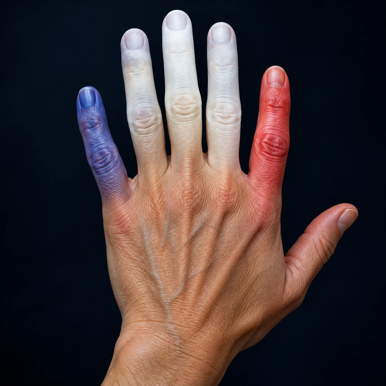
The triphasic color change is the hallmark of Raynaud's and reflects what is happening at the vascular level:
- WHITE (pallor): Vasospasm is at its peak. Blood flow is cut off almost completely. The fingers turn white, cold, and numb — sometimes painfully so. This is the ischemic phase.
- BLUE (cyanosis): The small amount of blood still present in the vessels becomes deoxygenated as the tissues extract what oxygen remains. The fingers turn blue or purple. This is the deoxygenation phase.
- RED (erythema/hyperemia): When the vasospasm releases — either because you warm up or the trigger resolves — blood rushes back into the previously starved vessels. This hyperemic reperfusion causes the fingers to turn red, often accompanied by burning, throbbing, and tingling. This is the rewarming phase.
Not every patient shows all three phases. A biphasic change (white then red, skipping the blue) is common. Some patients only notice white and blue without the red flush. A typical episode lasts anywhere from a few minutes to an hour, resolving completely once the hands are warmed. In primary Raynaud's, there is no lasting tissue damage between episodes.
Primary vs. Secondary Raynaud's — A Critical Distinction
This distinction is arguably the most important thing to understand about Raynaud's. The two types look similar from the outside but have completely different implications for your health.
Primary Raynaud's (Raynaud's Disease)
Primary Raynaud's has no identifiable underlying cause — the vasospasm is the disease itself, driven by genetic predisposition to vasomotor instability. It is common: a 2015 pooled analysis of 33 studies (33,733 people) found primary Raynaud's in 4.85% of the general population, with about 0.25% of people developing it each year. Key features:
- Demographics: More common in women — in the same 2015 analysis, women had about 1.65 times the odds of men. A family history was the strongest risk factor (odds ratio 16.6); migraine (odds ratio 4.02), manual work (2.66), cardiovascular disease (1.69) and smoking (1.27) were also linked. Onset is typically in the teens to early thirties.
- Symmetry: Episodes affect both hands symmetrically. If one hand is dramatically worse than the other, that asymmetry is a red flag for secondary Raynaud's.
- Normal investigations: antinuclear antibody (ANA) is negative or only weakly positive. Nailfold capillaroscopy is completely normal. Blood tests show no evidence of autoimmune disease.
- No tissue damage: Because the problem is vasospasm alone — there is no fixed obstruction of the vessels — blood flow recovers completely with rewarming. No digital ulcers, no pitting scars, no gangrene.
- Family history: Often positive; first-degree relatives frequently have the same tendency.
- Course: Generally benign and stable. May improve with age. Does not progress to organ damage.
- Treatment goal: Symptom management — keeping episodes infrequent and tolerable.
Secondary Raynaud's (Raynaud's Phenomenon)
Secondary Raynaud's is caused by an identifiable underlying disease. The term "Raynaud's phenomenon" (rather than "disease") technically refers to this secondary form. It is clinically far more important because the underlying disease — not the vasospasm itself — may be the real threat to your health. Key features:
- Can occur at any age and does not reliably follow the young-female pattern of primary Raynaud's.
- May be asymmetric — one hand or foot more severely affected, or single digits more involved than others.
- Tissue loss is possible: When an underlying disease causes structural damage to the vessel wall in addition to vasospasm, blood flow may not recover completely. This leads to digital ulcers (painful open sores on the fingertips), pitting scars, and in severe cases, gangrene requiring amputation.
- Abnormal nailfold capillaroscopy: Capillary loop architecture is distorted — see the dedicated section below.
- Positive autoimmune labs: antinuclear antibody, specific antibodies (anti-Scl-70, anti-centromere, anti-dsDNA, etc.) depending on the underlying disease.
- Critical timing: Raynaud's may be the first symptom of an underlying autoimmune disease, appearing months to years before the other manifestations become apparent — this is especially true for scleroderma (systemic sclerosis).
- Treatment strategy: Address the underlying disease first; manage the vasospasm as part of that treatment.
Causes of Secondary Raynaud's
Connective Tissue Diseases (Most Important Category)
Scleroderma (systemic sclerosis, SSc) is the connective tissue disease most closely associated with Raynaud's — 90 to 95% of SSc patients have it. Crucially, Raynaud's is often the very first symptom of scleroderma, preceding the skin thickening, lung fibrosis, and other manifestations by months to years. When a young person has new-onset Raynaud's and abnormal nailfold capillaroscopy, clinicians look actively for scleroderma. In SSc, the vessel wall itself becomes fibrotic and narrowed (not just vasospasm), which is why digital ulcers and pitting scars develop. Specific antibodies: anti-Scl-70 (topoisomerase-I) is associated with diffuse cutaneous SSc; anti-centromere antibody is associated with limited cutaneous SSc (the old "CREST syndrome").
Lupus (systemic lupus erythematosus, SLE) causes Raynaud's in 30 to 40% of patients. It is usually milder than in scleroderma and rarely leads to digital ulcers in isolation, though the overall vascular disease of lupus can complicate the picture.
Sjögren's Syndrome carries approximately 30% Raynaud's prevalence — often an underappreciated association. Dry eyes and dry mouth together with Raynaud's are the combination that leads to testing for Sjögren's.
Mixed Connective Tissue Disease (MCTD) has one of the highest Raynaud's rates of any CTD — up to 85% of patients. The hallmark antibody is anti-U1-RNP.
Dermatomyositis and Polymyositis both carry Raynaud's in a subset of patients, particularly those with anti-synthetase antibodies (anti-Jo-1 and related).
Rheumatoid Arthritis is associated with Raynaud's less commonly than the above, but the association is real in someone with joint disease and episodic finger color changes.
Arterial and Mechanical Causes
Buerger's disease (thromboangiitis obliterans) causes inflammatory occlusion of medium and small vessels in young smokers, producing ischemia of the digits that can be mistaken for Raynaud's but is actually fixed arterial obstruction combined with vasospasm.
Atherosclerotic peripheral artery disease (PAD) occurs in older patients with cardiovascular risk factors. The digital ischemia is driven by fixed vessel stenosis rather than vasospasm, though the two can coexist.
Thoracic outlet syndrome involves compression of the subclavian artery (or brachial plexus) by a cervical rib, an abnormal muscle (scalene or pectoralis minor), or post-traumatic scarring. Raynaud's-like symptoms are provoked or worsened by elevating the arm. Doppler ultrasound and provocative maneuvers (Adson's test, elevated arm stress test) can identify this.
Vibration white finger (hand-arm vibration syndrome, HAVS) is an occupational disease in workers who use vibrating tools — chain saws, jackhammers, grinders — for years. Chronic vibration damages both the vessel wall and the peripheral nerves, causing vasospasm alongside numbness. It is a legally recognized occupational injury in many countries.
Hypothenar hammer syndrome causes Raynaud's-like symptoms in the ring and little fingers due to thrombosis of the ulnar artery from repetitive palm trauma (common in carpenters, mechanics, baseball catchers). Ulnar artery occlusion can be confirmed with Allen's test and Doppler ultrasound.
Drug-Induced Raynaud's
Beta-blockers — especially non-selective agents like propranolol — block beta-2 receptors in peripheral blood vessels, leaving alpha-1 vasoconstrictor activity unopposed. Even a patient with only mild underlying Raynaud's tendency can develop severe episodes on a beta-blocker. A 2012 meta-analysis of 13 studies (1,012 people) found Raynaud's in 14.7% of people taking beta-blockers. Where a beta-blocker is needed for another condition, clinicians often choose a cardioselective agent (metoprolol, bisoprolol) at the lowest effective dose.
Ergotamine (used for migraines) causes intense vasoconstriction of peripheral vessels as a side effect of its activity at serotonin and alpha-adrenergic receptors. Even therapeutic doses can provoke severe Raynaud's in susceptible individuals.
Sympathomimetics — decongestants (pseudoephedrine), amphetamines, cocaine — directly stimulate alpha-adrenergic receptors, causing peripheral vasoconstriction. Cocaine, in particular, can cause catastrophic digital ischemia.
Chemotherapy agents — bleomycin and fluorouracil (5-FU) are the most recognized offenders; they cause direct vascular endothelial damage that leads to vasospasm and can persist long after chemotherapy ends.
Cyclosporine, the immunosuppressant used in transplantation and autoimmune disease, causes Raynaud's through its effects on endothelin (a potent vasoconstrictor) and nitric oxide (a vasodilator).
Hematological Causes
Cryoglobulinemia — immunoglobulins (usually immunoglobulin M (IgM) rheumatoid factor against immunoglobulin G (IgG)) that precipitate in the cold, physically blocking small vessels. The most common cause is chronic hepatitis C infection. Cryoglobulins are best detected by drawing blood into a pre-warmed tube and keeping it warm until processed.
Polycythemia vera causes hyperviscosity of blood, slowing flow through small vessels and making them more susceptible to vasospasm and occlusion in the cold.
Cold agglutinins and paraproteinemia (as in multiple myeloma or Waldenström's macroglobulinemia) can cause similar vessel obstruction in the cold.
Hypothyroidism
This is often overlooked. Thyroid hormone regulates vascular tone and sympathetic reactivity. Hypothyroid patients have reduced cardiac output, lower metabolic rate, and increased peripheral vascular resistance — all of which worsen Raynaud's. Correcting hypothyroidism with thyroid hormone replacement may substantially reduce Raynaud's episodes. A simple thyroid-stimulating hormone (TSH) blood test is commonly included in a Raynaud's workup.
Clinical Presentation — The Triphasic Color Change
Understanding the triphasic color change in detail helps patients recognize their episodes and communicate them to their doctor, and helps clinicians distinguish Raynaud's from other causes of cold hands.
Phase 1: Pallor (White)
An attack begins with sudden, sharply demarcated whitening of the digits. The onset can be almost instantaneous — reaching into a freezer, stepping outside on a cold morning, or even a moment of acute anxiety can trigger it. The fingers feel cold, numb, and in some cases painful. The whiteness is striking because it is sharply bordered: the vasospasm typically starts at a definite line partway up the finger rather than fading gradually. This sharp demarcation is diagnostically important — it distinguishes Raynaud's from the non-specific, diffusely cold hands that nearly everyone experiences in the cold.
During this phase, the tissues are ischemic — they are not receiving enough oxygenated blood. If you press a white finger and release it, the capillary refill is dramatically delayed.
Phase 2: Cyanosis (Blue/Purple)
As the vasospasm continues, the small amount of blood trapped in the vessels becomes deoxygenated as the tissues extract what little oxygen is available. The hemoglobin turns from oxyhemoglobin (red) to deoxyhemoglobin (blue), and the fingers take on a dusky blue or purple hue. The numbness usually continues; some patients experience a deeper aching pain during this phase.
Phase 3: Erythema (Red)
When the vasospasm releases — on entering a warm room, running warm (not hot — too sudden can be uncomfortable) water over the hands, or simply waiting long enough — the vessels dilate rapidly and blood floods back into the previously starved tissue. This reactive hyperemia causes the fingers to turn bright red. The sudden return of blood is often accompanied by a burning, throbbing, or tingling sensation that can be quite uncomfortable. Some patients find the rewarming phase more painful than the ischemic phase.
Triggers
- Cold: Any cold — cold air, cold water, cold food and drinks (holding a cold glass), air conditioning, reaching into a refrigerator or freezer, swimming in cold water.
- Emotional stress: The sympathetic nervous system activates in response to stress and anxiety, releasing norepinephrine that causes peripheral vasoconstriction — the same mechanism as cold. Stress-triggered attacks can occur in a warm environment.
- Smoking: Nicotine is a direct vasoconstrictor. Even patients with primary Raynaud's (no underlying disease) will have more frequent and more severe attacks if they smoke. Stopping smoking removes this trigger.
Distribution
Attacks typically start at the fingertips and can spread to involve the whole digit or multiple fingers simultaneously. In primary Raynaud's, the involvement is bilateral and roughly symmetric. Toes are involved in about 40% of patients. Ear tips, the tip of the nose, and nipples are affected in a smaller proportion. Asymmetric involvement — one hand dramatically worse than the other, or only certain fingers on one hand — suggests secondary Raynaud's with a unilateral cause such as thoracic outlet syndrome or hypothenar hammer syndrome.
In secondary Raynaud's, episodes may be more severe (longer duration, more complete blood flow cutoff), more frequent, and may not resolve completely with rewarming if there is a fixed vessel obstruction superimposed on the vasospasm.
Nailfold Capillaroscopy — The Key Diagnostic Tool
Nailfold capillaroscopy is one of the most underused and most powerful bedside tests in medicine for evaluating Raynaud's. It is inexpensive, non-invasive, takes only a few minutes, and can immediately distinguish primary from secondary Raynaud's in many patients — and even predict which autoimmune disease may be present years before it becomes clinically apparent.
How It Is Done
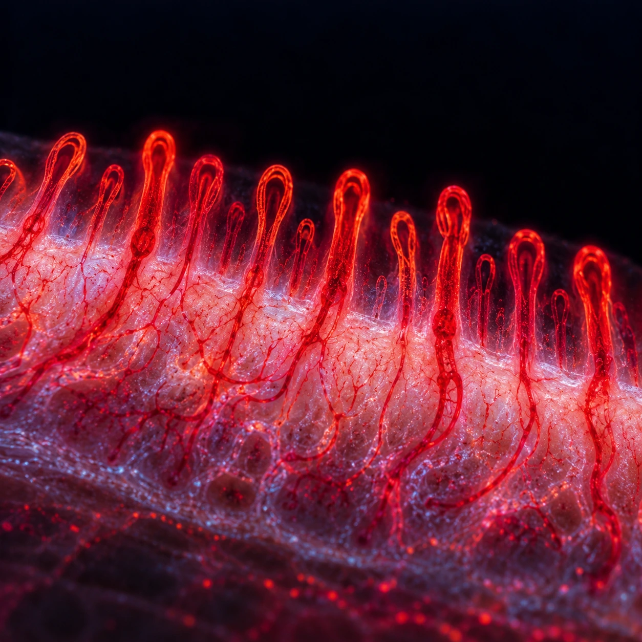
A drop of immersion oil is placed at the base of the fingernail, and the capillaries in the nailfold skin (the skin that runs along the base of the nail) are examined with a dermatoscope, a widefield microscope, or in specialized centers a video capillaroscope at 200x magnification. The ring finger and middle finger are most commonly examined (they have the most consistent capillary patterns). A normal exam takes only a minute per hand.
Normal Pattern (Primary Raynaud's)
Normal nailfold capillaries form regular, evenly spaced U-shaped hairpin loops arranged in parallel rows. Density is uniform — there are no gaps where capillaries are absent. No hemorrhages (small red dots from broken capillaries). This pattern is seen in people without underlying connective tissue disease, and its presence strongly supports a primary Raynaud's diagnosis.
Scleroderma Pattern (Secondary Raynaud's, Especially SSc)
The following abnormalities, alone or in combination, constitute the "scleroderma pattern" and indicate secondary Raynaud's — most often systemic sclerosis or another connective tissue disease:
- Giant capillary loops: Dramatically enlarged, dilated loop tips, often 10 times the normal size, indicating that the remaining capillaries are compensating for the loss of their neighbors by dilating.
- Avascular areas (capillary dropout): Gaps in the capillary architecture where loops have been lost entirely. This reflects irreversible vascular damage.
- Capillary hemorrhages: Small red dots or streaks next to the capillaries where fragile, enlarged loops have ruptured.
- Bushy or branching capillaries: Abnormal neovascularization — the body attempting to regrow vessels to replace those that have been lost.
An "early scleroderma pattern" — enlarged loops only, without dropout yet — may precede the diagnosis of systemic sclerosis by years. This is why capillaroscopy is commonly recommended for all patients with Raynaud's at their first specialist visit: finding the early pattern allows close monitoring and early treatment before organ damage occurs.
Any scleroderma pattern on capillaroscopy, regardless of antibody status, leads to a thorough workup for underlying connective tissue disease.
Diagnosis and Laboratory Workup
The diagnosis of Raynaud's itself is clinical — made from the history and description of the triphasic color change in response to cold or stress. The workup is designed to determine whether the Raynaud's is primary or secondary.
History and Physical Examination
Key questions include: age at onset; sex; whether episodes are bilateral and symmetric; whether there has been any tissue loss (ulcers, pitting scars); what the specific triggers are; complete medication list (beta-blockers, ergotamines, chemotherapy); occupational history (vibrating tools, repetitive palm trauma); and any symptoms of connective tissue disease (dry eyes, dry mouth, rash, joint swelling, skin thickening, shortness of breath, swallowing difficulty).
The examination looks for digital ulcers or pitting scars (an immediate red flag for secondary Raynaud's), skin tightening, joint inflammation, and asymmetry of involvement. Nailfold capillaroscopy can be done at the bedside.
Nailfold Capillaroscopy
As described above, this is the single most informative non-blood test for distinguishing primary from secondary Raynaud's. A normal result strongly supports primary Raynaud's; an abnormal scleroderma pattern leads to further investigation.
Blood Tests
- antinuclear antibody (ANA): The screening test for autoimmune connective tissue disease. A positive antinuclear antibody at significant titer (typically 1:160 or higher) prompts specific antibody testing: anti-Scl-70 (scleroderma, diffuse), anti-centromere (limited SSc/CREST), anti-dsDNA (lupus), anti-SSA/SSB (Sjögren's), anti-U1-RNP (MCTD), anti-Jo-1 and related (myositis).
- Complete blood count (CBC): To detect polycythemia (elevated hematocrit suggesting polycythemia vera).
- Cryoglobulins: Drawn in a pre-warmed tube; if positive, test for hepatitis B and C.
- Cold agglutinins: If cryoglobulinemia or paraproteinemia is suspected.
- thyroid-stimulating hormone (TSH): Exclude hypothyroidism — straightforward, inexpensive, and often overlooked.
- Erythrocyte sedimentation rate (ESR) and C-reactive protein (CRP): Non-specific markers of inflammation that may support a CTD diagnosis.
- Hepatitis B and C serology: Hepatitis C is the most common cause of mixed cryoglobulinemia, which is one of the more treatable secondary causes.
Vascular Studies
Doppler/duplex ultrasound of the upper limb vessels is useful when fixed arterial occlusion is suspected (asymmetric symptoms, absent pulses, suspicion of thoracic outlet syndrome or hypothenar hammer syndrome). It can quantify flow in the radial, ulnar, and digital arteries and detect aneurysm formation at the ulnar artery hook (hypothenar hammer syndrome).
Medication Review
A careful review of all current medications is part of the workup. Stopping a causative drug (beta-blocker, ergotamine) may resolve or substantially reduce Raynaud's without any other treatment.
Treatment — Lifestyle and Medications
Lifestyle Measures (The Cornerstone of Treatment)
For primary Raynaud's, lifestyle measures alone are often sufficient. For secondary Raynaud's, they are an important adjunct to medical treatment.
Warming the whole body, not just the hands. This is described as the most important and most counterintuitive lifestyle principle. When core body temperature drops even slightly, the body responds with sympathetic activation and peripheral vasoconstriction — the hands then cool down even if they are wearing gloves. Wearing a hat (which prevents significant heat loss), layering the torso with an insulated vest or thermal underlayer, and warming the core first is more effective than gloves in isolation. Chemical hand warmers in gloves or pockets provide supplemental local heat.
Reducing cold exposure. Measures people describe include wearing gloves when reaching into the refrigerator or freezer (not just outdoors); requesting that air conditioning be moderated in workplaces; carrying a light cardigan for air-conditioned restaurants and cinemas; using insulated cups for cold drinks; and selecting jobs that minimize cold exposure if Raynaud's is severe.
Stopping smoking. Nicotine directly constricts peripheral blood vessels. Even in primary Raynaud's — where the blood vessels are otherwise healthy — smoking significantly increases the frequency and severity of attacks. There is no safe level of smoking for someone with Raynaud's.
Biofeedback and relaxation techniques. Because emotional stress can trigger attacks through sympathetic activation, biofeedback (learning to consciously raise peripheral finger temperature using a thermal feedback device) has been tested. In the largest trial of primary Raynaud's, the Raynaud's Treatment Study (313 people, followed for one year), finger-temperature biofeedback did not reduce attacks more than a sham (control) biofeedback, while slow-release nifedipine cut colour-chart-confirmed attacks by 66% compared with a dummy pill; 15% of people stopped nifedipine because of side effects. Mindfulness, progressive muscle relaxation, and cognitive behavioral approaches to stress management are also used.
Medication review. Drugs that can cause or worsen Raynaud's are commonly stopped or substituted: non-selective beta-blockers replaced with cardioselective agents or another blood-pressure drug (angiotensin-converting enzyme inhibitor (ACE inhibitor), calcium channel blocker, or angiotensin receptor blocker (ARB)); ergotamine stopped if another migraine preventive is available; stimulants and decongestants reviewed. ACE inhibitors are not neutral here: a 2021 Cochrane review pooling three trials of captopril or enalapril in primary Raynaud's found a possible small increase in attacks per week compared with placebo (low-certainty evidence).
Calcium Channel Blockers (First-Line Pharmacotherapy)
Calcium channel blockers (CCBs) are the best-evidenced and most widely used drugs for Raynaud's. They work by relaxing the smooth muscle in the walls of blood vessels, preventing the vasospasm that causes episodes.
Nifedipine extended-release (30–60 mg once daily) has the strongest evidence base. In the Raynaud's Treatment Study of primary Raynaud's (313 people), it cut confirmed attacks by 66% compared with placebo. In a 2001 meta-analysis of 8 trials in scleroderma-related Raynaud's (109 patients), calcium channel blockers reduced attacks by a mean of 8.3 over two weeks and severity by 35% — an effect the authors called moderate at best. It is taken daily as a preventive medication, not just during acute attacks. Side effects include headache, facial flushing, ankle edema, and hypotension — most common in the first few weeks and often improving with time. The extended-release formulation is better tolerated than immediate-release.
Amlodipine (5–10 mg once daily) and felodipine (5–10 mg once daily) are alternatives with equivalent efficacy and once-daily dosing, which aids adherence. Some patients tolerate one dihydropyridine CCB better than another, so switching within the class is a common approach when side effects are troublesome.
Phosphodiesterase Type 5 (PDE5) Inhibitors
PDE5 inhibitors — best known as erectile dysfunction drugs — promote vasodilation through the cyclic GMP/nitric oxide pathway in blood vessel walls. They are particularly effective in secondary Raynaud's, especially the scleroderma-related form.
Sildenafil (25–50 mg twice daily) and tadalafil (20 mg daily) both have evidence from randomized trials in SSc-related Raynaud's. They reduce the frequency and severity of attacks, and in some studies have been shown to reduce digital ulcer healing time and help prevent new ulcers. Side effects include headache, flushing, and in men, visual changes at higher doses. They can be combined with iloprost infusion for severe disease.
Topical Vasodilators
Glyceryl trinitrate (GTN, nitroglycerin) gel or patch releases nitric oxide locally, relaxing vessel smooth muscle in the area where it is applied. In practice it is applied to the forearm (not directly to the fingertips — the concentration would be too high and cause intense headache and paradoxical irritation). A 0.5% GTN gel (available commercially or compounded by a pharmacy) is applied to the forearm before expected cold exposure. The evidence is weak: a 2021 Cochrane review of four trials of topical nitroglycerin in primary Raynaud's found no evidence of an effect on the number of attacks per week (very low-certainty evidence). A 2009 randomized trial of 219 people with primary or secondary Raynaud's, using a 0.9% nitroglycerin gel (MQX-503) applied at the start of each attack for 4 weeks, found a small improvement in the Raynaud's Condition Score (14.3% vs 1.3% with placebo) but no difference in how often or how long attacks lasted; side effects were similar to placebo. The major limiting side effect is headache, which affects the majority of users and limits long-term adherence.
Alpha-1 Adrenergic Blockers
Prazosin (0.5–1 mg twice daily, titrating slowly to reduce hypotension risk) reduces sympathetic vasoconstrictor tone. It is a second-line option for patients who do not respond to or tolerate CCBs. First-dose hypotension and dizziness are the main side effects — for this reason the first dose is commonly taken lying down at night.
Selective Serotonin Reuptake Inhibitors
Fluoxetine has modest evidence in primary Raynaud's, likely through reduction of serotonin-mediated vasoconstriction (serotonin causes constriction of peripheral vessels). Its role is modest compared to CCBs, but it may be useful in patients who also have anxiety or depression — treating both conditions simultaneously.
What reviews of the other drugs found. A 2021 Cochrane review of 15 trials (635 people) of vasodilator drugs other than calcium channel blockers in primary Raynaud's found small studies, limited data and evidence of very low to moderate certainty; the authors concluded the evidence is insufficient to support vasodilators and suggests they may even worsen the condition.
Treatment of Secondary Raynaud's and Digital Ulcers
When Raynaud's is secondary to a connective tissue disease — particularly systemic sclerosis — the stakes are higher. Digital ulcers are painful, slow to heal, vulnerable to infection (including osteomyelitis of the fingertip bones), and in severe cases can lead to amputation. Specialist treatments beyond CCBs are used in this setting.
A 2019 network meta-analysis of 58 randomized trials (3,867 patients, 91.5% with secondary Raynaud's) compared 15 drug classes. PDE5 inhibitors (sildenafil-type drugs) and calcium channel blockers were the most effective options, with moderate effect and a low level of evidence, and both led to more serious side effects or early drop-outs than placebo (incidence rate ratios 3.30 and 3.13). For attack severity, selective serotonin reuptake inhibitors and oral prostacyclin receptor agonists also beat placebo (very low and low level of evidence). The authors concluded that current evidence does not support any other drug in secondary Raynaud's.
Iloprost Intravenous Infusion (Standard of Care for Severe SSc-Related Raynaud's)
Iloprost is a synthetic prostacyclin analog — prostacyclin is the body's most potent naturally occurring vasodilator and platelet aggregation inhibitor. Intravenous iloprost is the standard of care for severe Raynaud's and digital ulcers in systemic sclerosis.
It is administered as an inpatient infusion over three to five consecutive days, typically every three to six months (or more frequently if ulcers develop). A large multicenter randomized trial (Wigley et al., Ann Intern Med 1994) established that iloprost infusion significantly reduces the frequency and severity of Raynaud's attacks and promotes healing of existing digital ulcers compared to placebo. In that trial of 131 patients, weekly attacks fell 39.1% with iloprost vs 22.2% with placebo, and 92% of people on iloprost had at least one side effect during the infusion vs 57% on placebo. It does not cure the underlying disease but provides a meaningful period of improved perfusion. The main side effects are headache, flushing, nausea, and hypotension during the infusion — managed by slow titration of the infusion rate.
Bosentan (Endothelin Receptor Antagonist)
Bosentan is an oral drug that blocks endothelin — one of the body's most potent vasoconstrictors — at both endothelin receptor subtypes. The RAPIDS-2 trial (Matucci-Cerinic et al., Ann Rheum Dis 2011) was a large randomized double-blind placebo-controlled study showing that bosentan significantly reduced the number of new digital ulcers appearing in SSc patients over the treatment period: across 188 patients over 24 weeks, people on bosentan developed 1.9 new ulcers on average vs 2.7 on placebo (30% fewer). Ankle and leg swelling and raised liver enzymes were linked to bosentan in the trial. Importantly, it did not speed up healing of ulcers that were already present at the start of treatment. Bosentan is therefore most useful as a preventive treatment in patients who keep developing new ulcers despite other therapy. It is approved for this indication in Europe. Liver toxicity is the main risk — monthly liver function tests are required during treatment. It is also teratogenic (causes serious birth defects) and contraindicated in pregnancy.
Riociguat (Soluble Guanylate Cyclase Stimulator)
Riociguat works through the same nitric oxide/cGMP pathway as PDE5 inhibitors but at a different point in the chain — it stimulates the enzyme that produces cGMP directly, even without nitric oxide as a signal. Its only Raynaud's trial so far, the 2018 DIGIT pilot study (20 patients, a single 2 mg dose), found improved finger blood flow in some patients, with large differences from person to person; it has not been tested in longer trials, and the authors called for long-term study. It is already established in pulmonary arterial hypertension (which commonly complicates SSc) and may offer dual benefit in SSc patients with both pulmonary hypertension and severe Raynaud's.
Digital Sympathectomy (Surgical)
In patients with severe digital ischemia — ulcers, threatened amputation — refractory to all medical therapy, surgical digital sympathectomy (stripping the sympathetic nerve fibers that surround the digital arteries) can provide relief. The benefit is real but time-limited: sympathetic reinnervation occurs over months to years, and symptoms gradually return. It is a last resort when other treatments have failed and tissue loss is imminent or ongoing.
Wound Care for Digital Ulcers
Digital ulcers require careful wound management. The principles are: keep the wound clean and moist using non-adherent dressings; protect the ulcer from trauma with padding; monitor for signs of infection (increasing redness, warmth, purulent discharge, fever). Infection is treated with antibiotics covering Staphylococcus aureus — oral antibiotics for superficial infection, intravenous antibiotics and bone biopsy for suspected osteomyelitis of the distal phalanx. Aggressive surgical debridement is generally not done unless necrotic tissue is clearly demarcated and the risk of spreading infection outweighs the risk of healing disruption.
Prognosis
Primary Raynaud's
The long-term outlook for primary Raynaud's is excellent. The vast majority of patients manage their condition well with a combination of lifestyle modifications and, where needed, a calcium channel blocker. There is no risk of tissue loss, organ damage, or shortened life expectancy from primary Raynaud's itself. Symptoms may fluctuate with the seasons (worse in winter, better in summer) and often improve with age. The condition does not in itself progress to a more serious disease.
The one important caveat: in a 1998 pooled analysis of 10 studies (639 people followed for 2,531 patient-years), 12.6% of people first labelled as primary Raynaud's later developed a secondary disease, nearly all connective tissue diseases — on average 2.8 years after study entry and 10.4 years after their Raynaud's began. An abnormal nailfold capillary pattern at entry was the best predictor (47% of people with it went on to a secondary disease); a positive antinuclear antibody predicted much less well (30%). This risk is higher in patients who have a positive antinuclear antibody at first presentation (even with an otherwise normal workup) or who develop any abnormality on follow-up nailfold capillaroscopy. For this reason, a follow-up visit one to two years after diagnosis is often suggested, even in apparently primary cases.
Secondary Raynaud's
The prognosis depends almost entirely on the underlying disease. Raynaud's associated with Sjögren's syndrome or MCTD tends to be milder and carries less risk of tissue loss. Raynaud's in systemic sclerosis carries significant morbidity from digital ulcers — estimates suggest that 30 to 50% of SSc patients with Raynaud's develop at least one digital ulcer over the course of their disease. Early treatment with iloprost infusions and bosentan has substantially reduced the rates of ulcer progression and amputation in specialist centers compared to historical controls.
Red Flags That Warrant Specialist Investigation
- Male sex (primary Raynaud's is rare in men; secondary cause is more likely)
- Age greater than 30 at onset
- Severe or asymmetric episodes
- Any tissue loss — digital ulcers, pitting scars, gangrene
- Abnormal nailfold capillaroscopy (any scleroderma pattern)
- Positive antinuclear antibody at meaningful titer
- Systemic symptoms — joint swelling, rash, skin tightening, dry eyes/mouth, shortness of breath on exertion
Any of these features is a usual reason for referral to a rheumatologist (a specialist in joint and autoimmune disease) for full evaluation, even if the initial presentation appeared to be primary Raynaud's.
Key Research Papers
- Wigley FM, Flavahan NA. Raynaud's Phenomenon. N Engl J Med. 2016;375(6):556–565. — PubMed PMID: 27509103
- Garner R, Kumari R, Lanyon P, Doherty M, Zhang W. Prevalence, risk factors and associations of primary Raynaud's phenomenon: systematic review and meta-analysis of observational studies. BMJ Open. 2015;5(3):e006389. — PubMed PMID: 25776043
- Herrick AL. The pathogenesis, diagnosis and treatment of Raynaud phenomenon. Nat Rev Rheumatol. 2012;8(8):469–479. — PubMed PMID: 22782008
- Cutolo M, Sulli A, Smith V. How to perform and interpret capillaroscopy. Best Pract Res Clin Rheumatol. 2013;27(2):237–248. — PubMed PMID: 23731933
- Matucci-Cerinic M, Denton CP, Furst DE, et al. Bosentan treatment of digital ulcers related to systemic sclerosis: results from the RAPIDS-2 randomised, double-blind, placebo-controlled trial. Ann Rheum Dis. 2011;70(1):32–38. — PubMed PMID: 20805294
- Fries R, Shariat K, von Wilmowsky H, Böhm M. Sildenafil in the treatment of Raynaud's phenomenon resistant to vasodilatory therapy. Circulation. 2005;112(19):2980–2985. — PubMed PMID: 16275885
- Baumhäkel M, Böhm M. Recent achievements in the management of Raynaud's phenomenon. Vasc Health Risk Manag. 2010;6:207–214. — PubMed PMID: 20407628
- Thompson AE, Shea B, Welch V, Fenlon D, Pope JE. Calcium-channel blockers for Raynaud's phenomenon in systemic sclerosis. Arthritis Rheum. 2001;44(8):1841–1847. — PubMed PMID: 11508437
- Cooke JP, Marshall JM. Mechanisms of Raynaud's disease. Vasc Med. 2005;10(4):293–307. — PubMed PMID: 16444858
- Lambova SN, Müller-Ladner U. The role of capillaroscopy in differentiation of primary and secondary Raynaud's phenomenon in rheumatic diseases: a review of the literature and two case reports. Rheumatol Int. 2009;29(11):1263–1271. — PubMed PMID: 19547979
- Chung L, Shapiro L, Fiorentino D, et al. MQX-503, a novel formulation of nitroglycerin, improves the severity of Raynaud's phenomenon: a randomized, controlled trial. Arthritis Rheum. 2009;60(3):870–877. — PubMed PMID: 19248104
- Wigley FM, Wise RA, Seibold JR, et al. Intravenous iloprost infusion in patients with Raynaud phenomenon secondary to systemic sclerosis. A multicenter, placebo-controlled, double-blind study. Ann Intern Med. 1994;120(3):199–206. — PubMed PMID: 7506013
- [No authors listed]. Comparison of sustained-release nifedipine and temperature biofeedback for treatment of primary Raynaud phenomenon. Results from a randomized clinical trial with 1-year follow-up. Arch Intern Med. 2000;160(8):1101–1108. — PubMed PMID: 10789602
- Spencer-Green G. Outcomes in primary Raynaud phenomenon: a meta-analysis of the frequency, rates, and predictors of transition to secondary diseases. Arch Intern Med. 1998;158(6):595–600. — PubMed PMID: 9521223
- Su KY, Sharma M, Kim HJ, Kaganov E, Hughes I, Abdeen MH, Ng JHK. Vasodilators for primary Raynaud's phenomenon. Cochrane Database Syst Rev. 2021;5(5):CD006687. — PubMed PMID: 33998674
- Khouri C, Lepelley M, Bailly S, et al. Comparative efficacy and safety of treatments for secondary Raynaud's phenomenon: a systematic review and network meta-analysis of randomised trials. Lancet Rheumatol. 2019;1(4):e237–e246. — PubMed PMID: 38229380
- Huntgeburth M, Kießling J, Weimann G, Wilberg V, Saleh S, Hunzelmann N, Rosenkranz S. Riociguat for the Treatment of Raynaud's Phenomenon: A Single-Dose, Double-Blind, Randomized, Placebo-Controlled Cross-Over Pilot Study (DIGIT). Clin Drug Investig. 2018;38(11):1061–1069. — PubMed PMID: 30238433
- Mohokum M, Hartmann P, Schlattmann P. The association of Raynaud syndrome with β-blockers: a meta-analysis. Angiology. 2012;63(7):535–540. — PubMed PMID: 22261435
Search PubMed for more: Raynaud's phenomenon treatment Raynaud's scleroderma digital ulcers nailfold capillaroscopy Raynaud's
Connections
- Cardiology
- Buerger's Disease (Thromboangiitis Obliterans)
- Peripheral Artery Disease
- Atherosclerosis
- Carotid Artery Stenosis
- Postural Orthostatic Tachycardia Syndrome (Dysautonomia)
- Lupus (Systemic Lupus Erythematosus)
- Sjögren's Syndrome
- Mixed Connective Tissue Disease
- Systemic Sclerosis (Scleroderma)
- All Conditions
- Rheumatology
- Cryotherapy Safety, Risks and Contraindications
- Discussed in depth on these pages: Raynaud's Disease