Cardiac Tamponade
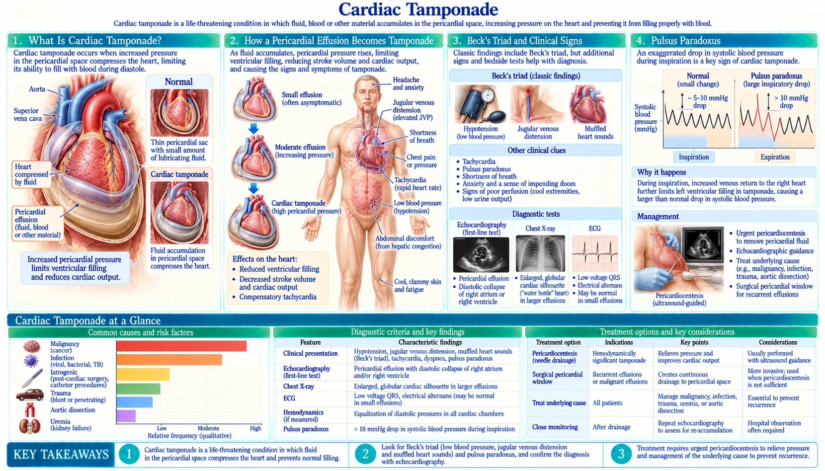
Table of Contents
- Cardiac Tamponade Explained
- How a Pericardial Effusion Becomes Tamponade
- Beck's Triad and Clinical Signs
- Pulsus Paradoxus
- Kussmaul's Sign: The Key Distinction from Constrictive Pericarditis
- Electrocardiogram and Echocardiography Findings
- Common Causes of Pericardial Effusion and Tamponade
- Treatment: Pericardiocentesis
- Post-Procedure Care and Monitoring
- Key Research Papers
- Connections
- Featured Videos
Cardiac Tamponade Explained
Cardiac tamponade is a life-threatening emergency in which fluid accumulates in the pericardial sac — the fibrous envelope surrounding the heart — and compresses the heart so severely that it can no longer fill adequately between beats. As cardiac output collapses, every organ in the body is starved of blood, leading rapidly to cardiogenic shock and death if the fluid is not drained.
Tamponade is not defined by the volume of fluid but by the pressure it creates. A slowly accumulating effusion (e.g., from cancer or hypothyroidism) may grow to 1,000–2,000 mL without causing tamponade because the pericardium stretches gradually. A sudden bleed of only 150–200 mL into a non-distensible pericardium — as in aortic dissection or cardiac trauma — can cause tamponade within minutes.
How a Pericardial Effusion Becomes Tamponade
The pericardium is a double-walled sac with a thin layer of fluid (normally 15–50 mL) that lubricates heart movement. It is relatively inelastic. When fluid accumulates faster than the pericardium can stretch, the intrapericardial pressure rises.
The critical physiological event in tamponade is equalization of filling pressures: as pericardial pressure rises, it begins to approach — and then match — the diastolic pressures inside each cardiac chamber. When intrapericardial pressure equals or exceeds right atrial (RA) pressure, the RA collapses during diastole. When it rises further to match right ventricular (RV) diastolic pressure, the RV collapses. This external compression prevents the right side of the heart from accepting venous return. Less blood enters the right heart → less gets pumped to the lungs → less arrives in the left heart → cardiac output falls.
A second mechanism is exaggerated ventricular interdependence. In tamponade the heart is enclosed in a fixed volume of fluid. When the right ventricle expands during inspiration (as venous return increases), the interventricular septum bows leftward into the left ventricle, reducing left ventricular filling. This is the anatomical basis for pulsus paradoxus (see below).
As cardiac output falls, compensatory mechanisms activate: heart rate rises (tachycardia), systemic vascular resistance increases (vasoconstriction), and catecholamines surge. These responses can transiently maintain blood pressure but are eventually overwhelmed — the terminal event is cardiovascular collapse.
Beck's Triad and Clinical Signs
In 1935, cardiac surgeon Claude Beck described three physical examination findings that, together, point to acute cardiac tamponade:
- Hypotension — systolic blood pressure is low and falling as cardiac output declines. In early or subacute tamponade, the blood pressure may still be maintained by compensatory tachycardia and vasoconstriction.
- Muffled (distant) heart sounds — the fluid surrounding the heart dampens the transmission of heart sounds to the chest wall. The sound is like hearing the heart through a wet pillow.
- Jugular venous distension (JVD) — as the right heart cannot accept venous return, blood backs up in the systemic veins. The external jugular veins and internal jugular pulsations become visibly prominent with the patient at 45°. Central venous pressure is elevated.
Beck's triad is present in its complete classic form in only 10–40% of cases, particularly in the acute traumatic setting. In subacute or chronic tamponade (e.g., from malignancy), patients often present with progressive dyspnea, fatigue, and weight gain — symptoms that mimic heart failure — long before frank hemodynamic collapse. Other clinical findings include: tachycardia (a fast pulse, found in about 77% of people with tamponade in a 2007 review), narrowed pulse pressure (<25 mmHg), and cool extremities from vasoconstriction.
A 2007 review of the bedside signs (Roy and colleagues, 8 studies) found that most people with tamponade are short of breath (87–89%), have a fast pulse (77%), have pulsus paradoxus (82%) or have raised neck-vein pressure (76%), but a minority have none of these. In one study within that review, a pulsus paradoxus above 10 mmHg in a person with fluid around the heart made tamponade about 3 times more likely (likelihood ratio 3.3), while 10 mmHg or less made it very unlikely (likelihood ratio 0.03). The review notes that confirming tamponade still needs further testing.
Pulsus Paradoxus
Pulsus paradoxus is defined as an inspiratory drop in systolic blood pressure of more than 10 mmHg. Despite the name ("paradox"), it is actually an exaggeration of a normal physiological phenomenon — normally, inspiration causes a small drop of <10 mmHg in systolic blood pressure (BP) because the expanding lungs temporarily pool blood in the pulmonary vasculature, transiently reducing left heart filling.
In tamponade, this normal respiratory variation is amplified by the exaggerated ventricular interdependence described above: during inspiration, the right ventricle expands and its septum bows left, squeezing the left ventricle and causing a dramatic fall in left ventricular (LV) stroke volume and systolic blood pressure.
How Pulsus Paradoxus Is Measured
- The blood pressure cuff is inflated above systolic pressure.
- It is slowly deflated while the patient breathes normally.
- The pressure at which Korotkoff sounds first become audible is noted — they appear only during expiration.
- Deflation continues; the pressure at which sounds are heard throughout both inspiration and expiration is noted.
- The difference between these two pressures is the pulsus paradoxus. More than 10 mmHg is abnormal.
A pulsus >25 mmHg is highly specific for severe tamponade. Note that pulsus paradoxus can be absent in tamponade complicated by severe aortic regurgitation, atrial septal defect, or severe LV dysfunction (the left ventricle is already too stiff to show additional inspiratory reduction).
Pulsus paradoxus is also seen in conditions other than tamponade: severe asthma, tension pneumothorax, large pulmonary embolism, and constrictive pericarditis (though the sign is less reliable in constrictive pericarditis).
Kussmaul's Sign: The Key Distinction from Constrictive Pericarditis
Cardiac tamponade and constrictive pericarditis can both present with JVD, dyspnea, and hemodynamic compromise. A single physical finding helps distinguish them: Kussmaul's sign.
Kussmaul's sign is a paradoxical rise (or failure to fall) in jugular venous pressure (JVP) with inspiration. Normally, inspiration lowers intrathoracic pressure and draws venous blood into the chest, reducing JVP. In constrictive pericarditis, the scarred, rigid pericardium cannot expand to accommodate the increased venous return that inspiration creates — so JVP rises instead of falling.
- Cardiac tamponade: Kussmaul's sign is ABSENT. In tamponade, the pericardium is not rigid — it is distended with fluid. Inspiration still draws venous blood into the right heart, causing the normal inspiratory JVP fall. The problem in tamponade is extrinsic compression, not inability to expand.
- Constrictive pericarditis: Kussmaul's sign is PRESENT. The calcified or fibrotic pericardial shell cannot expand with inspiration, so venous blood has nowhere to go — JVP rises.
This distinction is clinically critical because the two conditions have very different treatments: tamponade requires immediate fluid drainage; constrictive pericarditis requires pericardiectomy (surgical removal of the pericardium) after appropriate evaluation. A JVP that rises on inspiration in a person with pericardial disease points first toward constrictive pericarditis.
The pericardial knock — a high-pitched early diastolic sound heard in constrictive pericarditis when the ventricle fills rapidly and hits the rigid pericardium — is another distinguishing feature absent in tamponade.
Electrocardiogram and Echocardiography Findings
Electrocardiogram (ECG)
The electrocardiogram in cardiac tamponade shows two classic findings:
- Low-voltage QRS — all QRS complexes are small (<5 mm in limb leads, <10 mm in precordial leads) because the pericardial fluid insulates the heart and dampens the electrical signal reaching the chest electrodes.
- Electrical alternans — the QRS height and/or axis alternates every other beat (or in a fixed pattern). This occurs because the heart swings back and forth within the fluid-filled pericardial sac with each beat, changing its spatial orientation relative to the body surface electrodes. Electrical alternans is not always present but is highly specific for large pericardial effusion with tamponade physiology when seen.
Sinus tachycardia is common as a compensatory response (a fast pulse was found in about 77% of people with tamponade in a 2007 review). PR depression and diffuse ST elevation (concave upward, in the saddle pattern) may be present if the effusion is inflammatory in origin (pericarditis).
Echocardiography — The Definitive Bedside Test
Bedside echocardiography (echo) is the most important diagnostic tool for cardiac tamponade. Key findings:
- Pericardial effusion — identified as an echo-free (dark) space around the heart. Circumferential effusions are more likely to cause tamponade than loculated ones.
- Diastolic collapse of the right atrium (RA) — the RA wall inverts (buckles inward) during diastole when pericardial pressure exceeds RA pressure. This is an early and sensitive sign of hemodynamic compromise. Duration of RA collapse >30% of the cardiac cycle strongly predicts tamponade.
- Diastolic collapse of the right ventricle (RV) — RV free wall indentation during diastole; more specific for tamponade than RA collapse. The RV is more muscular than the RA so it collapses later, signaling more advanced compression.
- Exaggerated respiratory variation in valve flow velocities — Doppler shows >25% variation in mitral inflow E-wave velocity and >40% variation in tricuspid inflow with respiration, the echo correlate of pulsus paradoxus.
- Inferior vena cava (IVC) plethora — a dilated IVC (>2.1 cm) that does not collapse with inspiration (<50% collapse) reflects elevated right atrial pressure; found in tamponade and also right heart failure.
- "Swinging heart" — visual back-and-forth motion of the heart within the effusion, the anatomical basis for electrical alternans.
Point-of-care ultrasound (POCUS) performed at the bedside by the treating physician has transformed tamponade diagnosis — it allows immediate confirmation within minutes of clinical suspicion.
Common Causes of Pericardial Effusion and Tamponade
Any condition that causes pericardial inflammation, direct cardiac injury, or fluid accumulation near the heart can cause an effusion. When the effusion accumulates fast enough or large enough, tamponade results:
- Malignancy — one of the most common causes of large pericardial effusions in hospitalized patients. In a Mayo Clinic series of drainages from 1979 to 2000, heart surgery replaced cancer as the leading cause of effusions needing drainage in the later years, and the two together with catheter-procedure injuries accounted for nearly 70% of drainages. Lung cancer, breast cancer, lymphoma, leukemia, and melanoma commonly metastasize to the pericardium. Effusions may be hemorrhagic.
- Idiopathic or viral pericarditis — the most common cause overall in outpatients; usually inflammatory, responds to nonsteroidal anti-inflammatory drugs (NSAIDs) and colchicine; tamponade is uncommon but occurs with large effusions.
- Uremic pericarditis — in patients with advanced renal failure (typically creatinine >10 mg/dL or on dialysis); the fluid is often hemorrhagic; dialysis is the treatment of the uremia, but pericardiocentesis is needed if tamponade develops.
- Post-cardiac surgery (post-pericardiotomy syndrome) — immune-mediated inflammatory response 1–6 weeks after cardiac surgery; can cause effusion and tamponade; loculated effusions may compress only one chamber, making diagnosis harder.
- Aortic dissection (Type A) — blood tracks backward from the dissection into the pericardial space; this is the most lethal cause of tamponade (mortality >90% without emergency surgery). The effusion is purely hemorrhagic. Pericardiocentesis is relatively contraindicated in aortic dissection tamponade — draining the blood re-opens the communication and accelerates bleeding; emergency cardiac surgery is the definitive treatment.
- Cardiac trauma — blunt chest trauma (motor vehicle accidents, falls), penetrating trauma (stab wounds, gunshot wounds), or iatrogenic injury (cardiac catheterization, pacemaker lead perforation, electrophysiology procedures) can cause rapid hemorrhagic tamponade.
- Hypothyroidism — severe myxedema causes slowly accumulating serous effusions that can become very large without causing tamponade due to the gradual stretching of the pericardium; treated with thyroid hormone replacement.
- Autoimmune diseases — systemic lupus erythematosus (SLE), rheumatoid arthritis, and scleroderma can cause inflammatory pericarditis and effusions.
- Bacterial pericarditis — purulent (pus-filled) effusions from bacterial infection; requires drainage and antibiotics; associated with high mortality.
- Radiation therapy — mediastinal radiation (for lymphoma, lung cancer, esophageal cancer) can cause pericarditis and effusion months to years after treatment.
Treatment: Pericardiocentesis
Cardiac tamponade is treated by physically removing the fluid — called pericardiocentesis. It is a life-saving procedure, and hospitals treat it as urgent.
Pre-Procedure Preparation
- Hospital teams usually place an intravenous (IV) line and may give a cautious fluid bolus (250–500 mL normal saline) to temporarily increase preload and support blood pressure while preparing for pericardiocentesis; standard practice is not to hold up the drainage for fluids.
- Breathing machines (positive pressure ventilation) raise the pressure inside the chest and can trigger cardiovascular collapse in tamponade, so teams keep them to a minimum where possible.
- A bedside echo is set up to guide needle placement.
Subxiphoid (Xiphoid-Approach) Pericardiocentesis
The classic blind approach enters below the xiphoid process at a 45° angle directed toward the left shoulder. The needle is advanced while applying gentle negative pressure with the syringe; aspiration of bloody or serous fluid confirms entry. An electrocardiogram lead attached to the needle detects ST elevation or current-of-injury if the myocardium is contacted, the signal to withdraw the needle slightly.
This approach is now largely replaced by echo-guided pericardiocentesis in non-emergency situations.
Echo-Guided Pericardiocentesis
The preferred modern approach uses real-time echocardiography to identify the largest, most accessible fluid pocket and confirm needle position before aspiration. The apical or subcostal window is commonly used. In a Mayo Clinic registry of 1,127 echo-guided drainages in 977 patients over 21 years (1979–2000), the procedure worked 97% of the time; 4.7% had a complication (1.2% serious, 3.5% minor). Leaving a catheter in for extended drainage rose from 23% of cases in the first period to 75% in the last, while recurrences and the need for heart surgery fell significantly. A drainage catheter (pigtail catheter) is left in the pericardial space for 24–48 hours to ensure complete drainage and allow monitoring for reaccumulation.
Surgical Drainage
Surgical pericardial drainage (via a subxiphoid pericardial window, left anterior thoracotomy, or median sternotomy) is indicated when:
- Pericardiocentesis is technically impossible or has failed
- The effusion is loculated or posterior (echo-guided drainage may not be feasible)
- Aortic dissection is the cause (emergency surgery is the primary treatment)
- Purulent pericarditis requires surgical debridement
- A pericardial window is needed to prevent recurrence (especially in malignancy)
In malignant effusions, a pericardial window that drains fluid into the pleural space or peritoneal cavity prevents reaccumulation without requiring indefinite catheter drainage. Intrapericardial sclerotherapy (instillation of bleomycin or cisplatin) is an alternative for recurrent malignant effusions.
In 2025 the European Society of Cardiology published new guidelines that cover pericarditis and myocarditis together (Schulz-Menger and colleagues), a decade after its 2015 guideline on pericardial diseases.
Post-Procedure Care and Monitoring
After pericardiocentesis, haemodynamic improvement is often dramatic and immediate — blood pressure rises, heart rate slows, and JVD resolves as cardiac output is restored. The underlying cause of the effusion still has to be found and treated:
- Pericardial fluid analysis — the drained fluid is usually tested for cell count and differential, glucose, lactate dehydrogenase (LDH), protein (Light's criteria adapted for pericardial fluid), culture and sensitivity, cytology (for malignancy), acid-fast bacilli (AFB) smear and culture (for tuberculosis), and specific antibody tests as indicated.
- Echocardiogram after drainage — checks for residual effusion, left and right ventricular (LV/RV) function, and wall motion abnormalities (which might indicate concomitant myocarditis or takotsubo cardiomyopathy).
- Monitoring for reaccumulation — especially in malignant, uremic, or inflammatory etiologies; the pigtail catheter drainage over 24–48 hours reduces risk of early reaccumulation.
- Anti-inflammatory therapy — if the cause is pericarditis (viral/idiopathic), standard practice is nonsteroidal anti-inflammatory drugs (ibuprofen 600 mg three times a day or aspirin 750–1000 mg three times a day) plus colchicine. In the ICAP trial (240 adults with acute pericarditis), colchicine 0.5 mg twice a day (once a day for people weighing 70 kg or less) for 3 months, added to aspirin or ibuprofen, cut ongoing or returning pericarditis from 37.5% to 16.7%, symptoms still present at 72 hours from 40.0% to 19.2%, and recurrences per patient from 0.52 to 0.21, with no serious adverse events. In the earlier COPE trial (120 patients with a first attack), 8.3% stopped colchicine because of diarrhea, and steroid treatment in the first attack was linked to more recurrences.
- Post-pericardiotomy syndrome after cardiac surgery — fever, pleuritic chest pain, and new effusion 1–6 weeks post-op; treated with nonsteroidal anti-inflammatory drugs ± colchicine; usually self-limiting.
Key Research Papers
The following citations are from published peer-reviewed literature on cardiac tamponade, pericardial effusion, and pericardiocentesis.
- Adler Y, Charron P, Imazio M, et al. 2015 ESC Guidelines for the diagnosis and management of pericardial diseases. Eur Heart J. 2015;36(42):2921-2964. — PubMed PMID: 26320112
- Schulz-Menger J, Collini V, Gröschel J, et al. 2025 ESC Guidelines for the management of myocarditis and pericarditis. Eur Heart J. 2025;46(40):3952-4041. — PubMed PMID: 40878297
- Imazio M, Adler Y. Management of pericardial effusion. Eur Heart J. 2013;34(16):1186-1197. — PubMed PMID: 23125278
- Search PubMed (Cardiac tamponade: pathophysiology, clinical features, and management)
- Tsang TS, Enriquez-Sarano M, Freeman WK, et al. Consecutive 1127 therapeutic echocardiographically guided pericardiocenteses: clinical profile, practice patterns, and outcomes spanning 21 years. Mayo Clin Proc. 2002;77(5):429-436. — PubMed PMID: 12004992
- Roy CL, Minor MA, Brookhart MA, Choudhry NK. Does this patient with a pericardial effusion have cardiac tamponade? JAMA. 2007;297(16):1810-1818. — PubMed PMID: 17456823
- Imazio M, Bobbio M, Cecchi E, et al. Colchicine in addition to conventional therapy for acute pericarditis: results of the COlchicine for acute PEricarditis (COPE) trial. Circulation. 2005;112(13):2012-2016. — PubMed PMID: 16186437
- Imazio M, Brucato A, Cemin R, et al. A randomized trial of colchicine for acute pericarditis (ICAP). N Engl J Med. 2013;369(16):1522-1528. — PubMed PMID: 23992557
- Reddy PS, Curtiss EI, O'Toole JD, Shaver JA. Cardiac tamponade: hemodynamic observations in man. Circulation. 1978;58(2):265-272. — PubMed PMID: 668074
- Maisch B, Seferović PM, Ristić AD, et al. Guidelines on the diagnosis and management of pericardial diseases executive summary; the Task Force on the Diagnosis and Management of Pericardial Diseases of the European Society of Cardiology. Eur Heart J. 2004;25(7):587-610. — PubMed PMID: 15120056
- Burazor I, Imazio M, Markel G, Adler Y. Malignant pericardial effusion. Cardiology. 2013;124(4):224-232. — PubMed PMID: 23571453
- Fitch MT, Nicks BA, Pariyadath M, McGinnis HD, Manthey DE. Videos in clinical medicine. Emergency pericardiocentesis. N Engl J Med. 2012;366(12):e17. — PubMed PMID: 22435385
- McDonagh TA, Metra M, Adamo M, et al. 2021 ESC Guidelines for the diagnosis and treatment of acute and chronic heart failure. Eur Heart J. 2021;42(36):3599-3726. — PubMed PMID: 34447992
Connections
- Cardiology
- Pericarditis
- Constrictive Pericarditis
- Heart Failure
- Cardiovascular Disease
- Myocardial Infarction
- Coronary Artery Disease
- Cardiomyopathy
- Ventricular Tachycardia
- HFpEF
- Atrial Fibrillation
- Valvular Heart Disease
- Pulmonary Embolism
- Chest Pain
- Shortness of Breath
- Hypotension
- Chronic Kidney Disease