Taurine for Bile Acids and Fat Digestion
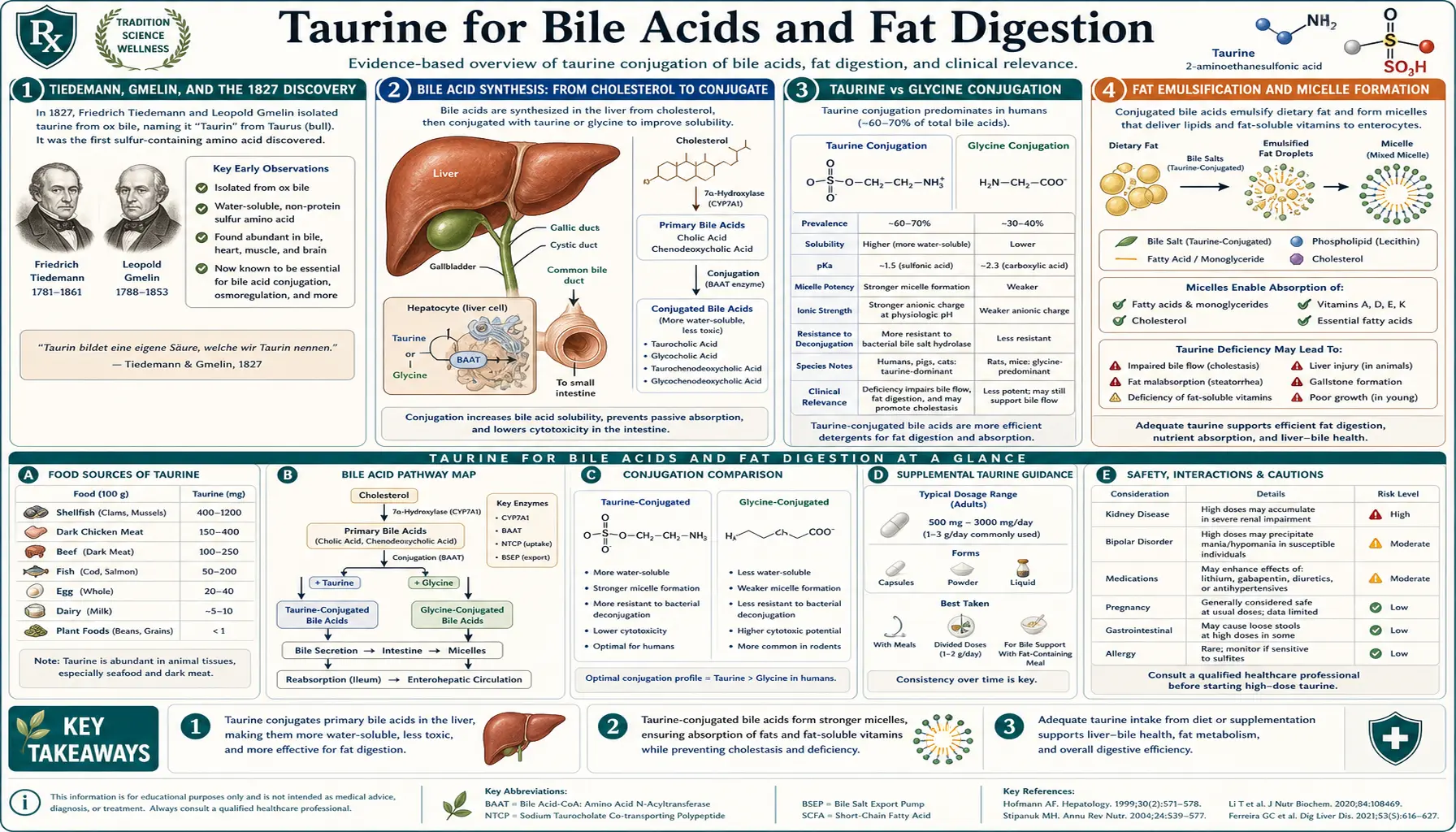
Taurine was first described in ox bile in 1827 by Friedrich Tiedemann and Leopold Gmelin, and the name taurine — from taurus, Latin for “bull” — came into use with H. Demarçay's 1838 study of bile. For a long time, conjugation with bile acids was taurine's only clearly recognized biochemical role (Gaull 1989). In the liver, a single enzyme joins primary bile acids to either glycine or taurine (Falany and colleagues 1994), forming bile salts such as taurocholic acid and taurochenodeoxycholic acid, the detergents that help emulsify dietary fat and absorb the fat-soluble vitamins A, D, E, and K. Because taurine carries a strongly acidic sulfonic acid group, its conjugates stay ionized at a lower pH than glycine conjugates. This deep-dive walks through bile acid synthesis, the taurine-versus-glycine conjugation choice, the enterohepatic circulation, bile acids as signaling molecules, and what is and is not known about taurine in cholestasis, gallbladder removal, and fat malabsorption.
Table of Contents
- Tiedemann, Gmelin, and the 1827 Discovery
- Bile Acid Synthesis: From Cholesterol to Conjugate
- Taurine vs Glycine Conjugation
- Fat Emulsification and Micelle Formation
- Absorption of Vitamins A, D, E, and K
- The Enterohepatic Circulation and ASBT
- Choleresis and Biliary Flow Dynamics
- Cholestasis: Intrahepatic and Extrahepatic
- After Gallbladder Removal
- FXR and TGR5: Bile Acids as Hormones
- Cholesterol Gallstones and Tauroursodeoxycholic Acid
- Doses Used in Bile-Acid Research
- Safety Findings
- Key Research Papers
- Connections
- Featured Videos
Tiedemann, Gmelin, and the 1827 Discovery
In 1827, the German physiologist Friedrich Tiedemann and the chemist Leopold Gmelin, working in Heidelberg, reported new constituents of ox bile in the journal Annalen der Physik (“Einige neue Bestandtheile der Galle des Ochsen”), among them the crystalline substance later called taurine. Standard accounts credit the chemist H. Demarçay with first using the name “taurine” in 1838, the year his study of the nature of bile (“Ueber die Natur der Galle”) appeared in the Annalen der Pharmacie. The name comes from taurus, Latin for bull or ox, after the ox bile in which the substance was found.
Tiedemann and Gmelin's discovery established the connection between bile and taurine that the molecule's name still records. They could not have known the biochemistry: taurine is not itself a bile acid but one of the two amino acids the liver attaches to bile acids (Falany and colleagues 1994).
For more than a century after the discovery, taurine's biological role was thought to be limited to bile acid conjugation. The molecule was dismissed as a dead-end product of sulfur metabolism, biologically inactive in its own right. Only in the second half of the twentieth century did the broader functions of taurine begin to be recognized — the cardiac role (see Cardiac Function), the retinal role (see Retinal Health), the osmolyte role (see Electrolyte Balance). But the bile acid conjugation function, the one Tiedemann and Gmelin glimpsed in 1827, remains one of taurine's most consequential biological activities.
Bile Acid Synthesis: From Cholesterol to Conjugate
Bile acids are synthesized in the liver from cholesterol via two main pathways:
- The classical (neutral) pathway begins with cholesterol 7-alpha-hydroxylase (CYP7A1), the rate-limiting enzyme of bile acid synthesis. Through a series of about 15 enzymatic steps, cholesterol is hydroxylated, side-chain shortened, and modified to produce the two primary human bile acids: cholic acid (a trihydroxy bile acid) and chenodeoxycholic acid (CDCA) (a dihydroxy bile acid). This pathway accounts for most bile acid synthesis under normal conditions.
- The alternative (acidic) pathway begins with sterol 27-hydroxylase (CYP27A1) and produces predominantly CDCA. It accounts for a smaller share under normal conditions.
The free primary bile acids cholic acid and CDCA are amphipathic molecules — they have a hydrophobic steroid backbone and a hydrophilic carboxylate side chain. They are mild detergents in this form, but they are not optimal: they remain weakly soluble at acidic pH, they are partially toxic to the bile duct epithelium, and they precipitate at calcium concentrations found in the gallbladder. To convert them into functional bile salts, the liver conjugates them with either taurine or glycine via the enzyme bile acid-CoA:amino acid N-acyltransferase (BAAT). The conjugation reaction:
Bile acid + Taurine (or Glycine) ⟶ Taurocholic acid / Taurochenodeoxycholic acid (or Glyco-conjugates) + H&sub2;O
The conjugation reaction adds an amide bond between the carboxylate of the bile acid and the amine of taurine (or glycine), producing a sulfonic acid (taurine) or carboxylic acid (glycine) terminal group. This dramatically increases polarity: the pKa of the sulfonic acid terminal in taurocholic acid is approximately 1.5, meaning it remains fully ionized (negatively charged) even at the acidic pH of the duodenum. The pKa of glycocholic acid is roughly 3.9, meaning a substantial fraction becomes protonated and less soluble at low pH.
The functional advantage of taurine conjugation is therefore most pronounced in the upper small intestine, where pH is most acidic (chyme just leaving the stomach). Taurine-conjugated bile salts remain fully active throughout the entire small intestine; glycine-conjugated bile salts are partially compromised at the duodenum and proximal jejunum.
Taurine vs Glycine Conjugation
In humans, a single liver enzyme, bile acid CoA:amino acid N-acyltransferase (BAAT), conjugates bile acids with either glycine or taurine (Falany and colleagues 1994). How dietary glycine and taurine affect bile acid conjugation in people was studied as early as 1959 (Sjövall 1959).
Several factors influence the taurine-to-glycine ratio of bile:
- Dietary taurine intake. Taurine comes from animal foods; plant foods contain none that is detectable (Laidlaw and colleagues 1990), and vegans have negligible taurine intake and lower plasma taurine (Laidlaw and colleagues 1988).
- Endogenous taurine synthesis. The body makes taurine from methionine and cysteine in the presence of vitamin B6 (Lourenço and Camilo 2002).
- Bile acid demand. High-fat meals draw on the bile salt pool, and chronic fat intake affects bile acid synthesis.
- Liver disease. Cholestatic liver disease changes bile composition in complex ways.
Both kinds of conjugate work as detergents. Because of the lower pKa of the sulfonic acid group, taurine conjugates are:
- More polar and more strongly anionic
- More fully ionized, and therefore more soluble, at the acidic pH of the upper small intestine
Clinical evidence that taurine supplements improve fat absorption comes from preterm infants and children with cystic fibrosis (Gaull 1989). Trials in healthy adults, vegans, or people after gallbladder removal are lacking.
Fat Emulsification and Micelle Formation
Dietary fat enters the small intestine as large droplets, dispersed by the mechanical action of gastric motility but still too large for the lipase enzymes of the pancreas to attack efficiently. Bile salts solve this problem by acting as detergents: their amphipathic structure (hydrophilic head, hydrophobic steroid body) allows them to align at the oil-water interface, breaking the large fat droplets into much smaller ones — a process called emulsification.
Once emulsified into droplets a few micrometers across, the fat presents a vastly larger surface area for pancreatic lipase to digest. Lipase cleaves triglycerides into free fatty acids and 2-monoglycerides. These products plus the bile salts plus cholesterol plus phospholipids assemble into mixed micelles — small (about 5 nanometer) spherical aggregates with the hydrophobic chains tucked into the center and the hydrophilic groups facing the aqueous lumen. The mixed micelle is the form in which fat-digestion products approach the intestinal epithelium for absorption.
At the brush border, the mixed micelle releases its contents into the unstirred water layer adjacent to the epithelial cell. Free fatty acids and monoglycerides diffuse across the brush-border membrane (partly via passive diffusion, partly via specific transporters like CD36 and FATP4). Inside the enterocyte, they are reassembled into triglycerides, packaged into chylomicrons with phospholipids, cholesterol esters, and apolipoprotein B-48, and released into the lymphatic system via the lacteal vessels of the villi. From the thoracic duct, the chylomicrons enter the bloodstream, where they distribute dietary fat to adipose tissue, muscle, and liver.
The bile salts themselves are left behind in the intestinal lumen. They continue downstream through the jejunum and ileum, where they are reabsorbed (see Enterohepatic Circulation below) and returned to the liver for re-use. The entire bile salt pool circulates 6 to 8 times per day — meaning the bile salts present at the start of a meal will help digest several subsequent meals before being lost.
Without adequate bile salt function, fat emulsification fails, micelle formation is impaired, and a substantial fraction of dietary fat passes undigested into the colon. This produces the syndrome of steatorrhea: bulky, greasy, foul-smelling, hard-to-flush stools, often associated with bloating, urgency, and weight loss despite normal caloric intake. Patients with bile acid problems frequently describe the floating stools that result from unabsorbed fat trapping gas, the oily film on the toilet water, and the cramping bloated discomfort an hour or two after fatty meals.
Absorption of Vitamins A, D, E, and K
The fat-soluble vitamins (A, D, E, K) are absorbed by the same mixed-micelle pathway as dietary fat. Inadequate bile salt function therefore produces, over time, deficiencies of all four fat-soluble vitamins — even when dietary intake is theoretically sufficient.
The clinical implications vary by vitamin:
- Vitamin A: deficiency presents earliest as night blindness (impaired rhodopsin regeneration), then progressing through xerophthalmia (dry conjunctiva), Bitot's spots, and ultimately corneal ulceration with permanent blindness in severe cases. See our Vitamin A Benefits hub for the full clinical picture.
- Vitamin D: deficiency drives bone loss (osteomalacia in adults, rickets in children), impaired calcium absorption, increased fracture risk, and a broad range of immune and metabolic consequences.
- Vitamin E: deficiency is uncommon as a clinical syndrome but produces hemolytic anemia, peripheral neuropathy, retinopathy, and ataxia in severe long-standing cases (typically in fat malabsorption syndromes like cystic fibrosis or abetalipoproteinemia).
- Vitamin K: deficiency presents as easy bruising, prolonged prothrombin time, and increased bleeding risk; in severe cases, intracranial hemorrhage in newborns or postoperative bleeding in adults.
Fat malabsorption syndromes — cystic fibrosis, advanced Crohn's disease, ileal resection, severe pancreatic insufficiency, cholestatic liver disease, bariatric surgery, and bile acid diarrhea after gallbladder removal — can lead to deficiencies of the fat-soluble vitamins. In children with cystic fibrosis and in preterm infants, taurine improved fat absorption (Gaull 1989).
The Enterohepatic Circulation and ASBT
The body conserves bile salts through one of physiology's most efficient recycling systems: the enterohepatic circulation. After bile salts complete their digestive work in the upper small intestine, they travel downstream to the terminal ileum, where they are almost quantitatively reclaimed by the apical sodium-dependent bile acid transporter (ASBT), also called the ileal bile acid transporter (IBAT, encoded by the gene SLC10A2) (Dawson and colleagues 2009).
ASBT is a sodium-coupled symporter that pumps conjugated bile acids back into ileal enterocytes against a concentration gradient. Each bile acid molecule is co-transported with one or two sodium ions, using the sodium gradient maintained by the basolateral Na+/K+-ATPase as the energy source. Inside the enterocyte, the bile acid is bound by the cytoplasmic ileal bile acid binding protein (I-BABP), shuttled across the cell, and exported into the portal blood by the basolateral heterodimer OSTα-OSTβ. From the portal blood, the bile acid travels to the liver, where hepatocytes extract it via the sinusoidal sodium-taurocholate cotransporting polypeptide (NTCP) and re-secrete it into bile via the bile salt export pump (BSEP).
This efficient transport through the hepatocyte and the ileal enterocyte maintains a pool of bile salts that cycles repeatedly between the liver and intestine (Hofmann and Hagey 2008).
The small fraction of bile salts that escape ileal reabsorption pass into the colon. There, intestinal bacteria deconjugate them (removing the taurine or glycine), and the resulting free bile acids may be further modified into secondary bile acids (deoxycholic acid from cholic acid; lithocholic acid from CDCA) by bacterial 7-alpha-dehydroxylation. The free and secondary bile acids in the colon are partially reabsorbed by passive diffusion, partially metabolized, and partially excreted in the stool. Daily fecal bile acid loss is replaced by new synthesis in the liver.
Disruptions to the enterohepatic circulation produce characteristic clinical syndromes:
- Ileal resection or severe Crohn's disease of the terminal ileum: loss of ASBT-mediated reabsorption produces bile acid diarrhea (excess bile acids reach the colon and act as detergents on colonic mucosa, producing watery diarrhea) and progressive bile salt pool depletion (the liver cannot synthesize fast enough to keep up with losses), leading to fat malabsorption.
- Bile acid sequestrants (cholestyramine, colesevelam): these drugs bind bile acids in the gut, preventing reabsorption. Used therapeutically for hypercholesterolemia (to deplete the bile salt pool and force the liver to make more bile acids from cholesterol) and for bile acid diarrhea.
- Cholecystectomy: without a gallbladder to concentrate and time-release bile, bile salts continuously trickle into the duodenum even between meals. This wastes some of the bile pool when no food is present and produces inadequate concentration during fatty meals. See the Cholecystectomy section below.
Choleresis and Biliary Flow Dynamics
Choleresis is the technical term for stimulated bile flow — the rate at which bile leaves the liver per unit time. Bile flow is driven primarily by the secretion of bile salts themselves: as bile salts are pumped from hepatocyte cytoplasm into the canalicular space (the tiny channels between adjacent hepatocytes that drain into the bile ductules), water follows osmotically, producing the bile fluid. This is called bile salt-dependent flow. The remainder is bile salt-independent flow, driven by canalicular secretion of bicarbonate, glutathione, and other osmotically active solutes.
Bile acids themselves generate bile flow (Chiang 2009). Claims that taurine conjugates stimulate bile flow more strongly than glycine conjugates in people have not been tested in the clinical studies cited on this page.
Conditions associated with reduced bile flow include:
- Cholestatic liver disease (primary biliary cholangitis, primary sclerosing cholangitis)
- Drug-induced cholestasis (estrogens, anabolic steroids, certain antibiotics, some chemotherapy agents)
- Pregnancy cholestasis
- Total parenteral nutrition cholestasis
- Cystic fibrosis liver disease
The first and most established medical treatment for cholestasis is the natural bile acid ursodeoxycholic acid (UDCA), whose limited efficacy in various cholestatic conditions has driven research into newer agents (Beuers and colleagues 2015). Tauroursodeoxycholic acid (TUDCA) is the taurine conjugate of UDCA (Vang and colleagues 2014); it is a different compound from free taurine.
Cholestasis: Intrahepatic and Extrahepatic
Cholestasis is the impairment of bile flow, either at the level of the hepatocyte and bile canaliculus (intrahepatic) or at the level of the bile ducts and biliary tree (extrahepatic). Clinical presentation depends on the cause and chronicity:
- Intrahepatic cholestasis: primary biliary cholangitis (PBC, autoimmune destruction of small bile ducts), drug-induced cholestasis, pregnancy cholestasis (often associated with itching in the third trimester), total parenteral nutrition cholestasis, cystic fibrosis liver disease, sepsis-associated cholestasis
- Extrahepatic cholestasis: gallstones obstructing the common bile duct, pancreatic head tumor compressing the bile duct, primary sclerosing cholangitis (PSC, fibrotic destruction of medium and large bile ducts), biliary atresia (in infants), cholangiocarcinoma
Clinical features of cholestasis include itching (often severe, often worst at night), fatigue, jaundice, pale stools, dark urine, and the laboratory findings of elevated alkaline phosphatase and gamma-glutamyl transferase (GGT) with relatively preserved AST/ALT in mild cases.
Management of cholestasis centres on the underlying cause (removing an obstructing stone, treating primary biliary cholangitis, withdrawing an offending drug, or managing cholestasis of pregnancy until delivery), with UDCA the most established drug (Beuers and colleagues 2015). Free taurine is not an established treatment for cholestasis.
For intrahepatic cholestasis of pregnancy, a randomized, double-blind trial in 130 women compared UDCA (1 g/day for 3 weeks), dexamethasone, and placebo. UDCA significantly reduced ALT and bilirubin; in the 34 women with serum bile acids of 40 µmol/L or more, it also reduced itching by 75% and bile acids by 79%, but it did not change fetal complication rates (Glantz and colleagues 2005). No trial of taurine in cholestasis of pregnancy was found.
After Gallbladder Removal
Cholecystectomy — surgical removal of the gallbladder — is one of the most common abdominal operations, usually performed for symptomatic gallstones. The procedure is generally well tolerated, but a minority of patients report persistent or new digestive symptoms after surgery, sometimes called “postcholecystectomy syndrome” or “PCS.”
The reason: without a gallbladder to concentrate bile and time-release it in response to meals, bile salts trickle continuously from the liver into the duodenum, regardless of whether food is present. This produces two related problems:
- Postprandial bile salt inadequacy. When a fatty meal arrives in the duodenum, the bile that flows in is not concentrated enough to handle the fat load efficiently. The patient experiences bloating, mild nausea, and incomplete digestion of the fat. Over time, mild fat-soluble vitamin insufficiency can develop.
- Interprandial bile reflux and bile acid diarrhea. Between meals, bile salts continue to trickle into the duodenum unnecessarily. Some reflux into the stomach, contributing to bile gastritis (burning upper abdominal pain, especially in the morning). Some pass downstream into the colon, where they act as detergents on colonic mucosa, producing the bile acid diarrhea pattern (urgent, often watery, sometimes orange-tinged stools, frequently within an hour of eating).
Standard medical management of postcholecystectomy syndrome, as usually described, includes:
- Dietary modification: smaller, more frequent meals; reduced fat per meal; avoiding very-high-fat meals (which overwhelm the trickle of bile)
- Bile acid sequestrants (cholestyramine, colesevelam) for bile acid diarrhea
- Pancreatic enzyme replacement if pancreatic insufficiency is suspected
- Proton pump inhibitor for bile gastritis
Taurine has not been tested as a treatment for postcholecystectomy symptoms in a published controlled trial.
FXR and TGR5: Bile Acids as Hormones
Bile acids are not just digestive detergents. Over the past two decades, they have been recognized as endogenous ligands for two distinct receptor systems with broad metabolic effects: the nuclear receptor FXR (farnesoid X receptor) and the G-protein-coupled receptor TGR5.
FXR is a nuclear transcription factor expressed in liver, intestine, kidney, adrenal, and immune cells. When bound by conjugated bile acids (with chenodeoxycholic acid derivatives being the most potent endogenous agonists), FXR regulates the transcription of genes involved in:
- Bile acid synthesis (negative feedback — FXR activation reduces further bile acid production)
- Bile acid transport (regulating ASBT, NTCP, BSEP)
- Lipid metabolism (reducing hepatic triglyceride synthesis)
- Glucose metabolism (improving insulin sensitivity)
- Inflammation (anti-inflammatory effects on hepatic and intestinal macrophages)
Obeticholic acid, a synthetic analog of CDCA that activates FXR, was developed as a treatment for primary biliary cholangitis; FXR is among the receptors under investigation as targets in cholestatic disorders (Beuers and colleagues 2015).
TGR5 is a G-protein-coupled receptor expressed in enteroendocrine L cells, brown adipose tissue, gallbladder smooth muscle, immune cells, and the central nervous system. TGR5 activation by conjugated bile acids:
- Stimulates GLP-1 secretion from L cells (improving glucose handling)
- Increases brown adipose tissue thermogenesis (modestly increasing energy expenditure)
- Anti-inflammatory effects on intestinal and hepatic macrophages
- Modulates gallbladder filling
Bile acids are therefore signaling molecules as well as digestive detergents (Chiang 2009; Hofmann and Hagey 2008). In mice lacking TGR5 (Gpbar1), metabolic effects differed by sex: female knockout mice had lower plasma cholesterol and triglycerides, while male knockout mice on a high-fat diet had more fatty liver (Vassileva and colleagues 2010).
Whether taurine intake changes bile acid signaling in people has not been studied in the trials cited here. In randomized trials, taurine supplementation lowered total and LDL cholesterol and triglycerides modestly (Nie and colleagues 2026); the mechanism was not established.
Cholesterol Gallstones and Tauroursodeoxycholic Acid
Cholesterol gallstones form when cholesterol becomes supersaturated in bile, exceeding the capacity of the bile salt — phospholipid — cholesterol mixed micelle system to keep it in solution. The result is precipitation of cholesterol crystals in the gallbladder, which can grow into stones large enough to obstruct bile flow.
Several factors contribute to cholesterol supersaturation:
- Genetic predisposition (cholesterol gallstones run in families)
- Obesity and rapid weight loss (paradoxically, both increase risk)
- Female sex (estrogen increases cholesterol secretion into bile)
- Pregnancy
- Increasing age
- Diet and intestinal transit; slower transit has been linked to more deoxycholic acid and greater cholesterol saturation of bile (Marcus and Heaton 1986)
- Sluggish gallbladder emptying
- Inadequate bile salt pool
Once gallstones form, the standard treatment is cholecystectomy for symptomatic stones. For asymptomatic stones, watchful waiting is the usual approach. For patients who are poor surgical candidates or who specifically want to avoid surgery, oral bile acid therapy with ursodeoxycholic acid (UDCA, ursodiol) can dissolve cholesterol stones in some suitable cases (small, cholesterol-rich stones with a patent cystic duct and functioning gallbladder); bile acids are used therapeutically to decrease the cholesterol saturation of bile (Hofmann and Hagey 2008). The mechanism: UDCA reduces cholesterol secretion into bile and increases bile salt pool, shifting the equilibrium away from cholesterol supersaturation.
Tauroursodeoxycholic acid (TUDCA) is the taurine conjugate of UDCA, which is approved by the U.S. Food and Drug Administration for certain cholestatic liver diseases. A 2014 review describes both UDCA and TUDCA as:
- Potent inhibitors of apoptosis, in part by interfering with the mitochondrial pathway of cell death
- Inhibitors of oxygen-radical production
- Reducers of endoplasmic reticulum stress
The same review reports that TUDCA has been studied as an anti-apoptotic agent in models of neurodegenerative diseases and some retinal disorders, and that a growing number of pre-clinical and clinical studies point to potential benefits (Vang and colleagues 2014). TUDCA is a different compound from free taurine, and results for one do not transfer to the other.
Doses Used in Bile-Acid Research
- Infant formula: taurine is added to many infant formulas, and supplementation improved fat absorption in preterm infants and children with cystic fibrosis (Gaull 1989).
- Cholestasis of pregnancy: UDCA 1 g/day for 3 weeks in the 2005 trial (Glantz and colleagues 2005); this is UDCA, not taurine.
- Adults with bile-acid conditions: no trial-established dose of free taurine.
The doses above describe studies, not recommendations.
A 2008 risk assessment found strong evidence for the absence of adverse effects in normal healthy adults at supplemental taurine intakes up to 3 g/day (Shao and Hathcock 2008).
Safety Findings
- Bile acid sequestrants. Cholestyramine, colesevelam, and colestipol bind bile acids in the gut and prevent their reabsorption; their interaction with supplemental taurine has not been studied in the trials cited here.
- Cholestasis and pregnancy. The trial evidence in cholestasis of pregnancy concerns UDCA, not taurine (Glantz and colleagues 2005).
- Who the safety data cover. The 2008 Observed Safe Level applies to normal healthy adults (Shao and Hathcock 2008); it does not address liver or kidney disease or pregnancy. Decisions about supplements alongside other medicines belong with a clinician.
Key Research Papers
- Tiedemann F, Gmelin L. Einige neue Bestandtheile der Galle des Ochsen. Annalen der Physik. 1827;85(2):326-337. — doi:10.1002/andp.18270850214
- Demarçay H. Ueber die Natur der Galle. Annalen der Pharmacie. 1838;27(3):270-291. — doi:10.1002/jlac.18380270304
- Hofmann AF, Hagey LR. Bile acids: chemistry, pathochemistry, biology, pathobiology, and therapeutics. Cellular and Molecular Life Sciences. 2008;65(16):2461-83. — doi:10.1007/s00018-008-7568-6 · PubMed PMID: 18488143
- Falany CN, Johnson MR, Barnes S, Diasio RB. Glycine and taurine conjugation of bile acids by a single enzyme. Molecular cloning and expression of human liver bile acid CoA:amino acid N-acyltransferase. Journal of Biological Chemistry. 1994;269(30):19375-9. — PubMed PMID: 8034703
- Sjövall J. Dietary glycine and taurine on bile acid conjugation in man; bile acids and steroids 75. Proceedings of the Society for Experimental Biology and Medicine. 1959;100(4):676-8. — doi:10.3181/00379727-100-24741 · PubMed PMID: 13645682
- Dawson PA, Lan T, Rao A. Bile acid transporters. Journal of Lipid Research. 2009;50(12):2340-57. — doi:10.1194/jlr.R900012-JLR200 · PubMed PMID: 19498215
- Chiang JY. Bile acids: regulation of synthesis. Journal of Lipid Research. 2009;50(10):1955-66. — doi:10.1194/jlr.R900010-JLR200 · PubMed PMID: 19346330
- Vassileva G, Hu W, Hoos L, Tetzloff G, Yang S, Liu L, et al. Gender-dependent effect of Gpbar1 genetic deletion on the metabolic profiles of diet-induced obese mice. Journal of Endocrinology. 2010;205(3):225-32. — doi:10.1677/JOE-10-0009 · PubMed PMID: 20354075
- Beuers U, Trauner M, Jansen P, Poupon R. New paradigms in the treatment of hepatic cholestasis: from UDCA to FXR, PXR and beyond. Journal of Hepatology. 2015;62(1 Suppl):S25-37. — doi:10.1016/j.jhep.2015.02.023 · PubMed PMID: 25920087
- Vang S, Longley K, Steer CJ, Low WC. The Unexpected Uses of Urso- and Tauroursodeoxycholic Acid in the Treatment of Non-liver Diseases. Global Advances in Health and Medicine. 2014;3(3):58-69. — doi:10.7453/gahmj.2014.017 · PubMed PMID: 24891994
- Crocenzi FA, Mottino AD, Cao J, Veggi LM, Pozzi EJ, Vore M, et al. Estradiol-17beta-D-glucuronide induces endocytic internalization of Bsep in rats. American Journal of Physiology. Gastrointestinal and Liver Physiology. 2003;285(2):G449-59. — doi:10.1152/ajpgi.00508.2002 · PubMed PMID: 12702498
- Marcus SN, Heaton KW. Intestinal transit, deoxycholic acid and the cholesterol saturation of bile--three inter-related factors. Gut. 1986;27(5):550-8. — doi:10.1136/gut.27.5.550 · PubMed PMID: 3699564
- Glantz A, Marschall HU, Lammert F, Mattsson LA. Intrahepatic cholestasis of pregnancy: a randomized controlled trial comparing dexamethasone and ursodeoxycholic acid. Hepatology. 2005;42(6):1399-405. — doi:10.1002/hep.20952 · PubMed PMID: 16317669
- Gaull GE. Taurine in pediatric nutrition: review and update. Pediatrics. 1989;83(3):433-42. — PubMed PMID: 2645571
- Lourenço R, Camilo ME. Taurine: a conditionally essential amino acid in humans? An overview in health and disease. Nutrición Hospitalaria. 2002;17(6):262-70. — PubMed PMID: 12514918
- Laidlaw SA, Shultz TD, Cecchino JT, Kopple JD. Plasma and urine taurine levels in vegans. American Journal of Clinical Nutrition. 1988;47(4):660-3. — doi:10.1093/ajcn/47.4.660 · PubMed PMID: 3354491
- Laidlaw SA, Grosvenor M, Kopple JD. The taurine content of common foodstuffs. Journal of Parenteral and Enteral Nutrition. 1990;14(2):183-8. — doi:10.1177/0148607190014002183 · PubMed PMID: 2352336
- Nie Z, Liu Y, Zhang M, Wu C, Cao Q, Xu J, et al. Effects of Oral Taurine Supplementation on Cardiometabolic Risk Factors: A Meta-analysis and Systematic Review of Randomized Clinical Trials. Nutrition Reviews. 2026;84(10):2028-2051. — doi:10.1093/nutrit/nuaf220 · PubMed PMID: 41275513
- Shao A, Hathcock JN. Risk assessment for the amino acids taurine, L-glutamine and L-arginine. Regulatory Toxicology and Pharmacology. 2008;50(3):376-99. — doi:10.1016/j.yrtph.2008.01.004 · PubMed PMID: 18325648
PubMed Topic Searches
- PubMed: Taurine and bile acid conjugation
- PubMed: Tauroursodeoxycholic acid (TUDCA)
- PubMed: Bile acid pool in vegan / vegetarian diets
- PubMed: Postcholecystectomy syndrome and bile acids
- PubMed: FXR and TGR5 bile acid receptors
- PubMed: Fat-soluble vitamin absorption and bile acids
- PubMed: Enterohepatic circulation and ASBT
Connections
- All Amino Acids
- Taurine Overview
- Taurine Benefits Hub
- Taurine for Cardiac Function
- Taurine for Electrolyte Balance
- Taurine for Retinal Health
- Glycine (Alternative Conjugator)
- Cysteine (Taurine Precursor)
- Vitamin A
- Vitamin D
- Vitamin E
- Vitamin K
- Vitamin B6 (P5P, for taurine synthesis)
- Liver Cleansing
- Organ Meats (taurine-rich)
- Beef
- History
- Sources