⚠ Evidence warning: Clinical trials or other evidence do not support the statement that D-serine in human spinal fluid and postmortem brain tissue falls with age. This page keeps it as a report of what people, websites or other media say.
Menin Protein and Brain Aging
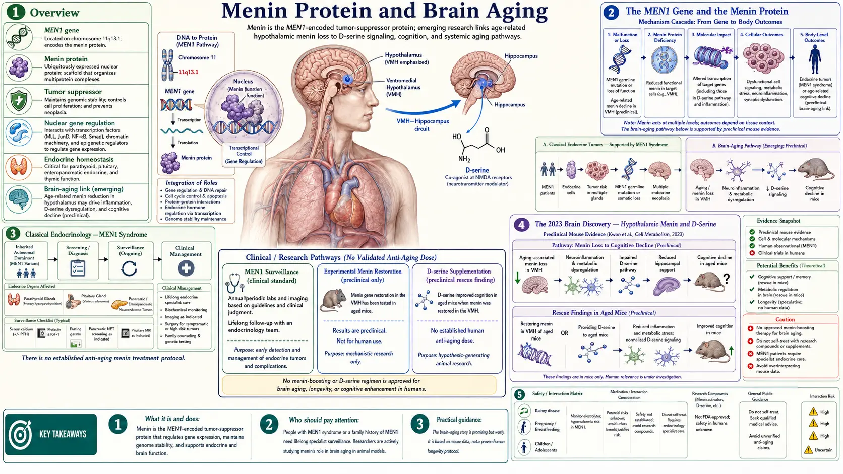
Table of Contents
- Overview
- The MEN1 Gene and the Menin Protein
- Classical Endocrinology — MEN1 Syndrome
- The 2023 Brain Discovery — Hypothalamic Menin and D-Serine
- D-Serine, N-Methyl-D-Aspartate Receptor Co-Agonism, and Cognition
- Menin in Microglia and Neuroinflammation
- Menin Restoration Strategies
- Translational Status — Where the Evidence Actually Is
- Practical Implications Today
- Key Research Papers
- Connections
- Featured Videos
Overview
Menin is a 610-amino-acid scaffold protein encoded by the MEN1 gene on chromosome 11q13. For nearly three decades after its 1997 identification, menin was studied almost exclusively as a tumor suppressor responsible for multiple endocrine neoplasia type 1 (MEN1) syndrome — an inherited condition characterized by tumors of the parathyroid glands, pancreatic islets, and anterior pituitary. The protein had a clear endocrine-tumor identity and a relatively small but dedicated research community working on its molecular biology and on small-molecule inhibitors of the bond between menin and the mixed-lineage leukemia (MLL) protein as targeted leukemia therapies.
That picture began to shift in March 2023, when a mouse study from Xiamen University in China (first author Lige Leng, senior author Jie Zhang) reported that menin in a specific population of hypothalamic neurons declines with age in mice, and that restoring menin in that brain region partially reverses several systemic features of aging — including impairments in cognition, bone mass, skin thickness and metabolic rhythm — and extends lifespan. The proposed mechanism centered on D-serine, a co-agonist at the N-methyl-D-aspartate (NMDA) glutamate receptor whose production is supported by hypothalamic menin signaling. According to this hypothesis, hypothalamic menin loss leads to reduced D-serine availability, which weakens N-methyl-D-aspartate-receptor function in distant brain regions involved in learning and memory and contributes to age-related cognitive decline.
The hypothalamic-menin story is genuinely exciting because it links three previously separate domains: a well-characterized endocrine tumor-suppressor protein, a well-characterized neuromodulator (D-serine), and a high-priority but mechanistically vague clinical problem (cognitive aging). It also offers a relatively concrete intervention concept: D-serine itself is an inexpensive amino acid that has been used in human trials at multi-gram daily doses for schizophrenia and cognitive endpoints. The translational distance between mouse hypothalamic neurons and a clinically useful intervention is still substantial, but the conceptual pathway is one of the more tractable in modern brain-aging research.
This page summarizes what menin is, what we have learned about its newly described role in brain aging, where the evidence is firm, and where it is preliminary. The goal is to help readers interpret news coverage of "menin restoration" claims without either dismissing the underlying biology or overstating how close it is to a clinical product.
The MEN1 Gene and the Menin Protein
The MEN1 gene was cloned in 1997 by Chandrasekharappa and colleagues, a National Institutes of Health team at the National Human Genome Research Institute whose authors included Francis Collins. Positional cloning of the responsible gene on 11q13 had been a multi-year effort across several international groups; the 1997 Science paper identified a 10-exon gene encoding a 610-amino-acid protein that was named menin. The gene is broadly expressed across tissues, but its loss-of-function consequences are most visible in endocrine cells, where it functions as a tumor suppressor in a classical two-hit Knudson sense: a germline loss-of-function allele predisposes to tumors that arise after somatic loss of the remaining wild-type allele.
At the molecular level, menin is a nuclear scaffold protein. It has no enzymatic activity of its own. Instead, it serves as an assembly platform that brings together other functional proteins into multiprotein complexes. The best-characterized of these is its participation in the MLL/KMT2A (lysine methyltransferase 2A) histone methyltransferase complex, which deposits trimethylation marks on histone H3 lysine 4 (H3K4) at the promoters of target genes. By holding MLL near specific chromatin sites, menin contributes to the gene-expression programs that those marks support — including, importantly, expression of the cyclin-dependent kinase inhibitors p27 (CDKN1B) and p18 (CDKN2C), which restrain endocrine-cell proliferation.
Menin also interacts directly with the tumor suppressor p53 and with several transcription factors involved in transforming growth factor beta (TGF-beta) and Wnt signaling. Loss of menin destabilizes p53 activity, removes the p27/p18 brakes on the cell cycle, and disinhibits Wnt-driven proliferation in endocrine progenitor cells. The net effect in pancreatic islet beta cells, parathyroid chief cells, and pituitary anterior-lobe cells is unchecked clonal expansion — the cellular basis of MEN1 syndrome.
Outside the MLL complex, menin participates in DNA damage response pathways and in regulation of the JunD transcription factor. JunD is unusual among the activator protein 1 (AP-1) family in being preferentially anti-proliferative; menin enhances JunD's anti-proliferative effect, so loss of menin removes this brake as well. These interactions are part of what makes menin a multi-function scaffold rather than a single-pathway protein, and they help explain why changes in menin are tied to conditions ranging from endocrine neoplasia, where losing menin leads to tumors, to leukemia, where cells carrying MLL fusion proteins depend on menin and removing it stops their cancer behavior in laboratory experiments, to, now apparently, hypothalamic dysfunction in mice.
Classical Endocrinology — MEN1 Syndrome
Multiple endocrine neoplasia type 1, also called Wermer syndrome, is the inherited disorder for which the MEN1 gene is named. It affects approximately 1 in 30,000 people and is inherited as an autosomal dominant condition with high penetrance: by age 50, more than 90 percent of carriers show clinical or biochemical evidence of disease. The classical triad is tumors of the parathyroid glands, the endocrine pancreas, and the anterior pituitary. Other manifestations include adrenocortical tumors, foregut carcinoid tumors, lipomas, angiofibromas, and collagenomas.
Primary hyperparathyroidism is usually the earliest manifestation, often presenting in the second or third decade of life with multi-gland parathyroid hyperplasia rather than the single adenoma typical of sporadic disease. Surgical management is more complex for the same reason. Pancreatic neuroendocrine tumors in MEN1 are frequently multiple and small; they include functional gastrinomas (causing Zollinger-Ellison syndrome), insulinomas, and a substantial fraction of non-functional but still potentially metastatic lesions. Pituitary tumors are most often prolactinomas, with growth-hormone- and adrenocorticotropic hormone-secreting tumors representing smaller subsets.
From a brain-aging perspective, the most important lesson of MEN1 syndrome is that menin loss in adult endocrine cells produces clonal expansion and tumorigenesis. This means that any "menin restoration" intervention aimed at the central nervous system would need to be highly targeted — ideally to specific hypothalamic neurons — rather than systemic. Globally raising menin in all tissues is biologically implausible as a longevity strategy because the tumor-suppressor function is already saturated in most adult cells, and globally lowering menin would, in some tissue compartments, be frankly oncogenic.
The flip side of the same observation is that menin loss is therapeutically useful in a different cellular context: certain MLL-rearranged leukemias depend on the menin–MLL interaction for their abnormal gene-expression programs. Disrupting this interaction with small-molecule menin inhibitors (revumenib was approved by the U.S. Food and Drug Administration (FDA) in late 2024 for relapsed/refractory KMT2A-rearranged acute leukemia) starves the leukemic cells of the transcriptional support they need. On October 24, 2025, the FDA also approved revumenib for relapsed or refractory acute myeloid leukemia with a susceptible NPM1 gene change in patients 1 year and older with no satisfactory alternative treatment, and on November 13, 2025, it approved a second menin inhibitor, ziftomenib, for the same use in adults. In the trial behind ziftomenib, 22% of 92 people reached complete remission or near-complete remission; differentiation syndrome, a drug reaction tied to leukemia cells maturing, occurred in 25% (15% grade 3, none grade 4 or 5). With revumenib in NPM1 disease, 23.4% of 64 adults reached the same response, lasting a median of 4.7 months. The FDA notices list differentiation syndrome, QTc interval prolongation (a heart-rhythm effect) and embryo-fetal toxicity among the warnings for both drugs. This pharmacology cuts in the opposite direction of the brain-aging hypothesis, which would want more menin function in hypothalamic neurons. Both can be true at the same time, in different tissue compartments, because menin's function is determined by the partner proteins available to it in each cell type.
The 2023 Brain Discovery — Hypothalamic Menin and D-Serine
The reframing of menin as a brain-aging protein emerged from a 2023 mouse study in PLOS Biology by a team at Xiamen University in China (Leng and colleagues). It sits on top of a separate line of work: the laboratory of Dongsheng Cai at the Albert Einstein College of Medicine has spent more than a decade investigating the hypothalamus as a regulator of systemic aging, building on earlier observations that hypothalamic inflammation accelerates aging phenotypes and that hypothalamic stem cells and microRNAs can influence lifespan in mice.
The 2023 report described neurons carrying the marker SF-1 in the ventromedial nucleus of the mouse hypothalamus whose menin protein levels decline with age; menin did not fall in astrocytes or microglia there. The authors reported that deleting menin in this region, in mice bred without it in those neurons and by gene-transfer injection in middle-aged mice, produced phenotypes that resembled accelerated aging — including impairments in spatial memory, reduced muscle fiber size, increased hypothalamic inflammation, changes in skin and bone, and a shorter lifespan. Conversely, restoring menin in the ventromedial hypothalamus of 20-month-old mice using viral-vector gene delivery extended lifespan and improved several of these measures, including performance on memory tasks.
The proposed mechanism centered on the amino acid D-serine. Hypothalamic menin loss was shown to lower the expression of enzymes involved in D-serine synthesis and turnover, with a net reduction in central D-serine availability. Because D-serine is a key co-agonist at N-methyl-D-aspartate-type glutamate receptors throughout the brain — including in hippocampus and cortex — reduced D-serine availability impairs N-methyl-D-aspartate-receptor function and the synaptic plasticity it supports. Restoring menin in the hypothalamus, or supplementing D-serine directly in the drinking water of aged mice, partially reversed the cognitive deficits in their reported models. The authors describe the D-serine effect as limited to cognitive improvement, leaving aging features outside the brain unchanged.
This work has been received with cautious enthusiasm by the brain-aging field. The strengths are the use of orthogonal interventions (genetic, viral, and dietary) that converge on the same readouts, and the identification of a specific molecule (D-serine) that is already known to be biologically active at N-methyl-D-aspartate receptors and that falls with age in the hippocampus of aged rats. The limitations — appropriately acknowledged by the authors and by commentators — are that the work is in mice, that the magnitude of cognitive improvement is modest, and that the proposed route, which the authors describe as impaired D-serine release by a neural circuit running from the ventromedial hypothalamus to the hippocampus, is so far described in mouse experiments.
D-Serine, N-Methyl-D-Aspartate Receptor Co-Agonism, and Cognition
The mammalian central nervous system contains substantial concentrations of D-serine, the right-handed stereoisomer of the dietary amino acid L-serine. D-serine is synthesized in the brain from L-serine by the enzyme serine racemase, and it is degraded by D-amino acid oxidase. Once thought to be a microbial-only metabolite, D-serine is now recognized as a major physiological co-agonist at the N-methyl-D-aspartate glutamate receptor, binding at the glycine-modulatory site and required — together with glutamate — for N-methyl-D-aspartate-receptor channel opening.
N-methyl-D-aspartate receptors are central to most current models of learning and memory. They permit calcium entry into postsynaptic neurons when both glutamate and a co-agonist (glycine or D-serine) are present, triggering downstream signaling cascades that strengthen synapses (long-term potentiation, LTP) or weaken them (long-term depression, LTD). The dependence on a co-agonist makes N-methyl-D-aspartate-receptor function exquisitely sensitive to the concentration of available D-serine or glycine, and chronic reductions in either reduce the plasticity ceiling of the network.
D-serine levels in cerebrospinal fluid and in postmortem brain tissue are said in some writing on this topic to decline with age in humans, though no human paper showing this was found for this page. What was read: lower hippocampal D-serine in aged rats (Billard 2015), lower D-serine in the blood serum of 5 adults aged 83 to 94 than of 5 aged 22 to 26 (Leng 2023), and a 2021 review noting "some controversy" while describing the proposal that D-serine falls in normal aging (Orzylowski 2021). Several lines of evidence link reduced D-serine availability to cognitive aging and to the cognitive features of schizophrenia. Clinical trials of D-serine supplementation in schizophrenia, typically at doses of 30 to 60 mg per kilogram per day (which translates to roughly 2 to 4 grams daily for an average adult), have shown modest improvements on negative symptoms and cognitive measures in some but not all studies. A dose-escalation study gave 42 people with schizophrenia or schizoaffective disorder 30, 60 or 120 mg per kilogram per day for 4 weeks on top of their usual medicine; thinking scores improved significantly at 60 mg per kilogram or more and not at 30. It was open-label, meaning there was no placebo group (Kantrowitz 2010). The amino acid is generally well tolerated at these doses in published trials, with the main concern being theoretical: at supra-physiological exposure, D-amino acid oxidase activity in the kidney could generate hydrogen peroxide and cause renal damage, an effect documented in rats at very high doses. A 2021 cross-species safety review reports that doses above 500 mg per kilogram in rats very commonly cause acute kidney tubular damage within hours, that this has not been reported in other species including mice and rabbits, and that across published human studies one person had abnormal kidney values related to D-serine, which resolved within a few days of stopping (Meftah 2021).
In Alzheimer's disease the direction runs opposite to the simple "too little D-serine" idea. A meta-analysis of 7 studies and more than 1,186 people found D-serine higher in the blood and spinal fluid of people with Alzheimer's disease than in comparison groups (standardized mean difference 0.679, 95% confidence interval 0.335 to 1.022) (Chang 2020). Whether extra D-serine helps or harms an aging brain is not settled.
For the menin hypothesis, the practical importance of D-serine is that it provides a downstream, pharmacologically accessible target. Even if direct manipulation of hypothalamic menin in the human brain remains years away, D-serine itself is an off-the-shelf molecule, and modest reductions in its central availability with age are a measurable target. Whether D-serine supplementation produces cognitive benefits in non-schizophrenic older adults at risk of cognitive decline is a question that would benefit from dedicated, adequately powered trials.
Menin in Microglia and Neuroinflammation
Beyond its role in hypothalamic neurons, menin participates in epigenetic programming of immune cells, including microglia — the central nervous system's resident innate immune population. Menin's partnership with the MLL/KMT2A H3K4 methyltransferase complex deposits activating chromatin marks at the promoters of specific inflammatory and metabolic genes, helping to define the transcriptional state of myeloid cells.
During aging, microglia drift from a homeostatic, surveillance-oriented state toward a "primed" or "senescent" state characterized by increased baseline expression of inflammatory cytokines, reduced phagocytic clearance of debris, and exaggerated responses to secondary insults. This drift is one component of the broader concept of inflammaging — the chronic, low-grade, sterile inflammation that accompanies organismal aging across multiple tissues. In the brain, primed microglia are implicated in the pathophysiology of Alzheimer's disease, Parkinson's disease, vascular cognitive impairment, and post-operative or post-illness delirium.
The intersection with menin is conceptually rich but mechanistically still being mapped. The same MLL/menin axis that controls developmental gene expression in endocrine progenitors also tunes inflammatory gene expression in mature myeloid cells. Whether age-related changes in microglial menin contribute to microglial priming, and whether the proposed hypothalamic-menin decline alters peripheral signals that feed into microglial state, are open questions that the field has begun to investigate. Reviews in 2023 and 2024 have framed the menin–MLL axis as one of several epigenetic nodes that could plausibly integrate metabolic, hormonal, and inflammatory inputs in brain aging.
The practical takeaway for readers is that "menin in brain aging" is unlikely to be a single, unitary mechanism. The hypothalamic-D-serine story is the cleanest current narrative, but it sits inside a larger landscape in which menin contributes to gene-expression programs in multiple cell types, including the immune cells whose drift over time shapes the brain's inflammatory tone.
Menin Restoration Strategies
If the hypothalamic-menin hypothesis is broadly correct, several intervention strategies become conceptually plausible. None is currently a clinical product, and all sit at different distances from human application.
- D-serine supplementation — The most accessible downstream rescue. D-serine is an inexpensive amino acid that has been used in human trials at multi-gram daily doses for schizophrenia. The proposed brain-aging dose would likely fall in a similar or somewhat lower range, but dedicated trials in non-schizophrenic older adults have not been completed.
- N-methyl-D-aspartate-receptor modulators with co-agonist activity — D-cycloserine is a partial agonist at the N-methyl-D-aspartate glycine site and has been used at low doses (typically 50–100 mg) in trials of cognitive enhancement in extinction learning and exposure therapy. Sub-anesthetic ketamine, which acts as an N-methyl-D-aspartate antagonist, has been studied for depression and post-traumatic stress disorder; its relationship to the menin/D-serine pathway is mechanistically inverse but clinically informative.
- Gene-therapy concepts — The mouse studies that motivated the hypothesis used adeno-associated virus (AAV) vectors delivered into the hypothalamus to raise menin expression in specific neurons. Direct hypothalamic AAV delivery in humans is technically feasible for other indications but is invasive and would face substantial safety review, particularly given the tumor-suppressor role of menin in non-target tissues.
- Lifestyle inputs to hypothalamic stress — The Cai laboratory has previously linked hypothalamic inflammation and stress to aging phenotypes, and downstream this connects to interventions that reduce systemic inflammatory load: caloric moderation, regular aerobic and resistance exercise, adequate sleep, glycemic control, and avoidance of chronic alcohol exposure. None of these is specific to menin, but each may slow the hypothalamic-stress trajectory that the menin story sits inside.
- Mechanistic target of rapamycin (mTOR) and nicotinamide adenine dinucleotide (NAD+) context — Rapamycin (a mechanistic target of rapamycin inhibitor) and nicotinamide adenine dinucleotide precursors (nicotinamide riboside, nicotinamide mononucleotide) are the two most-studied small molecules in mainstream longevity research. Their relationship to the menin pathway is not directly established but is biologically plausible: mechanistic target of rapamycin signaling controls protein-synthesis programs that include scaffold proteins like menin, and nicotinamide adenine dinucleotide availability constrains the activity of sirtuins and poly(ADP-ribose) polymerases (PARPs) that participate in the same chromatin-modification universe.
The conservative read is that D-serine supplementation is the closest-to-actionable concept, and even there the clinical data outside schizophrenia are thin. The other strategies are research-stage and are not products.
Translational Status — Where the Evidence Actually Is
It is worth being explicit about the evidence hierarchy for this topic, because it sits at a stage of the research cycle where overstatement is easy and corrosive.
- Robust: menin is a real scaffold protein, encoded by MEN1, and a bona fide tumor suppressor in endocrine tissue. The menin–MLL interaction is a validated drug target in MLL-rearranged leukemia. D-serine is a real N-methyl-D-aspartate co-agonist. N-methyl-D-aspartate-receptor function is central to learning and memory and is impaired in cognitive aging.
- Reasonably supported but still preclinical: menin expression in hypothalamic neurons declines with age in mice; reducing hypothalamic menin in middle-aged mice produces phenotypes resembling accelerated aging; restoring menin or supplementing D-serine partially reverses some of these phenotypes in mouse models, with effects on memory tasks, body composition, and tissue integrity. D-serine levels fall with age in the hippocampus of aged rats.
- Hypothesis-level: D-serine levels decline with age in human cerebrospinal fluid (blood serum was lower in one comparison of 5 older and 5 younger adults); the same hypothalamic-menin decline drives meaningful systemic and cognitive aging in humans; restoring central menin function or D-serine availability will produce clinically detectable cognitive benefit in older adults; menin-based interventions can be made safe in humans given the tumor-suppressor concerns.
- Not established: any consumer supplement product currently being marketed as a "menin restoration" intervention has clinical evidence of efficacy or has been demonstrated to raise central menin levels in humans. Several products in this category are reported to exist; no peer-reviewed human data behind their claims appear in the sources read for this page.
This division matters because the popular-science framing has moved faster than the human trial evidence. The mouse studies are genuinely interesting and the underlying biology is plausible, but the translational distance is real.
Practical Implications Today
No human trial of a menin-pathway treatment appears in the sources read for this page. Writing on brain aging points instead to habits already studied for brain health. The same broad set of inputs shows up across the menin literature, the hypothalamic-aging literature, and the longevity literature more generally:
- Sleep and glymphatic clearance — Whether the brain's clearance system for protein aggregates and metabolic waste works mainly during sleep is now disputed. A 2013 mouse study reported that natural sleep or anesthesia enlarged the space between brain cells by 60% and increased beta-amyloid clearance (Xie 2013); a 2024 mouse study found that brain clearance is markedly reduced, not increased, during sleep and anesthesia (Miao 2024). Writing on brain aging describes chronic sleep restriction or fragmentation as reducing clearance and accelerating the accumulation of aggregation-prone proteins implicated in dementia, and commonly names seven to nine hours of regular sleep as a high-yield, low-cost habit.
- Exercise and hypothalamic effects — Aerobic and resistance exercise alter hypothalamic inflammatory signaling, improve insulin sensitivity (which itself is a hypothalamic input), and increase brain-derived neurotrophic factor (BDNF) in regions involved in learning and memory. The exercise literature is one of the few in brain aging where the human evidence is comparable in quality to the mouse evidence.
- Mechanistic target of rapamycin signaling and eating patterns — Chronic over-nutrition and persistently elevated insulin and insulin-like growth factor 1 (IGF-1) signaling drive mechanistic target of rapamycin activity beyond what evolved environments selected for. Longevity writers describe time-restricted eating windows, no late-evening meals and less chronic snacking as ways to keep this signaling axis closer to its physiological set point. Pharmacological mechanistic target of rapamycin inhibition with rapamycin is studied in formal clinical trials rather than as self-treatment.
- Nicotinamide adenine dinucleotide context — nicotinamide adenine dinucleotide precursors are widely available as supplements (nicotinamide riboside, nicotinamide mononucleotide), and human data on their cognitive and metabolic effects are still being accumulated. They are not menin pathway interventions but they sit in the same metabolic-epigenetic neighborhood and are the subject of ongoing research.
- Glycemic and metabolic control — Insulin resistance, type 2 diabetes, and hypertension are among the strongest modifiable risk factors for cognitive decline and dementia. Improvements in these systemic markers feed back to hypothalamic function and to the broader inflammatory tone of the brain.
- "Menin restoration" supplement claims — Some products are marketed with mouse-study citations as if they were human-trial data. The mouse data are interesting; they are not yet the basis for a clinical product, and no validated human assay for central menin status exists against which a claim to "raise menin levels" in humans could be measured.
The menin and D-serine work is one of the most interesting threads in current brain-aging research because it offers a concrete molecular target downstream of a poorly understood phenomenon. It is also early, and the evidence is still accumulating.
Key Research Papers
- Chandrasekharappa SC, Guru SC, Manickam P, et al. Positional cloning of the gene for multiple endocrine neoplasia-type 1. Science. 1997;276(5311):404-407. — PubMed PMID: 9103196 · doi:10.1126/science.276.5311.404
- Hughes CM, Rozenblatt-Rosen O, Milne TA, et al. Menin associates with a trithorax family histone methyltransferase complex and with the hoxc8 locus. Molecular Cell. 2004;13(4):587-597. — PubMed PMID: 14992727 · doi:10.1016/S1097-2765(04)00081-4
- Yokoyama A, Somervaille TC, Smith KS, Rozenblatt-Rosen O, Meyerson M, Cleary ML. The menin tumor suppressor protein is an essential oncogenic cofactor for MLL-associated leukemogenesis. Cell. 2005;123(2):207-218. — PubMed PMID: 16239140 · doi:10.1016/j.cell.2005.09.025
- Matkar S, Thiel A, Hua X. Menin: a scaffold protein that controls gene expression and cell signaling. Trends in Biochemical Sciences. 2013;38(8):394-402. — PubMed PMID: 23850066 · doi:10.1016/j.tibs.2013.05.005
- Thakker RV, Newey PJ, Walls GV, et al. Clinical practice guidelines for multiple endocrine neoplasia type 1 (MEN1). Journal of Clinical Endocrinology & Metabolism. 2012;97(9):2990-3011. — PubMed PMID: 22723327 · doi:10.1210/jc.2012-1230
- Mothet JP, Parent AT, Wolosker H, et al. D-serine is an endogenous ligand for the glycine site of the N-methyl-D-aspartate receptor. Proceedings of the National Academy of Sciences. 2000;97(9):4926-4931. — PubMed PMID: 10781100 · doi:10.1073/pnas.97.9.4926
- Wolosker H, Balu DT, Coyle JT. The rise and fall of the d-serine-mediated gliotransmission hypothesis. Trends in Neurosciences. 2016;39(11):712-721. — PubMed PMID: 27742076 · doi:10.1016/j.tins.2016.09.007
- Billard JM. D-serine in the aging hippocampus. Journal of Pharmaceutical and Biomedical Analysis. 2015;116:18-24. — PubMed PMID: 25740810 · doi:10.1016/j.jpba.2015.02.013
- Zhang G, Li J, Purkayastha S, et al. Hypothalamic programming of systemic ageing involving IKK-β, NF-κB and GnRH. Nature. 2013;497(7448):211-216. — PubMed PMID: 23636330 · doi:10.1038/nature12143
- Zhang Y, Kim MS, Jia B, et al. Hypothalamic stem cells control ageing speed partly through exosomal miRNAs. Nature. 2017;548(7665):52-57. — PubMed PMID: 28746310 · doi:10.1038/nature23282
- Thakur RK, Wang ES. The promise of menin inhibitors: from approval to triplet regimens. Hematology Am Soc Hematol Educ Program. 2025;2025(1):599-606. — PubMed PMID: 41347983 · doi:10.1182/hematology.2025000755
- Avanesov AS, Ma S, Pierce KA, et al. Age- and diet-associated metabolome remodeling characterizes the aging process driven by damage accumulation. eLife. 2014;3:e02077. — PubMed PMID: 24843015 · doi:10.7554/eLife.02077
- Leng L, Yuan Z, Su X, et al. Hypothalamic Menin regulates systemic aging and cognitive decline. PLoS Biology. 2023;21(3):e3002033. — PubMed PMID: 36928253 · doi:10.1371/journal.pbio.3002033
- Orzylowski M, Fujiwara E, Mousseau DD, Baker GB. An Overview of the Involvement of D-Serine in Cognitive Impairment in Normal Aging and Dementia. Frontiers in Psychiatry. 2021;12:754032. — PubMed PMID: 34707525 · doi:10.3389/fpsyt.2021.754032
- Chang CH, Kuo HL, Ma WF, Tsai HC. Cerebrospinal Fluid and Serum d-Serine Levels in Patients with Alzheimer's Disease: A Systematic Review and Meta-Analysis. Journal of Clinical Medicine. 2020;9(12):3840. — PubMed PMID: 33256147 · doi:10.3390/jcm9123840
- Kantrowitz JT, Malhotra AK, Cornblatt B, et al. High dose D-serine in the treatment of schizophrenia. Schizophrenia Research. 2010;121(1-3):125-130. — PubMed PMID: 20541910 · doi:10.1016/j.schres.2010.05.012
- Meftah A, Hasegawa H, Kantrowitz JT. D-Serine: A Cross Species Review of Safety. Frontiers in Psychiatry. 2021;12:726365. — PubMed PMID: 34447324 · doi:10.3389/fpsyt.2021.726365
- Xie L, Kang H, Xu Q, et al. Sleep drives metabolite clearance from the adult brain. Science. 2013;342(6156):373-377. — PubMed PMID: 24136970 · doi:10.1126/science.1241224
- Miao A, Luo T, Hsieh B, et al. Brain clearance is reduced during sleep and anesthesia. Nature Neuroscience. 2024;27(6):1046-1050. — PubMed PMID: 38741022 · doi:10.1038/s41593-024-01638-y
- Wang ES, Montesinos P, Foran J, et al. Ziftomenib in Relapsed or Refractory NPM1-Mutated AML. Journal of Clinical Oncology. 2025;43(31):3381-3390. — PubMed PMID: 40997296 · doi:10.1200/JCO-25-01694
- Arellano ML, Thirman MJ, DiPersio JF, et al. Menin inhibition with revumenib for NPM1-mutated relapsed or refractory acute myeloid leukemia: the AUGMENT-101 study. Blood. 2025;146(9):1065-1077. — PubMed PMID: 40332046 · doi:10.1182/blood.2025028357
External Authoritative Resources
- GeneReviews — Multiple Endocrine Neoplasia Type 1
- NINDS — Dementia and Cognitive Aging
- U.S. Food and Drug Administration — revumenib approval, October 24, 2025
- U.S. Food and Drug Administration — ziftomenib approval, November 13, 2025
- PubMed — menin hypothalamus aging (live search)
- PubMed — D-serine cognition aging (live search)
Connections
- All Remedies
- Brain Aging Overview
- Rapamycin
- Longevity Protocols
- Nicotinamide Adenine Dinucleotide / Nicotinamide Mononucleotide
- Gut-Brain Axis
- Alzheimer's Disease
- Parkinson's Disease
- Cancer (MEN1 syndrome context)
- Multiple Endocrine Neoplasia Type 1
- D-Serine
- Vitamin B12
- Vitamin D3
- Magnesium
- Berberine