Manganese — Benefits Deep Dive
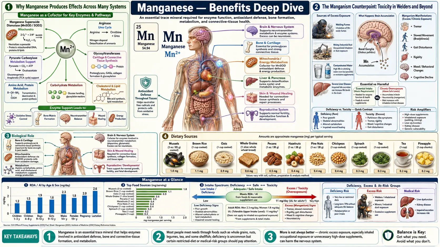
Manganese is the quiet workhorse of trace mineral biochemistry. The human body contains only 10-20 milligrams of it, yet it sits at the active site of four enzymes that are absolutely essential to life — the mitochondrial antioxidant manganese superoxide dismutase (MnSOD), the urea-cycle terminator arginase, the gluconeogenic gateway pyruvate carboxylase, and the brain's glutamate-recycling glutamine synthetase. Add in prolidase (collagen recycling) and the entire family of glycosyltransferases (proteoglycan assembly), and a single 2-milligram-per-day mineral turns out to be the cofactor for systems as diverse as mitochondrial defense, ammonia disposal, blood glucose regulation, neurotransmitter recycling, collagen turnover, and bone matrix assembly. Four benefit pages below explore the conditions where manganese produces the largest clinical effect — skeletal formation, antioxidant defense at the SOD2 level, wound healing through prolidase and glycosyltransferase action, and glucose metabolism through gluconeogenic enzyme support — and the counterpoint of manganism, the Parkinsonian neurotoxicity that defines the upper boundary of safe intake.
Deep-Dive Articles
Bone Formation
Manganese as the cofactor for glycosyltransferases that build chondroitin sulfate, keratan sulfate, and aggrecan in bone matrix; prolidase-mediated collagen recycling; MnSOD protection of osteoblasts; the Strause-Saltman 1994 trial showing trace-mineral plus calcium reduced postmenopausal spinal bone loss; perosis in poultry as the original deficiency syndrome; manganism as the upper safety boundary.
Read the deep dive →Antioxidant Mn-SOD (SOD2)
The mitochondrial antioxidant defender. Superoxide-to-hydrogen-peroxide dismutation chemistry at near-diffusion-limited rate. SOD2 knockout mouse mortality (death within the first 10 days in one mouse strain, from dilated cardiomyopathy and metabolic acidosis, and by 3 weeks in another). The Val16Ala (rs4880) polymorphism and elevated risk of cardiomyopathy, doxorubicin cardiotoxicity, diabetic complications, and neurodegeneration. SIRT3 regulation via lysine acetylation, the link to caloric restriction and nicotinamide adenine dinucleotide (NAD+) status, and the mitochondrial theory of aging.
Read the deep dive →Wound Healing
Manganese-activated prolidase as the dipeptidase that recycles proline from degraded collagen for new collagen synthesis — the inherited disorder of prolidase deficiency causes severe non-healing skin ulcers, demonstrating the enzyme's essential role. Glycosyltransferase-built proteoglycan ground substance for fibroblast and keratinocyte migration. Chondrocyte function in cartilage repair. MnSOD containment of the inflammatory-phase oxidative burst. Application to diabetic foot ulcers, burns, surgical recovery, and pressure ulcers.
Read the deep dive →Blood Sugar & Glucose Metabolism
Manganese-dependent gluconeogenic enzymes pyruvate carboxylase (the entry point) and phosphoenolpyruvate carboxykinase (PEPCK). Manganese deficiency and impaired insulin response from human and rodent studies. The U-shaped relationship between blood manganese and type 2 diabetes risk (both low and high are bad). Manganese-magnesium interplay in glucose homeostasis. MnSOD and the Brownlee mitochondrial-superoxide model of diabetic complications.
Read the deep dive →Table of Contents
- Deep-Dive Articles
- Why Manganese Produces Effects Across Many Systems
- The Manganism Counterpoint: Toxicity in Welders and Beyond
- Research Papers: Bone Formation & Connective Tissue
- Research Papers: MnSOD Antioxidant Defense
- Research Papers: Wound Healing & Prolidase
- Research Papers: Blood Sugar & Gluconeogenesis
- Research Papers: Cross-Cutting (Toxicity, Status, Homeostasis)
- External Authoritative Resources
- Connections
- Featured Videos
Why Manganese Produces Effects Across Many Systems
Most trace minerals serve as cofactors for one or a few enzymes. Manganese is unusual because it is the active-site metal for an unusually diverse set of metalloenzymes, and the activating cofactor for a whole family of glycosyltransferases. The biochemistry maps onto five different physiologic systems, each producing a distinct category of clinical effect.
- Mitochondrial antioxidant defense (MnSOD / SOD2) — manganese is the active-site metal of the mitochondrial superoxide dismutase, the single most critical enzyme defending against the reactive oxygen species generated by oxidative phosphorylation. Mice bred without the enzyme die young: in one strain within the first 10 days of life, with an enlarged, weakened heart (dilated cardiomyopathy), fat build-up in liver and muscle, and acid build-up in the blood (Li 1995); in another strain by 3 weeks of age, with severe anemia, degeneration of nerve cells in the brain and progressive movement problems (Lebovitz 1996). These are mice lacking the enzyme gene, not humans short of dietary manganese, but they show how much the enzyme matters. The Val16Ala polymorphism reducing MnSOD mitochondrial import is associated with elevated risk of cardiomyopathy, diabetic complications, and neurodegeneration. See the antioxidant deep-dive.
- Urea cycle (arginase) — arginase is a manganese-containing metalloenzyme that catalyzes the final step of urea synthesis, hydrolyzing arginine to ornithine and urea. This is the body's principal nitrogen-disposal pathway. Inadequate manganese impairs ammonia detoxification, particularly during high-protein intake or amino acid catabolism.
- Gluconeogenesis (pyruvate carboxylase, PEPCK) — two of the four committed gluconeogenic enzymes are manganese-dependent: pyruvate carboxylase (entry point) and phosphoenolpyruvate carboxykinase (second step). These enzymes also serve tricarboxylic acid (TCA) cycle anaplerosis, replenishing oxaloacetate that is drawn off for biosynthesis. Manganese deficiency impairs both glucose synthesis during fasting and oxidative adenosine triphosphate (ATP) production. See the blood-sugar deep-dive.
- Neurotransmitter recycling (glutamine synthetase) — glutamine synthetase in astrocytes converts glutamate to glutamine for re-export to neurons, sustaining the glutamate-glutamine cycle essential for excitatory neurotransmission. The enzyme is manganese-dependent. Disruption (by either deficiency or, paradoxically, toxic excess) contributes to neurologic dysfunction.
- Connective tissue (prolidase + glycosyltransferases) — manganese activates prolidase for collagen recycling and the entire family of glycosyltransferases for proteoglycan assembly. These enzymes are critical for bone matrix, cartilage, skin, and wound healing. See the bone-formation and wound-healing deep-dives.
The complication is that manganese has an unusually narrow window between enough and too much. Inadequate intake produces measurable but rarely dramatic dysfunction across the five systems above. Excessive intake (especially inhalational exposure) produces dramatic Parkinsonian neurodegeneration. Most healthy adults consuming a varied diet are well within the therapeutic range and require no supplementation. The Institute of Medicine set the adequate intake at 2.3 mg/day for adult men and 1.8 mg/day for adult women, and average United States intakes from food are about 2.1–2.3 mg/day for men and 1.6–1.8 mg/day for women (Linus Pauling Institute). Its Tolerable Upper Intake Level for adults is 11 mg/day, a deliberately conservative limit set because of manganese's nerve toxicity. Supplements in the 1–5 mg/day range appear in multi-mineral bone formulas and are used after bariatric surgery and in malabsorption syndromes.
Large population studies of diet and diabetes add a further piece. In a 2026 UK Biobank study of 165,194 adults, the highest fifth of dietary manganese intake was not significantly linked with lower type 2 diabetes risk than the lowest fifth (hazard ratio 0.91, 95% confidence interval 0.82–1.01), and showed no link with heart and blood-vessel disease or deaths from it. When the same authors pooled six prospective cohorts (over 270,000 people), each extra 1 mg a day of dietary manganese went with a 4% lower type 2 diabetes risk (relative risk 0.96, 95% confidence interval 0.94–0.99), possibly not in a straight line (Gebretsadik 2026). These are observational links in people eating food, not trials of supplements.
The Manganism Counterpoint: Toxicity in Welders and Beyond
No discussion of manganese benefits is complete without the counterpoint: manganese is a known neurotoxin, and chronic excess exposure produces a serious, often progressive, Parkinsonian disease called manganism. The condition was first described in 1837 by James Couper in workers grinding manganese dioxide and remains an active occupational health concern in the modern era.
- Occupational exposure populations — welders (especially metal inert gas (MIG) and tungsten inert gas (TIG) welding of manganese-containing steels), miners, steel workers, ferromanganese smelter workers, dry-cell battery manufacturers. The U.S. Centers for Disease Control and Prevention (CDC) and the Occupational Safety and Health Administration (OSHA) monitor occupational manganese exposure; permissible exposure limits are being progressively tightened as the evidence accumulates.
- Clinical syndrome — manganism develops insidiously over months to years of chronic inhalational exposure. Early symptoms include irritability, emotional lability, anorexia, and headache. Progression leads to bradykinesia, dystonia, postural instability, gait disorder (with characteristic "cock-walk"), and dysarthria. Later stages produce frank parkinsonism with rigidity, tremor, and cognitive decline. Unlike Parkinson's disease, manganism is largely unresponsive to levodopa therapy because the lesion is post-synaptic in the basal ganglia rather than the dopaminergic neuron loss of Parkinson's.
- Pathology — manganese accumulates selectively in the globus pallidus and other basal ganglia structures. The deposition is visible on T1-weighted brain magnetic resonance imaging (MRI) as hyperintensity in the globus pallidus, a finding useful for both diagnosis and detecting subclinical exposure in at-risk workers.
- Routes of high-risk exposure — inhalation (the dominant route, because olfactory uptake bypasses biliary excretion); contaminated drinking water (well water with manganese above U.S. Environmental Protection Agency (EPA) recommendations); long-term parenteral nutrition with manganese-containing formulas; hepatic insufficiency (cirrhosis or biliary obstruction impairs biliary excretion).
- Dietary intake is generally not a concern — the gastrointestinal absorption of manganese from food is tightly regulated (only 1-5% of dietary manganese is absorbed in adults with normal status), and the biliary excretion route handles any excess. Even relatively high dietary intake (10-15 mg/day from food) does not produce manganism in healthy individuals.
- Children are more vulnerable — infants and children absorb manganese at higher fractional rates and have less developed biliary excretion. Infant formulas (especially soy-based) provide much higher manganese than breast milk, and concerns have been raised about possible neurodevelopmental effects, though clinical evidence is limited.
- Treatment — primary intervention is removal from exposure. Chelation with ethylenediaminetetraacetic acid (EDTA), usually as its calcium disodium salt (CaNa2EDTA), has been used acutely but reverses symptoms only partially. Levodopa is largely ineffective. The neurologic damage tends to be permanent once established.
Two further findings sharpen the picture.
- Intravenous feeding — manganese poisoning is a well-recognised complication of long-term intravenous (parenteral) nutrition, more likely when bile flow is blocked (cholestasis) or doses are high. It usually shows as Parkinson-like symptoms, but can be found earlier as raised blood manganese or as a bright signal in the basal ganglia on T1-weighted MRI. The currently recommended adult intravenous dose is 55 micrograms a day, yet most multi-trace-element products supply more, and the feeding solutions are already contaminated with enough manganese to meet needs; deficiency in these patients is rare (Livingstone 2018).
- Children's development — a 2020 review and meta-analysis of 55 studies (13,388 children) found higher environmental manganese exposure linked to poorer cognitive and motor development, mostly in children under 6. A 10-fold rise in hair manganese went with a 2.51-point lower full-scale IQ (95% confidence interval −4.58 to −0.45), while the blood and drinking-water meta-analyses showed no significant IQ effect (Liu 2020). These are associations, not proof of cause.
The window, in short: dietary manganese from whole foods is the normal, necessary source; the adult upper limit is 11 mg/day from all sources; and the situations linked with toxicity are inhaled manganese, liver disease or blocked bile flow (which slow excretion), and long-term intravenous feeding.
Research Papers: Bone Formation & Connective Tissue
- Strause L, Saltman P, Smith KT et al. (1994). Spinal bone loss in postmenopausal women supplemented with calcium and trace minerals. Journal of Nutrition 124(7):1060-1064. 59 women (mean age 66), 2 years, double-blind and placebo-controlled; spinal bone density changed −3.53% on placebo, −1.89% on trace minerals only (zinc 15 mg, manganese 5 mg, copper 2.5 mg a day), −1.25% on calcium only (1,000 mg) and +1.48% on calcium plus trace minerals; only calcium plus trace minerals differed significantly from placebo, and manganese was not tested alone. — PubMed PMID: 8027856
- Saltman PD, Strause LG (1993). The role of trace minerals in osteoporosis. Journal of the American College of Nutrition 12(4):384-389. — PubMed PMID: 8409100
- Leach RM Jr (1971). Role of manganese in mucopolysaccharide metabolism. Federation Proceedings. — PubMed
- Leach RM, Muenster AM, Wien EM (1969). Studies on the role of manganese in bone formation: chondroitin sulfate synthesis in chick epiphyseal cartilage. Archives of Biochemistry and Biophysics. — PubMed
- Hurley LS, Keen CL (1987). Manganese. In: Trace Elements in Human and Animal Nutrition. — PubMed
- Palacios C (2006). The role of nutrients in bone health, from A to Z. Critical Reviews in Food Science and Nutrition. — PubMed
- Freeland-Graves JH, Lin PH (1991). Plasma uptake of manganese as affected by oral loads of manganese, calcium, milk, phosphorus, copper, and zinc. Journal of the American College of Nutrition 10(1):38-43. — PubMed PMID: 2010579
- Hurley LS (1981). Teratogenic aspects of manganese, zinc, and copper nutrition. Physiological Reviews. — PubMed
- Aschner JL, Aschner M (2005). Nutritional aspects of manganese homeostasis. Molecular Aspects of Medicine. — PubMed
Research Papers: MnSOD Antioxidant Defense
- Li Y et al. (1995). Dilated cardiomyopathy and neonatal lethality in mutant mice lacking manganese superoxide dismutase. Nature Genetics 11(4):376-381. — PubMed PMID: 7493016
- Lebovitz RM et al. (1996). Neurodegeneration, myocardial injury, and perinatal death in mitochondrial superoxide dismutase-deficient mice. Proceedings of the National Academy of Sciences of the USA (PNAS) 93(18):9782-9787. — PubMed PMID: 8790408
- Sutton A et al. (2003). The Ala16Val genetic dimorphism modulates the import of human MnSOD into rat liver mitochondria. Pharmacogenetics 13(3):145-157. — PubMed PMID: 12618592
- Qiu X et al. (2010). Calorie restriction reduces oxidative stress by SIRT3-mediated SOD2 activation. Cell Metabolism 12(6):662-667. — PubMed PMID: 21109198
- Tao R et al. (2010). Sirt3-mediated deacetylation of evolutionarily conserved lysine 122 regulates MnSOD activity in response to stress. Molecular Cell 40(6):893-904. — PubMed PMID: 21172655
- Bresciani G et al. (2015). Manganese superoxide dismutase and oxidative stress modulation. Advances in Clinical Chemistry. — PubMed
- Bag A, Bag N (2008). MnSOD target sequence polymorphism and cancer risk. Cancer Epidemiol Biomarkers Prev. — PubMed
- Wang Y, Branicky R, Noë A, Hekimi S (2018). Superoxide dismutases: dual roles in controlling ROS damage and regulating ROS signaling. Journal of Cell Biology 217(6):1915-1928. — PubMed PMID: 29669742
- Harman D (1956). Aging: a theory based on free radical and radiation chemistry. Journal of Gerontology. — PubMed
- Mohammedi K, Bellili-Muñoz N, Driss F et al. (2014). Manganese superoxide dismutase (SOD2) polymorphisms, plasma advanced oxidation protein products (AOPP) concentration and risk of kidney complications in subjects with type 1 diabetes. PLoS One 9(5):e96916. — PubMed PMID: 24819633
Research Papers: Wound Healing & Prolidase
- Lupi A, Tenni R, Rossi A, Cetta G, Forlino A (2008). Human prolidase and prolidase deficiency: an overview on the characterization of the enzyme involved in proline recycling and on the effects of its mutations. Amino Acids 35(4):739-752. — PubMed PMID: 18340504
- Kitchener RL, Grunden AM (2012). Prolidase function in proline metabolism and its medical applications. Journal of Applied Microbiology. — PubMed
- Phang JM, Liu W (2012). Proline metabolism and cancer. Frontiers in Bioscience. — PubMed
- Heyland DK et al. (2005). Antioxidant nutrients in the critically ill: systematic review. Intensive Care Medicine. — PubMed
- Berger MM, Shenkin A (2007). Trace element requirements in critically ill burned patients. Journal of Trace Elements in Medicine and Biology. — PubMed
- Stechmiller JK (2010). Understanding the role of nutrition and wound healing. Nutrition in Clinical Practice. — PubMed
- Posthauer ME et al. (2015). Pressure ulcer nutrition: NPUAP/EPUAP/PPPIA white paper. Advances in Skin & Wound Care. — PubMed
- Powell GF, Maniscalco RM (1976). Bound hydroxyproline excretion following gelatin loading in prolidase deficiency. Metabolism 25(5):503-508. — PubMed PMID: 772363
- Reiber GE et al. (1999). Causal pathways for diabetic foot ulcers. Diabetes Care. — PubMed
- Lupi A, Della Torre S, Campari E et al. (2006). Human recombinant prolidase from eukaryotic and prokaryotic sources. Expression, purification, characterization and long-term stability studies. FEBS Journal 273(23):5466-5478. — PubMed PMID: 17081196
Research Papers: Blood Sugar & Gluconeogenesis
- Brownlee M (2005). The pathobiology of diabetic complications: a unifying mechanism. Diabetes. — PubMed
- Du S, Wu X, Han T et al. (2018). Dietary manganese and type 2 diabetes mellitus: two prospective cohort studies in China. Diabetologia 61(9):1985-1995. Two cohorts (3,350 and 7,133 adults); higher dietary manganese went with lower type 2 diabetes incidence. — PubMed PMID: 29971528
- Gebretsadik GG, Yang B, Glenn AJ et al. (2026). Dietary manganese, type 2 diabetes, and cardiovascular disease: A UK Biobank cohort study and meta-analysis of over 270,000 individuals. Journal of Nutrition, Health and Aging 30(2):100754. — PubMed PMID: 41380425
- Walter RM Jr et al. (1991). Copper, zinc, manganese, and magnesium status and complications of diabetes mellitus. Diabetes Care 14(11):1050-1056. — PubMed PMID: 1797486
- Jomova K, Valko M (2011). Advances in metal-induced oxidative stress and human disease. Toxicology. — PubMed
- Rorsman P, Ashcroft FM (2018). Pancreatic beta-cell electrical activity and insulin secretion. Physiological Reviews. — PubMed
Research Papers: Cross-Cutting (Toxicity, Status, Homeostasis)
- Aschner JL, Aschner M (2005). Nutritional aspects of manganese homeostasis. Molecular Aspects of Medicine. — PubMed
- Horning KJ, Caito SW, Tipps KG, Bowman AB, Aschner M (2015). Manganese is essential for neuronal health. Annual Review of Nutrition. — PubMed
- Finley JW, Davis CD (1999). Manganese deficiency and toxicity: cause for concern? BioFactors. — PubMed
- Couper J (1837). Sur les effets du peroxide de manganese (first description of manganism). Journal de Chimie Médicale. — PubMed
- Olanow CW (2004). Manganese-induced parkinsonism and Parkinson's disease. Annals of the New York Academy of Sciences. — PubMed
- Livingstone C (2018). Manganese Provision in Parenteral Nutrition: An Update. Nutrition in Clinical Practice 33(3):404-418. — PubMed PMID: 28445108
- Liu W, Xin Y, Li Q et al. (2020). Biomarkers of environmental manganese exposure and associations with childhood neurodevelopment: a systematic review and meta-analysis. Environmental Health 19(1):104. — PubMed PMID: 33008482
- Lucchini RG, Martin CJ, Doney BC (2009). From manganism to manganese-induced parkinsonism: a conceptual model. NeuroMolecular Medicine. — PubMed
- Bowman AB et al. (2011). Role of manganese in neurodegenerative diseases. Journal of Trace Elements in Medicine and Biology. — PubMed
- Roth JA (2006). Homeostatic and toxic mechanisms regulating manganese uptake, retention, and elimination. Biological Research. — PubMed
- Greger JL (1999). Nutrition versus toxicology of manganese in humans. Neurotoxicology. — PubMed
- Institute of Medicine (2001). Dietary Reference Intakes for vitamin A, vitamin K, arsenic, boron, chromium, copper, iodine, iron, manganese, molybdenum, nickel, silicon, vanadium, and zinc. National Academy Press. — PubMed
External Authoritative Resources
- Linus Pauling Institute — Manganese Micronutrient Information Center — the most authoritative open-access scientific review of manganese biology, regularly updated
- U.S. National Institutes of Health (NIH) Office of Dietary Supplements — Manganese Fact Sheet (Health Professionals)
- Agency for Toxic Substances and Disease Registry (ATSDR) — Toxicological Profile for Manganese (U.S. Centers for Disease Control and Prevention document on occupational and environmental exposure)
- Occupational Safety and Health Administration (OSHA) — Manganese Compounds occupational exposure guidance
- U.S. National Institutes of Health Office of Dietary Supplements — Manganese (consumer fact sheet)
- PubMed — All research on manganese in human nutrition
Connections
- All Minerals
- Manganese (Main Page)
- Manganese for Bone Formation
- Manganese MnSOD Antioxidant Defense
- Manganese for Wound Healing
- Manganese for Blood Sugar
- Magnesium
- Calcium
- Calcium and Bone Health
- Zinc
- Copper
- Selenium
- Chromium
- Boron
- Silicon
- Phosphorus
- Vitamin D3
- Vitamin K2
- Vitamin C
- Osteoporosis
- Arthritis
- Parkinson's Disease
- Type 2 Diabetes
- Metabolic Syndrome
- Collagen
- Proline
- Oxidative Stress
- Copper-Iron Dysregulation
- Spinach
- Brown Rice
- Deficiency
- Toxicity
- History
- Sources