Iron – Essential Mineral for Human Health
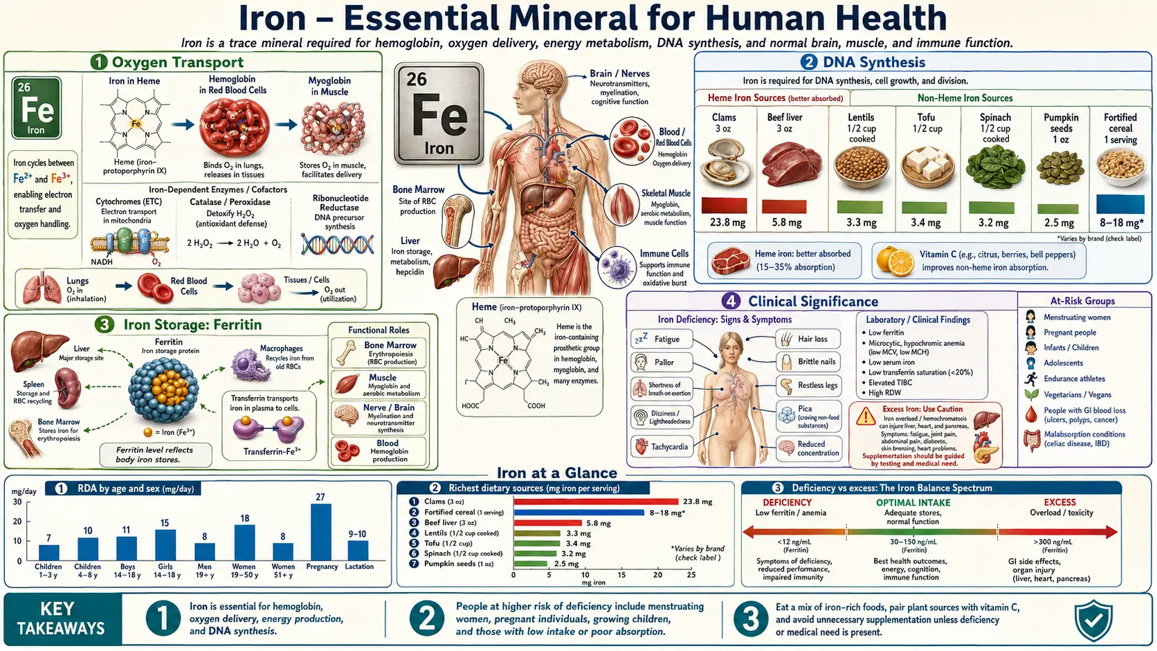
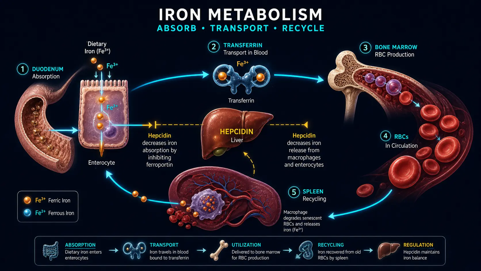
Table of Contents
- Oxygen Transport
- Energy Production
- Enzymatic Functions
- DNA Synthesis
- Immune Function
- Cognitive Development and Neurotransmitter Synthesis
- Iron Storage: Ferritin
- Iron Transport: Transferrin
- Heme Iron vs Non-Heme Iron
- Clinical Significance
- Key Research Papers
- Connections
- Featured Videos
Iron (Fe) is a transition metal and one of the most critical trace minerals in human physiology. The Institute of Medicine (IOM) states that a 75-kg adult man contains about 4 grams of iron, most of it incorporated into functional proteins (IOM 2001). Iron's ability to readily cycle between its ferrous (Fe2+) and ferric (Fe3+) oxidation states makes it indispensable for electron transfer reactions, oxygen binding, and a wide array of enzymatic processes. Because the body has no physiologic mechanism for excreting iron, iron balance is regulated by absorption (Hurrell 2010).
Oxygen Transport
Hemoglobin
Almost two-thirds of the body's iron is found in hemoglobin, the oxygen-carrying protein within circulating red blood cells (IOM 2001). Each hemoglobin molecule is a tetramer composed of four globin subunits, each containing a heme prosthetic group with a centrally coordinated iron atom in the ferrous (Fe2+) state. This iron atom reversibly binds molecular oxygen in the lungs and releases it in peripheral tissues according to their metabolic demands. The cooperative binding behavior of hemoglobin, described by the oxygen-hemoglobin dissociation curve, allows efficient oxygen loading at high partial pressures in the pulmonary capillaries and unloading at lower partial pressures in metabolically active tissues.
- Anaemia cut-offs: The 2024 WHO guideline defines anaemia in adults as a haemoglobin below 120 g/L in non-pregnant women and below 130 g/L in men aged 15–65 (WHO 2024). Laboratory reference ranges vary, and a result is read against the range printed on the report.
- Oxygen-carrying capacity: The theoretical oxygen-binding capacity of hemoglobin (Hüfner's factor) is 1.39 mL of oxygen per gram; values measured in human blood are lower (Shimizu 1986).
- Bohr effect: Increased carbon dioxide and decreased pH in metabolically active tissues shift the dissociation curve to the right, promoting oxygen release where it is most needed.
Myoglobin
Myoglobin is a monomeric heme protein found in skeletal and cardiac muscle tissue. According to the IOM, most of the roughly 15 percent of body iron that is neither in hemoglobin nor in stores is in muscle myoglobin and a variety of enzymes (IOM 2001). Unlike hemoglobin, myoglobin has a hyperbolic oxygen-binding curve, giving it a higher affinity for oxygen at low partial pressures. This property makes myoglobin well suited for accepting oxygen from hemoglobin at the tissue level and storing it within muscle cells for use during periods of intense metabolic activity or transient ischemia.
- Oxygen reservoir: Myoglobin serves as an intracellular oxygen buffer, releasing stored oxygen during muscle contraction when blood flow is temporarily occluded.
- Cardiac significance: Serum myoglobin has been studied as an early marker of heart attack; in 309 patients with chest pain, its diagnostic accuracy was highest 5 hours after symptoms began (de Winter 2000). Because myoglobin is also present in skeletal muscle, it is not specific to the heart.
Energy Production
Cytochromes and the Electron Transport Chain
Iron is a central component of the mitochondrial electron transport chain (ETC), where it participates in the sequential transfer of electrons from NADH and FADH2 to molecular oxygen, driving the generation of ATP through oxidative phosphorylation. Iron is present in two forms within the ETC:
- Heme-containing cytochromes: Cytochromes b, c1, c, a, and a3 contain iron within their heme prosthetic groups. Cytochromes b and c1 sit in Complex III, cytochrome c carries electrons from Complex III to Complex IV, and cytochromes a and a3 belong to cytochrome c oxidase (Complex IV), which catalyzes the final transfer of electrons to oxygen, reducing it to water.
- Iron-sulfur (Fe-S) clusters: Found in Complexes I, II, and III, iron-sulfur proteins contain iron atoms coordinated with inorganic sulfur. These clusters function as one-electron carriers, facilitating the stepwise transfer of electrons and contributing to proton translocation across the inner mitochondrial membrane.
A 2021 review notes that the clinical and functional impairments of iron deficiency can occur in the absence of anaemia (Pasricha 2021). In a randomized trial in 198 non-anaemic menstruating women with unexplained fatigue and a ferritin below 50 µg/L, 12 weeks of oral iron reduced fatigue scores more than placebo, with no significant effect on quality of life, depression or anxiety (Vaucher 2012).
Citric Acid Cycle Enzymes
The enzyme aconitase, which catalyzes the interconversion of citrate and isocitrate in the tricarboxylic acid (TCA) cycle, requires an iron-sulfur cluster ([4Fe-4S]) for its catalytic activity. Loss of iron from this cluster inactivates the enzyme and impairs the flow of metabolites through the cycle, further diminishing energy production.
Enzymatic Functions
Beyond oxygen transport and energy metabolism, iron serves as a cofactor or structural component in a diverse array of enzymes:
- Catalase: A heme-containing enzyme that decomposes hydrogen peroxide (H2O2) into water and oxygen, protecting cells from oxidative damage.
- Peroxidases: Including myeloperoxidase in neutrophils, which generates hypochlorous acid as part of the innate antimicrobial defense system.
- Ribonucleotide reductase: A di-iron enzyme essential for the conversion of ribonucleotides to deoxyribonucleotides, a rate-limiting step in DNA synthesis (see below).
- Prolyl and lysyl hydroxylases: Iron-dependent enzymes required for collagen biosynthesis, catalyzing the hydroxylation of proline and lysine residues needed for collagen's structural stability and cross-linking.
- Cytochrome P450 enzymes: A large superfamily of heme-containing monooxygenases involved in drug metabolism, steroid hormone biosynthesis, bile acid formation, and the detoxification of xenobiotics in the liver.
- Tryptophan hydroxylase and phenylalanine hydroxylase: Non-heme iron-dependent enzymes involved in the synthesis of serotonin and tyrosine, respectively.
DNA Synthesis
Iron plays an indispensable role in DNA replication and repair through the enzyme ribonucleotide reductase (RNR). This enzyme catalyzes the reduction of all four ribonucleoside diphosphates to their corresponding deoxyribonucleoside diphosphates, providing the building blocks required for DNA synthesis. The active site of class I RNR contains a di-iron center that generates a stable tyrosyl radical essential for catalysis. Iron depletion reduces RNR activity, slowing DNA replication and contributing to the impaired cell proliferation seen in iron deficiency states, particularly in rapidly dividing cells such as hematopoietic precursors and intestinal epithelium.
- Cell cycle effects: In cell-culture studies, iron chelation blocks dividing cells at the G1/S phase of the cell cycle; one study in cultured human CD4+ T cells found this block and pointed to increased levels of the cell-cycle inhibitor p21 as a possible mechanism (Tewari 2020).
- DNA repair: Iron-sulfur clusters are found in DNA polymerases and in enzymes of several DNA repair processes that maintain genomic integrity (Fuss 2015).
Immune Function
Iron is required for the proper function of both the innate and adaptive arms of the immune system. However, the relationship between iron and immunity is complex because iron is also an essential nutrient for most pathogenic microorganisms.
- Neutrophil bactericidal activity: Myeloperoxidase, a heme enzyme in neutrophil granules, generates reactive oxygen species that kill ingested bacteria. In a study of 32 children with iron deficiency anaemia and 29 healthy children, a smaller share of neutrophils and monocytes in the anaemic group showed an oxidative burst (Ekiz 2005).
- Lymphocyte proliferation: T-cell and B-cell expansion depends on iron-requiring enzymes such as ribonucleotide reductase, and an iron chelator inhibited the proliferation of cultured human CD4+ T cells (Tewari 2020). The same study of children with iron deficiency anaemia found no difference in the distribution of T-lymphocyte subgroups (Ekiz 2005).
- Nutritional immunity: The host sequesters iron from invading pathogens by reducing serum iron levels through hepcidin-mediated mechanisms, upregulating lactoferrin in mucosal secretions, and increasing ferritin synthesis to lock iron in storage. This strategy, known as nutritional immunity, limits microbial access to this essential growth factor.
- Iron overload and infection risk: Conversely, excess iron can favour some bacteria. The 2011 AASLD hemochromatosis guideline notes reports of Vibrio vulnificus infection in people with hereditary hemochromatosis who ate raw shellfish (Bacon 2011), and case reports describe Yersinia enterocolitica liver abscesses (Höpfner 2001) and Listeria monocytogenes meningitis with reduced phagocyte function (van Asbeck 1982) in people with iron overload.
Cognitive Development and Neurotransmitter Synthesis
Brain Iron and Neurodevelopment
The developing brain has a particularly high demand for iron. Iron is required for myelination of neuronal axons, synaptogenesis, and the synthesis of monoamine neurotransmitters. Iron deficiency is common in pregnant women and infants worldwide. A 2006 review reports that infants with iron deficiency anaemia test lower in cognitive, motor, social-emotional and neurophysiologic development than comparison infants, that iron therapy does not consistently improve developmental outcome, with long-term differences observed, and that randomized trials of infant iron supplementation indicate adverse effects can be prevented or reversed when iron is given earlier in development or before the deficiency becomes severe or chronic (Lozoff 2006).
- Myelination: Oligodendrocytes, the myelin-producing cells of the central nervous system, are the principal brain cells that stain for iron under normal conditions. Decreased dietary iron is associated with hypomyelination, and the hypomyelination can persist long after the systemic iron deficiency has been corrected (Todorich 2009).
- Animal studies: In rodent models, iron deficiency during gestation and lactation altered brain metabolism, neurotransmitters, myelination and gene and protein profiles, both before and after iron repletion at weaning (Lozoff 2006).
Neurotransmitter Synthesis
Iron serves as a cofactor for key enzymes in the biosynthesis of monoamine neurotransmitters:
- Tyrosine hydroxylase: Catalyzes the rate-limiting step in the synthesis of dopamine and norepinephrine, converting tyrosine to L-DOPA. This enzyme requires ferrous iron and tetrahydrobiopterin as cofactors.
- Tryptophan hydroxylase: Catalyzes the rate-limiting step in serotonin synthesis, converting tryptophan to 5-hydroxytryptophan. Also iron-dependent.
- Monoamine oxidase: Breaks down dopamine, serotonin, and norepinephrine. Monoamine oxidases A and B are flavoproteins that carry a covalently bound FAD cofactor rather than iron (Edmondson 2004).
Disruption of these iron-dependent pathways is one proposed explanation for the lower developmental test scores reported in infants with iron deficiency anaemia (Lozoff 2006).
Iron Storage: Ferritin
Ferritin is the principal intracellular iron storage protein, found in virtually all cell types but most abundantly in hepatocytes, macrophages of the reticuloendothelial system, and bone marrow. Each ferritin molecule is a hollow shell of 24 subunits (in vertebrates a mixture of H and L chains) whose cavity can store up to 4,500 iron(III) atoms as an inorganic mineral core (Harrison 1996).
- H-subunit (heavy chain): Possesses ferroxidase activity, catalyzing the oxidation of Fe2+ to Fe3+ for incorporation into the mineral core. Predominates in tissues with high iron utilization such as the heart and brain.
- L-subunit (light chain): Facilitates iron nucleation and long-term storage. Predominates in tissues with primary storage functions such as the liver and spleen.
- Serum ferritin: A small amount of ferritin is released into the bloodstream. The British Society of Gastroenterology's 2021 guideline describes serum ferritin as the single most useful marker of iron deficiency: a ferritin below 15 µg/L (the same as 15 ng/mL) indicates absent iron stores and one below 30 µg/L generally indicates low stores; because ferritin rises with inflammation, an apparently normal ferritin can occur with iron deficiency when inflammation is present, and transferrin saturation can help (Snook 2021). At the high end, the 2011 AASLD hemochromatosis guideline treats a ferritin above 200 ng/mL in women or above 300 ng/mL in men as elevated, which may suggest iron overload, and notes that a variety of conditions unrelated to iron overload can also raise it (Bacon 2011).
- Hemosiderin: When iron stores are high, ferritin molecules aggregate and undergo partial lysosomal degradation to form hemosiderin, a water-insoluble iron complex visible on tissue biopsy with Prussian blue staining.
Iron Transport: Transferrin
Transferrin is a glycoprotein synthesized primarily by the liver that serves as the principal plasma iron transport protein. Each transferrin molecule has two high-affinity binding sites for ferric iron (Fe3+), and the share of these sites occupied is the transferrin saturation. The AASLD guideline gives 20 to 50 percent for normal subjects (Bacon 2011); in U.S. survey data cited by the IOM, median transferrin saturation was 26 to 30 percent in men and 21 to 24 percent in women (IOM 2001).
- Transferrin receptor 1 (TfR1): Expressed on the surface of most cells, with particularly high density on erythroid precursors in the bone marrow. The transferrin-TfR1 complex is internalized by receptor-mediated endocytosis. Acidification of the endosome releases iron from transferrin, and the apotransferrin-TfR1 complex is recycled to the cell surface.
- Transferrin receptor 2 (TfR2): Expressed predominantly in hepatocytes, where it acts as an iron sensor that modulates hepcidin expression in response to circulating transferrin saturation levels.
- Soluble transferrin receptor (sTfR): A truncated form of TfR1 released into plasma, the concentration of which reflects total erythropoietic activity and tissue iron demand. Elevated sTfR is a useful marker of iron deficiency, particularly in the setting of concurrent inflammation where ferritin levels may be falsely elevated.
- Hepcidin: Described as the main regulator of systemic iron homeostasis (Pasricha 2021), hepcidin is a peptide hormone produced by the liver. It binds ferroportin, the iron exporter on absorptive enterocytes, macrophages, hepatocytes and placental cells, causing ferroportin to be internalized and degraded and so decreasing iron export (Nemeth 2004); hepcidin and ferroportin thereby control intestinal absorption, the release of recycled iron from macrophages and the release of stored iron from hepatocytes (Ganz 2013). Hepcidin is feedback-regulated by plasma iron and iron stores, induced by inflammatory signals including interleukin-6, and negatively regulated by the activity of red-cell precursors (Ganz 2013); in mice, anemia and hypoxia sharply decreased liver hepcidin gene expression (Nicolas 2002).
Heme Iron vs Non-Heme Iron
Dietary iron exists in two chemical forms that differ markedly in their absorption mechanisms and bioavailability:
- Heme iron: Derived from hemoglobin and myoglobin in animal-source foods (red meat, poultry, fish). Heme is absorbed mainly in the proximal small intestine. How it crosses the gut wall is not fully settled: a 2005 study reported HCP1 as the intestinal heme transporter (Shayeghi 2005), but a 2006 study established that the same protein's major function is proton-coupled folate transport and renamed it PCFT/HCP1 (Qiu 2006). The IOM describes heme iron as highly bioavailable and little affected by dietary factors, and used a conservative estimate of 25 percent for overall heme-iron absorption when setting requirements (IOM 2001).
- Non-heme iron: Found in plant foods, dairy, eggs, and iron-fortified products. Non-heme iron must be reduced from Fe3+ to Fe2+ by duodenal cytochrome b (Dcytb) before transport via the divalent metal transporter 1 (DMT1). Phytate, polyphenols, calcium, ascorbic acid and muscle tissue have repeatedly been shown to influence iron absorption in single-meal isotope studies, while in multimeal studies with a varied diet the effect of single components has been more modest; the nature of the meat-related enhancing factor remains unresolved (Hurrell 2010). In a study of complete diets, the enhancing effect of vitamin C on non-heme iron absorption was far less pronounced than in single meals (Cook 2001). From intake data and isotope studies, overall iron bioavailability has been estimated at 14 to 18 percent for mixed diets and 5 to 12 percent for vegetarian diets in people with no iron stores (Hurrell 2010).
For a detailed comparison of these two forms, see Heme vs Non-Heme Iron.
Clinical Significance
Both too little and too much iron cause serious problems. The WHO has recognised iron deficiency anaemia as the most common nutritional deficiency in the world (Kumar 2022); iron deficiency progresses from depleted stores to anemia, while iron overload deposits free iron in the liver, heart, and pancreas through conditions such as hereditary hemochromatosis. For a full, patient-friendly guide to the symptoms, causes, and treatment of each, see Iron-Deficiency Anemia: Symptoms, Causes, and Recovery and Iron Overload (High Iron): Symptoms, Causes, and Risks.
Recommended Dietary Allowances
The Recommended Dietary Allowances and the Tolerable Upper Intake Level below are those set by the Institute of Medicine in 2001 (IOM 2001).
- Adult men (19+ years): 8 mg/day
- Adult premenopausal women (19-50 years): 18 mg/day
- Pregnant women: 27 mg/day
- Postmenopausal women (51+ years): 8 mg/day
- Infants (7-12 months): 11 mg/day
- Children (1-3 years): 7 mg/day
- Tolerable Upper Intake Level (adults; ages 14 and older, including pregnancy and lactation): 45 mg/day, based on gastrointestinal distress as the adverse effect
Key Research Papers
- Institute of Medicine (US) Panel on Micronutrients (2001). Iron. In: Dietary Reference Intakes for Vitamin A, Vitamin K, Arsenic, Boron, Chromium, Copper, Iodine, Iron, Manganese, Molybdenum, Nickel, Silicon, Vanadium, and Zinc. Washington, DC: National Academies Press — NCBI Bookshelf NBK222309
- World Health Organization (2024). Guideline on haemoglobin cutoffs to define anaemia in individuals and populations. Geneva: World Health Organization (WHO Guidelines Approved by the Guidelines Review Committee) — PubMed PMID: 38530913
- Snook J, Bhala N, Beales ILP, et al. (2021). British Society of Gastroenterology guidelines for the management of iron deficiency anaemia in adults. Gut 70(11):2030-2051 — PubMed PMID: 34497146
- Bacon BR, Adams PC, Kowdley KV, Powell LW, Tavill AS; American Association for the Study of Liver Diseases (2011). Diagnosis and management of hemochromatosis: 2011 practice guideline by the American Association for the Study of Liver Diseases. Hepatology 54(1):328-343 — PubMed PMID: 21452290
- Hurrell R, Egli I (2010). Iron bioavailability and dietary reference values. American Journal of Clinical Nutrition 91(5):1461S-1467S — PubMed PMID: 20200263
- Cook JD, Reddy MB (2001). Effect of ascorbic acid intake on nonheme-iron absorption from a complete diet. American Journal of Clinical Nutrition 73(1):93-98 — PubMed PMID: 11124756
- Ganz T (2013). Systemic iron homeostasis. Physiological Reviews 93(4):1721-1741 — PubMed PMID: 24137020
- Nemeth E, Tuttle MS, Powelson J, et al. (2004). Hepcidin regulates cellular iron efflux by binding to ferroportin and inducing its internalization. Science 306(5704):2090-2093 — PubMed PMID: 15514116
- Nicolas G, Chauvet C, Viatte L, et al. (2002). The gene encoding the iron regulatory peptide hepcidin is regulated by anemia, hypoxia, and inflammation. Journal of Clinical Investigation 110(7):1037-1044 — PubMed PMID: 12370282
- Shayeghi M, Latunde-Dada GO, Oakhill JS, et al. (2005). Identification of an intestinal heme transporter. Cell 122(5):789-801 — PubMed PMID: 16143108
- Qiu A, Jansen M, Sakaris A, et al. (2006). Identification of an intestinal folate transporter and the molecular basis for hereditary folate malabsorption. Cell 127(5):917-928 — PubMed PMID: 17129779
- Harrison PM, Arosio P (1996). The ferritins: molecular properties, iron storage function and cellular regulation. Biochimica et Biophysica Acta 1275(3):161-203 — PubMed PMID: 8695634
- Pasricha SR, Tye-Din J, Muckenthaler MU, Swinkels DW (2021). Iron deficiency. Lancet 397(10270):233-248 — PubMed PMID: 33285139
- Kumar A, Sharma E, Marley A, Samaan MA, Brookes MJ (2022). Iron deficiency anaemia: pathophysiology, assessment, practical management. BMJ Open Gastroenterology 9(1):e000759 — PubMed PMID: 34996762
- Vaucher P, Druais PL, Waldvogel S, Favrat B (2012). Effect of iron supplementation on fatigue in nonanemic menstruating women with low ferritin: a randomized controlled trial. CMAJ 184(11):1247-1254 — PubMed PMID: 22777991
- Lozoff B, Georgieff MK (2006). Iron deficiency and brain development. Seminars in Pediatric Neurology 13(3):158-165 — PubMed PMID: 17101454
- Todorich B, Pasquini JM, Garcia CI, Paez PM, Connor JR (2009). Oligodendrocytes and myelination: the role of iron. Glia 57(5):467-478 — PubMed PMID: 18837051
- Edmondson DE, Mattevi A, Binda C, Li M, Hubálek F (2004). Structure and mechanism of monoamine oxidase. Current Medicinal Chemistry 11(15):1983-1993 — PubMed PMID: 15279562
- Ekiz C, Agaoglu L, Karakas Z, Gurel N, Yalcin I (2005). The effect of iron deficiency anemia on the function of the immune system. Hematology Journal 5(7):579-583 — PubMed PMID: 15692603
- Tewari D, Lloyd-Jones K, Hider RC, Collins H (2020). HPO iron chelator, CP655, causes the G1/S phase cell cycle block via p21 upregulation. Immunity, Inflammation and Disease 8(4):568-583 — PubMed PMID: 32865890
- Fuss JO, Tsai CL, Ishida JP, Tainer JA (2015). Emerging critical roles of Fe-S clusters in DNA replication and repair. Biochimica et Biophysica Acta 1853(6):1253-1271 — PubMed PMID: 25655665
- Höpfner M, Nitsche R, Rohr A, Harms D, Schubert S, Fölsch UR (2001). Yersinia enterocolitica infection with multiple liver abscesses uncovering a primary hemochromatosis. Scandinavian Journal of Gastroenterology 36(2):220-224 — PubMed PMID: 11252417
- van Asbeck BS, Verbrugh HA, van Oost BA, Marx JJ, Imhof HW, Verhoef J (1982). Listeria monocytogenes meningitis and decreased phagocytosis associated with iron overload. British Medical Journal (Clinical Research Edition) 284(6315):542-544 — PubMed PMID: 6800535
- Shimizu S, Enoki Y, Kohzuki H, Ohga Y, Sakata S (1986). Determination of Hüfner's factor and inactive hemoglobins in human, canine, and murine blood. Japanese Journal of Physiology 36(5):1047-1051 — PubMed PMID: 3560533
- de Winter RJ, Lijmer JG, Koster RW, Hoek FJ, Sanders GT (2000). Diagnostic accuracy of myoglobin concentration for the early diagnosis of acute myocardial infarction. Annals of Emergency Medicine 35(2):113-120 — PubMed PMID: 10650227
PubMed Topic Searches
Connections
- Fake Meat — Haem versus non-haem iron in meat and its alternatives, with the trial evidence
- All Minerals
- How Your Body Makes Red Blood Cells (EPO) — interactive animation
- Hemoglobin & the Oxygen Dissociation Curve — interactive animation
- Stomach Acid, pH & the PPI Trade-off — interactive animation
- Iron Absorption & the Hepcidin Gate — interactive animation
- Iron-Deficiency Anemia: Symptoms, Causes, and Recovery
- Iron Overload (High Iron): Symptoms, Causes, and Risks
- Iron Overload & Toxicity
- Iron Benefits Deep Dive
- Iron Deficiency Anemia
- Heme vs Non-Heme Iron
- Iron and Cognitive Performance
- Iron and Athletic Performance
- Anemia
- Hemochromatosis
- Copper
- Relationship Between Hemoglobin and Ceruloplasmin — why ceruloplasmin (the copper ferroxidase) is required to load iron into hemoglobin, and the view, argued by some practitioners, that "iron-deficiency anemia" is often functional copper deficiency.
- Zinc
- Vitamin C
- Vitamin B12
- Vitamin B6
- Fatigue
- Thalassemia
- Sickle Cell Disease
- Beef
- Eggs
- Complete Blood Count
- Whole Food Copper Sources
- Celiac Disease
- Selenium
- Kaelin, Ratcliffe & Semenza — the oxygen-sensing Nobel; iron is a required cofactor for the enzymes that do the sensing
- Blackstrap Molasses