Thalassemia
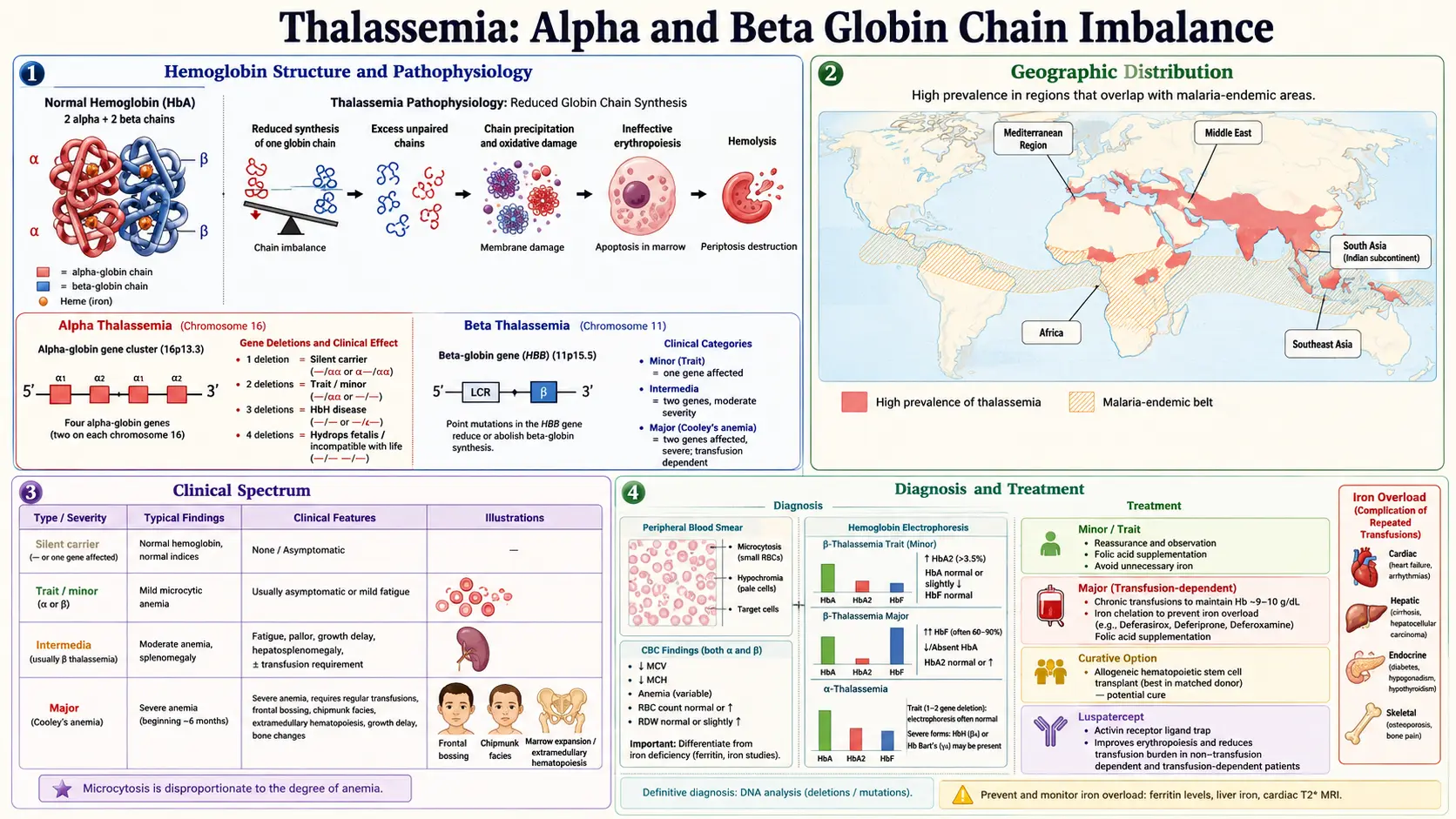
Table of Contents
- Table of Contents
- What Is Thalassemia?
- Alpha vs Beta Thalassemia
- Classification by Severity
- Genetics and Inheritance Patterns
- Geographic Prevalence
- Symptoms by Severity
- Diagnosis
- Conventional Treatment
- Supplement Research
- Iron Overload from Transfusions
- Nutrition and Iron Absorption
- Psychological Support
- Genetic Counseling
- Safety Findings
- Key Research Papers
- Connections
- Featured Videos
What Is Thalassemia?
Thalassemia is a group of inherited disorders of hemoglobin, the oxygen-carrying protein in red blood cells, in which one of the globin chains that make up hemoglobin is produced in reduced amounts or not at all. The name comes from the Greek thalassa (sea) and haima (blood) (Galanello 2010).
Adult hemoglobin is a four-part molecule built from two alpha-like and two beta-like globin chains (α2β2) (Harteveld 2010). In thalassemia the chains are produced out of balance, and the unpaired chains damage developing red cells. A 2022 review describes the result as ineffective red-cell production in the bone marrow, increased destruction of red cells (hemolysis) and disturbed iron balance, with a range of severity from close to normal to lifelong dependence on transfusions (Kattamis 2022).
Small trials of antioxidants and other supplements, which measured blood, iron and liver markers rather than long-term outcomes, are summarized in the Supplement Research section below.
Alpha vs Beta Thalassemia
Alpha Thalassemia
Alpha-globin is made from four genes, two on each copy of chromosome 16 (written αα/αα). Alpha thalassemia most often results from deletion of one (−α) or both (−−) alpha genes on a chromosome, and the degree of anemia depends roughly on how many alpha genes are affected (Harteveld 2010):
- One gene affected (−α/αα) — a “silent” carrier state; the 2010 review notes that molecular analysis is usually needed to confirm silent alpha thalassemia.
- Alpha thalassemia trait (two genes affected: −−/αα or −α/−α) — mild to moderate microcytic, hypochromic anemia without symptoms; it may be confused with iron-deficiency anemia when iron status is not measured.
- Hemoglobin H disease (three genes affected: −−/−α) — patients usually make less than 30% of the normal amount of alpha-globin; hemoglobin ranges from 2.6 to 13.3 g/dL, the spleen is usually enlarged, and jaundice, gallstones, leg ulcers, folic acid deficiency and acute hemolytic episodes with drugs and infections are described. HbH is made of four beta chains (β4) and is unstable.
- Hb Bart's hydrops fetalis (no working alpha genes: −−/−−) — the most severe form; affected infants almost always die in the womb (23–38 weeks) or shortly after birth (Harteveld 2010). A 2024 review reports that, with intensive perinatal care and transfusions given in the womb, an increasing number of infants now survive, and then need regular transfusions and iron chelation (Amid 2024).
Beta Thalassemia
Beta-globin is made from the HBB gene on chromosome 11; each person carries two copies. Most beta thalassemia mutations are point mutations, more rarely deletions, that either reduce (β+) or abolish (β0) beta-globin production (Galanello 2010):
- Beta thalassemia minor / trait (one copy affected) — usually without symptoms, sometimes with mild anemia
- Beta thalassemia intermedia — presents later, with milder anemia that by definition needs no transfusions or only occasional ones
- Beta thalassemia major (Cooley's anemia) — usually presents in the first two years of life with severe anemia that requires regular red-cell transfusions
The 2025 Thalassaemia International Federation (TIF) guidelines group patients for management by transfusion need rather than genotype alone: patients who receive six or more red-cell units over 6 months with a transfusion-free period of 6 weeks or less, or who receive frequent transfusions for more than a year, can be classed as transfusion-dependent thalassemia (TDT) (Musallam 2025).
Hemoglobin E / Beta Thalassemia
HbE is a beta-globin variant that behaves like a mild β+ thalassemia allele. Inherited together with a beta thalassemia allele, it causes HbE/β-thalassemia, an important cause of chronic childhood disease in Southeast Asia, in which HbE makes up 35% to 75% of the hemoglobin (Fucharoen 2000). Severity varies widely. A 2010 review describes three groups: a mild form (about 15% of cases in Southeast Asia) with hemoglobin of 9–12 g/dL that usually needs no treatment; a moderately severe form, the majority, with hemoglobin of 6–7 g/dL that behaves like thalassemia intermedia; and a severe form with hemoglobin as low as 4–5 g/dL that is treated like thalassemia major (Galanello 2010). Co-inheritance of alpha thalassemia lessened severity in patients with at least one mild beta thalassemia allele (Fucharoen 2000).
Classification by Severity
Thalassemia Trait (Minor)
Carriers have one affected and one normal gene. They are usually without symptoms but sometimes have mild anemia, with small red cells (low MCV), less hemoglobin per cell (low MCH) and, in beta thalassemia trait, a raised HbA2 level (Galanello 2010). Carrier status matters mainly for family planning (see Genetic Counseling).
Thalassemia Intermedia
At the severe end, patients present between the ages of 2 and 6 years and can survive without regular transfusions, though growth and development are slowed; at the mild end, people have only mild anemia and no symptoms until adult life. Expansion of the bone marrow, with blood formation outside the marrow, is common, and its consequences include bone and facial deformities, osteoporosis with fractures, and masses of blood-forming tissue. Gallstones, leg ulcers and blood clots (especially after removal of the spleen) are more common than in thalassemia major (Galanello 2010).
Thalassemia Major (Cooley's Anemia)
Thalassemia major presents between 6 and 24 months of age: infants fail to thrive and become progressively pale, and the spleen and liver enlarge. In the 2010 review, thalassemia major is characterized by hemoglobin below 7 g/dL with an MCV between 50 and 70 fL. Where patients are untreated or poorly transfused, the picture includes:
- Growth retardation, pallor, jaundice and poor musculature
- Enlarged liver and spleen (hepatosplenomegaly)
- Skeletal changes from bone-marrow expansion: deformities of the long bones of the legs and typical facial changes (bossing of the skull, prominent cheekbones, a depressed nasal bridge, and an enlarged upper jaw that exposes the upper teeth)
- Leg ulcers and masses of blood-forming tissue outside the marrow
The same review reports that people who are not regularly transfused usually die before the second or third decade of life, while survival of those regularly transfused and treated with appropriate chelation extends beyond age 40 (Galanello 2010). A 2022 review describes transfusion and chelation as having turned thalassemia major into a chronic disease with a prolonged life expectancy, though with substantial disease burden (Kattamis 2022).
Genetics and Inheritance Patterns
Beta thalassemia is inherited in an autosomal recessive pattern (rare dominant forms are also reported) (Galanello 2010):
- A child with thalassemia major or intermedia has inherited an affected beta-globin gene from both parents.
- When both parents are carriers, each pregnancy has a 25% chance of an affected child, a 50% chance of a carrier and a 25% chance of a child who is neither affected nor a carrier.
- Carriers are usually without symptoms.
Alpha thalassemia risks depend on which deletions each parent carries. When both parents carry an α0 mutation (both alpha genes missing from the same chromosome, written −−/αα), each pregnancy has a 25% risk of Hb Bart's hydrops fetalis; when one parent carries α0 and the other α+ (−α/αα), the risk of HbH disease is 25% (Harteveld 2010). A 2024 review describes Hb Bart's hydrops fetalis as the most common cause of hydrops fetalis in Southeast Asia (Amid 2024), and a 2005 review reported that in southern China and Southeast Asia two to three times more fetuses are affected by it than by beta thalassemia major or intermedia (Chui 2005).
Geographic Prevalence
A 2008 global analysis found that hemoglobin disorders are a significant health problem in 71% of 229 countries, which together include 89% of births worldwide. More than 330,000 affected infants are born each year (83% with sickle cell disorders, 17% with thalassemias), and around 7% of pregnant women carry beta or alpha-zero thalassemia or hemoglobin S, C, D Punjab or E (Modell and Darlison 2008).
- Beta thalassemia — prevalent in Mediterranean countries, the Middle East, Central Asia, India, southern China and the Far East, along the north coast of Africa and in South America. The highest carrier frequencies are reported in Cyprus (14%), Sardinia (10.3%) and Southeast Asia (Galanello 2010).
- Alpha thalassemia — especially frequent in Mediterranean countries, Southeast Asia, Africa, the Middle East and the Indian subcontinent; carriers are found at frequencies above 1% in all tropical and subtropical populations studied, and in some areas the carrier frequency may be as high as 80–90% (Harteveld 2010).
The high gene frequencies are thought to reflect protection of carriers against falciparum malaria (Galanello 2010; Harteveld 2010). Through migration, thalassemia is now found in almost every country (Galanello 2010), and the 2021 TIF guidelines note that health systems in Western countries that have received large numbers of migrant patients were not ready for their needs (Farmakis 2022).
Symptoms by Severity
Thalassemia Trait (Mild)
- Usually without symptoms; sometimes mild anemia (Galanello 2010)
- Low MCV and MCH on a blood count, which can be mistaken for iron deficiency (Harteveld 2010)
- For alpha thalassemia carriers, a 2010 review reports that complaints such as fatigue and shortness of breath are uncommon and almost certainly related to other disorders (Harteveld 2010)
Thalassemia Intermedia (Moderate)
As reported in a 2010 review (Galanello 2010):
- Enlarged spleen (splenomegaly)
- Gallstones, more common than in thalassemia major
- Bone changes — bone and facial deformities, osteoporosis with fractures of long bones
- Masses of blood-forming tissue outside the marrow, which can compress the spinal cord
- Leg ulcers
- Blood clots — deep vein thrombosis, portal vein thrombosis, stroke and pulmonary embolism, especially after splenectomy
- Iron overload even without transfusions, from increased intestinal iron absorption
Thalassemia Major (Severe)
As reported in a 2010 review (Galanello 2010):
- Severe anemia appearing between 6 and 24 months of age
- Failure to thrive, feeding problems, diarrhea, irritability and recurrent fever
- Progressive pallor and enlargement of the abdomen from an enlarged spleen and liver
- Craniofacial and long-bone changes from marrow expansion when transfusion is absent or inadequate
- Growth retardation and failure or delay of sexual maturation as complications of iron overload in children
- Iron overload complications later in life in the heart, liver and endocrine glands (see Iron Overload)
Diagnosis
The 2025 TIF guidelines call for thalassemia to be considered in everyone with hypochromic microcytic anemia, and for iron-deficiency anemia always to be excluded in the work-up (Musallam 2025).
Complete Blood Count (CBC)
- Low hemoglobin — below 7 g/dL in thalassemia major and between 7 and 10 g/dL in thalassemia intermedia (Galanello 2010)
- Low MCV (mean corpuscular volume) — small red cells
- Low MCH (mean corpuscular hemoglobin) — less hemoglobin per cell
- Red cell count — a raised red cell count with normal ferritin points toward alpha thalassemia carrier status (Harteveld 2010)
- Peripheral blood smear — small, pale and irregular red cells (microcytosis, hypochromia, anisocytosis, poikilocytosis) and, in affected individuals, nucleated red cells, whose number rises markedly after splenectomy (Galanello 2010)
Hemoglobin Electrophoresis
Separation and measurement of the hemoglobin fractions (by electrophoresis or HPLC) is the basis for identifying beta thalassemia carriers (Musallam 2025):
- Beta thalassemia trait — raised HbA2. Co-inherited delta thalassemia can bring HbA2 back to normal, and very mild “silent” mutations can give normal or borderline values (Galanello 2010).
- Beta thalassemia major — in β0 homozygotes, HbA is absent and HbF makes up 92–95% of the total; in β+ homozygotes and β+/β0 compound heterozygotes, HbA is 10–30% and HbF 70–90% (Galanello 2010).
- Alpha thalassemia — HbA2 is normal or slightly reduced; HbH is shown in HbH disease, and Hb Bart's in many newborns carrying alpha thalassemia (Harteveld 2010).
Genetic Testing
- DNA analysis identifies the specific mutations in the alpha- or beta-globin genes.
- The 2025 TIF guidelines state that molecular analysis is not required to confirm a beta thalassemia carrier but is necessary to confirm alpha thalassemia carrier status (Musallam 2025).
- Both disease-causing alleles must be identified before prenatal testing can be performed (Galanello 2010).
Iron Studies
- Serum ferritin separates thalassemia trait from iron deficiency; a 2010 review notes that alpha thalassemia trait may be confused with iron-deficiency anemia when iron status is not carefully assessed (Harteveld 2010).
Conventional Treatment
Blood Transfusions
Regular transfusion is the standard treatment for transfusion-dependent thalassemia. The 2021 TIF guidelines describe an effective regimen as one that gives good growth and development, good energy levels and sufficient suppression of blood formation inside and outside the marrow (Farmakis 2022). The 2025 TIF guidelines state (Musallam 2025):
- Transfusion every 2–5 weeks, targeting a pre-transfusion hemoglobin of 9.5–10.5 g/dL
- A higher pre-transfusion target of 10–11 g/dL (or as high as practicable) for patients with heart disease including pulmonary hypertension, significant blood formation outside the marrow, or inadequate marrow suppression at the lower level
- A pre-transfusion hemoglobin of 10 g/dL in pregnancy
- A post-transfusion hemoglobin below 13–15 g/dL
- Extended red-cell antigen typing before the first transfusion and units matched for C, c, E, e and Kell antigens, with leukodepleted red cells
Earlier, the 2021 edition gave transfusions every 2–4 weeks to keep pre-transfusion hemoglobin above 90–105 g/L, or up to 110–120 g/L with cardiac complications (Farmakis 2022). Risks include iron overload, red-cell antibodies and transfusion reactions.
Iron Chelation Therapy
Because the body has no mechanism to excrete excess iron, iron overload is inevitable with regular transfusion (Farmakis 2022). The 2025 TIF guidelines state that chelation improves survival and decreases the risk of heart failure and other iron-related illness, and that its optimal regimen depends on local prescribing information and the individual's iron profile (Musallam 2025). Three chelators are licensed:
- Deferoxamine — not absorbed by mouth. The US label describes slow subcutaneous infusion by portable pump over 8 to 12 hours, normally 5 to 7 times a week (US label, deferoxamine). Local reactions at the infusion site are the most frequent adverse effects (Galanello 2010).
- Deferasirox — taken by mouth once daily; the US label covers transfusional iron overload from age 2 and carries a boxed warning for renal failure, hepatic failure and gastrointestinal hemorrhage, each of which can be fatal (US label, deferasirox). In a phase 3 trial in patients aged 2 and older randomized to deferasirox (296) or deferoxamine (290), patients with liver iron of 7 mg/g dry weight or more had similar dose-dependent falls in liver iron on both drugs, but the trial's primary endpoint was not met in the overall population, possibly because patients with lower liver iron received proportionally lower deferasirox doses (Cappellini 2006).
- Deferiprone — taken by mouth as twice-daily or three-times-daily tablets; the US label covers transfusional iron overload from age 8 and carries a boxed warning for agranulocytosis and neutropenia: deferiprone can cause agranulocytosis leading to serious infections and death, and neutropenia may precede it. Agranulocytosis occurred in 1% of 642 patients with thalassemia syndromes in pooled trials, and the label calls for measuring the neutrophil count before and during treatment. The label also lists decreased plasma zinc (US label, deferiprone). In a 4-year study (531 patient-years), the rate of agranulocytosis was 0.2 and of milder neutropenia 2.8 per 100 patient-years, and neutropenia was more common in patients with an intact spleen (Cohen 2003). In a randomized trial of 61 patients with myocardial iron but no symptoms, myocardial T2* improved more on deferiprone than on deferoxamine over a year (27% vs 13%), as did left ventricular ejection fraction (3.1 vs 0.3 absolute percentage points); no agranulocytosis occurred in that trial (Pennell 2006). A 2010 review reports that deferoxamine and deferiprone used together achieve iron excretion that neither achieves alone (Galanello 2010).
Luspatercept
Luspatercept is a recombinant fusion protein that binds selected TGF-β superfamily ligands and is meant to improve red-cell maturation (Cappellini 2020). Its US label covers anemia in adults with beta thalassemia who require regular red-cell transfusions (US label, luspatercept). In the phase 3 BELIEVE trial, 336 adults with transfusion-dependent beta thalassemia were randomized 2:1 to luspatercept or placebo on top of supportive care. A reduction in transfusion burden of at least 33% (plus at least 2 units) during weeks 13 to 24 occurred in 21.4% versus 4.5%; during any 12-week interval, a reduction of at least 33% occurred in 70.5% versus 29.5% and of at least 50% in 40.2% versus 6.3%. Transient bone pain, joint pain, dizziness, high blood pressure and raised uric acid were more common with luspatercept (Cappellini 2020). The 2025 TIF guidelines recommend luspatercept for adults with TDT to reduce transfusion burden, and note more thromboembolic events with luspatercept than with placebo in BELIEVE, an average rise of 5 mmHg in blood pressure, and cases of blood-forming masses outside the marrow (Musallam 2025).
Bone Marrow Transplant (BMT)
Allogeneic hematopoietic stem cell transplantation replaces the patient's blood-forming cells with a donor's and is the long-established curative treatment (Galanello 2010; Kattamis 2022):
- In a European registry of 1,493 patients with thalassemia major transplanted between 2000 and 2010, 2-year overall survival was 88% and thalassemia-free survival 81%. Transplants from an HLA-identical sibling gave the best results (overall survival 91%, thalassemia-free survival 83%), and the threshold age for optimal outcomes was around 14 years, with overall survival of 90–96% before that age (Baronciani 2016).
- A 2010 review reports that approximately 25–30% of patients could have a matched sibling donor, and that chronic graft-versus-host disease of variable severity occurs in 5–8% (Galanello 2010).
- The 2025 TIF guidelines call for transplantation to be offered at an early age, before iron-overload complications develop, when an HLA-identical donor is available; state that a closely matched unrelated donor can be used; and limit haploidentical transplantation to experienced centers within clinical trials (Musallam 2025).
Gene Therapy
Two gene therapies are approved by the US Food and Drug Administration (FDA) for beta thalassemia. Both are made from the patient's own blood stem cells, which are collected, modified in a laboratory and given back once, after full myeloablative conditioning: busulfan for betibeglogene, and busulfan or another myeloablative regimen for exagamglogene, according to their labels (FDA label, betibeglogene autotemcel; FDA label, exagamglogene autotemcel).
- Betibeglogene autotemcel (beti-cel) — lentiviral gene addition. The FDA approved it on August 17, 2022 “for the treatment of adult and pediatric patients with ß-thalassemia who require regular red blood cell (RBC) transfusions” (FDA approval letter, August 17, 2022), describing it as the first cell-based gene therapy for this group (FDA product page). The stem cells are transduced with the BB305 lentiviral vector, which carries a modified beta-globin gene (βA-T87Q) (Locatelli 2022). In the label's two phase 3 studies (41 patients aged 4 to 34), 32 of 36 evaluable patients (89%) achieved transfusion independence, defined as a weighted average hemoglobin of at least 9 g/dL without transfusions for 12 months or more. In HGB-207, in patients with a non-β0/β0 genotype, transfusion independence occurred in 20 of 22 evaluable patients (91%), including 6 of 7 younger than 12, with an average hemoglobin of 11.7 g/dL during transfusion independence and no cancers observed (Locatelli 2022). In HGB-212, in patients with severe genotypes (β0/β0, β0/β+IVS-I-110 or β+IVS-I-110/β+IVS-I-110), 16 of 18 patients (89%) reached and maintained transfusion independence over a median follow-up of 47.9 months, with no serious adverse events attributed to beti-cel and no deaths (Kwiatkowski 2024). In long-term follow-up of 63 treated participants (median 5.9 years), transfusion independence was achieved by 37 of 41 phase 3 participants (90.2%) and 15 of 22 phase 1/2 participants (68.2%); 38 of 52 who achieved it had stopped iron chelation, and no malignancies or insertional oncogenesis were reported (Kwiatkowski 2026). The label lists a potential risk of lentiviral vector–mediated insertional oncogenesis and calls for monitoring for blood cancers for at least 15 years (FDA label, betibeglogene autotemcel).
- Exagamglogene autotemcel (exa-cel) — CRISPR-Cas9 gene editing. The stem cells are edited with CRISPR-Cas9 at the erythroid-specific enhancer of BCL11A to reactivate fetal hemoglobin production; no viral vector is used (Locatelli 2024). It was first approved by the FDA on December 8, 2023 for sickle cell disease with recurrent vaso-occlusive crises in patients 12 and older (FDA approval letter, December 8, 2023); the FDA approved it for transfusion-dependent beta thalassemia on January 16, 2024, “for the treatment of patients aged 12 years and older with transfusion-dependent ß-thalassemia (TDT)” (FDA approval letter, January 16, 2024). On July 1, 2026 the FDA approved an expansion of the indication to patients aged 2 to under 12 years (FDA approval letter, July 1, 2026); the current label covers patients aged 2 years and older with TDT, notes that it has not been studied in patients younger than 5, and supports use from 2 to under 5 years by extrapolation from trial data (FDA label, exagamglogene autotemcel; FDA product page). In the phase 3 CLIMB THAL-111 study of patients aged 12 to 35, 52 patients received exa-cel; among the 35 with enough follow-up, 32 (91%) achieved transfusion independence, with a mean total hemoglobin of 13.1 g/dL and fetal hemoglobin of 11.9 g/dL, and no deaths or cancers occurred (Locatelli 2024). In the label's pediatric study (ages 5 to under 12), 8 of 9 evaluable patients (89%) achieved transfusion independence; one patient died of hepatic veno-occlusive disease after treatment. The label also states that the risk of unintended off-target editing cannot be ruled out and that its clinical significance is unknown (FDA label, exagamglogene autotemcel).
The HGB-212 investigators describe beti-cel as a potentially curative option without the risks and limitations of allogeneic transplantation (Kwiatkowski 2024); both therapies, however, still require full myeloablative conditioning. The 2025 TIF guidelines describe gene therapy as an optimal option for patients aged 14 and older or without an HLA-identical family donor, while transplantation from an HLA-identical sibling remains the preferred curative option for children up to 14, and they note that beti-cel was then available only in the USA (Musallam 2025).
Splenectomy
The 2021 TIF guidelines report that current transfusion targets have considerably reduced the need for splenectomy, and that the guideline panel does not recommend it as a standard procedure because it is linked to pulmonary hypertension, silent brain infarcts, venous thrombosis and sepsis. The guidelines list three situations in which it is considered (Farmakis 2022):
- Increased blood requirement that prevents adequate control of iron with chelation
- Hypersplenism
- Symptomatic splenomegaly
The same guidelines advise against splenectomy in children under 5 because of a considerably greater risk of fulminant post-splenectomy sepsis, and list immunization against Streptococcus pneumoniae, Haemophilus influenzae type B and Neisseria meningitidis at least 2 weeks before the operation (Farmakis 2022). A 2010 review describes prevention of post-splenectomy sepsis as immunization, antibiotic prophylaxis and early antibiotic treatment for fever, and notes an increase in thromboembolic events after splenectomy (Galanello 2010).
Supplement Research
Most supplement studies in thalassemia are small, short trials, often from single centers, that measured oxidative-stress, iron or liver markers rather than survival or transfusion need. The 2021 TIF guidelines summarize nutritional supplementation as follows (Farmakis 2022), and the trials below add detail.
Folic Acid
The 2021 TIF guidelines state that folic acid supplements of up to 1 mg/day are needed for all patients with low hemoglobin levels and may be considered for all patients (Farmakis 2022). A 2010 review reports that untransfused patients and those on low transfusion regimens use more folate and may develop a relative deficiency, while patients on high transfusion regimens rarely do (Galanello 2010).
Vitamin E
Vitamin E is a fat-soluble antioxidant reported to be depleted in thalassemia as a result of iron overload (ElLaboudy 2025). The 2021 TIF guidelines describe a diet rich in vitamin E foods such as eggs and vegetable oils and state that prolonged use of supplements requires further research (Farmakis 2022); a 2010 review stated that its effectiveness and safety in thalassemia major had not been formally assessed (Galanello 2010). In a 3-month open-label trial in 78 adults with TDT, total oxidative stress fell with vitamin E (10 U/kg/day) but hemoglobin did not change (Haghpanah 2021). In a 12-month trial of 180 children with TDT on one of three chelators, those randomized to vitamin E 400 mg daily had lower liver iron, serum ferritin and transfusion index and higher hemoglobin and cardiac T2* than those on placebo (ElLaboudy 2025).
Antioxidant Studies
- N-acetyl cysteine (NAC) — in the same 3-month trial, total oxidative stress did not fall significantly in the NAC group (10 mg/kg/day), and hemoglobin did not change (Haghpanah 2021).
- Curcumin — in a 12-week double-blind trial in 68 patients with thalassemia major, curcumin 1,000 mg/day lowered non-transferrin-bound iron and the liver enzymes ALT and AST compared with placebo; hepcidin and the other measures, including ferritin, did not change (Mohammadi 2018).
- Green tea extract (EGCG) — in a 60-day placebo-controlled trial in 27 patients with TDT, the authors reported improved hemoglobin and lower plasma erythroferrone with green tea extract tablets (Settakorn 2023).
- Silymarin (milk thistle) — a 2021 meta-analysis found lower serum ferritin when silymarin was added to chelation (eight trials, 477 patients) but no significant change in liver iron (two trials, 106 patients), with very high heterogeneity, and concluded that there is no evidence that adding silymarin to standard chelators reduces iron load in TDT (Darvishi-Khezri 2021).
L-Carnitine
The 2021 TIF guidelines state that L-carnitine may be beneficial at 50 mg/kg/day, with caution in patients with thyroid dysfunction (Farmakis 2022). In an uncontrolled study of 30 patients given 50 mg/kg/day for 6 months, echocardiography showed no significant change, while nuclear imaging in 20 patients showed a rise in left ventricular ejection fraction from 58.25% to 63.95% (El-Beshlawy 2004). In a 6-month randomized trial of 60 patients, those given L-carnitine had less left ventricular dilatation and hypertrophy than controls (Shahidi 2020).
Vitamin D and Calcium
Osteoporosis is present in approximately 40–50% of patients with thalassemia major (Farmakis 2022). In a UK clinic of 99 adults with thalassemia major, 29.9% had vitamin D deficiency, 65.7% insufficiency and 12.4% optimal levels; vitamin D status was not associated with bone mass (Tzoulis 2014). The 2021 TIF guidelines recommend calcium and vitamin D supplements (2,000 IU/day of vitamin D) for all patients, with vitamin D levels measured every 6 months (Farmakis 2022).
Zinc
Zinc deficiency is among the factors listed for poor growth in thalassemia major (Farmakis 2022), and decreased plasma zinc has been observed with deferiprone (US label, deferiprone). In a controlled study of 32 patients with thalassemia major, of whom 40.6% were below the 10th height percentile with biochemical zinc deficiency, only those given zinc sulphate showed accelerated growth in height (Arcasoy 1987). In an 18-month double-blind trial of 42 patients aged 10 to 30 with low bone mass, zinc 25 mg/day gave greater gains in total-body bone mass than placebo (Fung 2013). The 2021 TIF guidelines state that zinc supplements may be given for deficiency, poor growth and reduced bone mass but are not recommended as routine for all patients (Farmakis 2022).
Iron Overload from Transfusions
Iron overload in thalassemia comes from transfusions and from increased absorption of iron from the gut; transfusion is the main cause in TDT, and gut absorption is more important in non-transfusion-dependent thalassemia (Farmakis 2022). The 2025 TIF guidelines state that uncontrolled transfusional iron overload increases the risks of heart failure, endocrine damage, liver cirrhosis and liver cancer (Musallam 2025). Cardiac disease from iron in the heart muscle is described as the most important life-limiting complication of iron overload in beta thalassemia (Galanello 2010).
Cardiac Iron Overload
- The 2021 TIF guidelines describe cardiovascular complications as the leading cause of death in thalassemia, with cardiovascular deaths falling markedly in recent cohorts with MRI-guided chelation (Farmakis 2022). In the UK, deaths from iron overload fell from 7.9 to 2.3 per 1,000 patient-years between 1980–1999 and 2000–2003; the authors attribute the fall mainly to identification of heart iron by T2* MRI and intensified chelation, alongside other improvements in care (Modell 2008).
- Heart iron is measured by cardiac MRI T2*. In the study that developed the method, among 106 patients with thalassemia major, all those with ventricular dysfunction had a myocardial T2* below 20 ms, and myocardial iron could not be predicted from serum ferritin or liver iron (Anderson 2001). In 652 patients from 21 UK centers, a cardiac T2* below 10 ms carried a relative risk of 160 for heart failure, and a value below 20 ms a relative risk of 4.6 for arrhythmia (Kirk 2009).
- The 2025 TIF guidelines state that chelation removes stored heart iron slowly (months or years) but can reverse iron-related heart dysfunction within weeks when 24-hour chelation cover is achieved (Musallam 2025).
Liver Iron Overload
- Cardiac iron accumulates later than liver iron and is rare before the age of 8 years (Musallam 2025).
- Liver iron concentration (LIC) is measured by MRI. The 2021 TIF guidelines give normal LIC as up to 1.8 mg/g dry weight, note that levels up to 7 mg/g are seen in some non-thalassemic populations without apparent harm, and link sustained levels above 15 mg/g to a worse prognosis, progression of liver fibrosis and abnormal liver function (Farmakis 2022).
- Hepatitis C or hepatitis B infection may further accelerate liver fibrosis in thalassemia (Farmakis 2022).
Endocrine Complications
Endocrine complications of iron overload include growth retardation, failure of sexual maturation, diabetes mellitus and insufficiency of the parathyroid, thyroid, pituitary and, less commonly, adrenal glands (Galanello 2010):
- Diabetes mellitus and impaired glucose tolerance — increasing with age, reported in 10% to 24% (Farmakis 2022)
- Hypogonadism — delayed or failed sexual maturation and subfertility
- Hypothyroidism
- Hypoparathyroidism
- Growth hormone deficiency — among the contributors to short stature (Farmakis 2022)
Nutrition and Iron Absorption
A 2010 review states that patients with thalassemia do not have specific dietary requirements unless they have special prescriptions, and that adding restrictions without a likely clear benefit is counterproductive for people who already carry a heavy treatment schedule (Galanello 2010).
What Guidelines and Reviews Report
- Iron in food — there is no evidence that iron-poor diets are useful in thalassemia major; the 2010 review names only foods very rich in iron (such as liver, and many baby foods, breakfast cereals and multivitamins with added iron) as the ones to leave out (Galanello 2010). The 2021 TIF guidelines recommend dietary iron restriction for patients on low transfusion regimens with low pre-transfusion hemoglobin (Farmakis 2022).
- Tea with meals — drinking a glass of black tea with meals reduces iron absorption from food, particularly in thalassemia intermedia (Galanello 2010).
- Calcium — the 2010 review describes a diet with adequate calcium (milk, cheese, dairy products, kale) but notes kidney stones in some adults and reserves calcium supplements for a clear indication (Galanello 2010); the 2021 TIF guidelines recommend calcium and vitamin D supplements for all patients and a diet high in calcium (Farmakis 2022).
- Vitamin C — vitamin C can increase iron absorption from the gut and labile iron, and the 2010 review describes it as potentially harmful in patients not receiving deferoxamine; given at the time of a deferoxamine infusion, it may increase the iron available for chelation (Galanello 2010). The 2021 TIF guidelines list vitamin C supplements with deferoxamine infusions (2–3 mg/kg/day) or for proven deficiency (Farmakis 2022).
- Refined carbohydrates — during adolescence and adult life, a diet low in highly refined carbohydrates may help prevent or delay impaired glucose tolerance or diabetes (Galanello 2010).
Psychological Support
The 2021 TIF guidelines note a general lack of large randomized trials on psychological support in thalassemia, with most published reports descriptive, and summarize the findings as follows (Farmakis 2022):
- Vulnerability — patients with thalassemia are vulnerable to psychological challenges.
- Adherence — psychological well-being affects adherence to chelation and hence survival.
- Treatment route — patient-reported outcomes favor oral over injected chelation.
- Cognition — clear intellectual and psychopathological problems are found in a very limited number of patients.
- Approaches — suggested benefits come from changes in institutional practice, patient group sessions, family therapy and patient chelation camps; the panel states that expert psychological support has to be available at all centers specializing in thalassemia care.
- Transition to adult care — the guidelines describe problems with the transition from pediatric to adult care in inherited chronic disease and the need for continuity of care.
Genetic Counseling
Prevention of beta thalassemia rests on carrier identification, genetic counseling and prenatal diagnosis (Galanello 2010):
- Carrier screening — a 2017 obstetric committee opinion states that information about carrier screening should be provided to every pregnant woman, that screening ideally takes place before pregnancy, and that a patient may decline any or all screening (Committee Opinion No. 691, 2017). A 2008 global analysis concluded that screening for hemoglobin disorders should form part of basic health services in most countries (Modell and Darlison 2008). Red-cell indices followed by measurement of hemoglobin fractions identify beta thalassemia carriers; alpha carriers need molecular testing (Musallam 2025).
- Partner testing — the same committee opinion states that the partner of a carrier should be offered testing, and that genetic counseling should be offered when both are carriers (Committee Opinion No. 691, 2017). Counseling covers the mode of inheritance, the risk of affected children, and the natural history and treatments of the disease (Galanello 2010).
- Prenatal diagnosis — DNA from fetal cells obtained by chorionic villus sampling at about 11 weeks or amniocentesis at about 15–18 weeks; preimplantation genetic diagnosis may be available when the family's mutations are known (Galanello 2010).
- Population programs — prevention programs combining carrier detection, molecular diagnosis, counseling and prenatal diagnosis operate in many countries in Europe, Asia and Australia, often drawing on the Sardinian experience (Cao 2013). In Sardinia, the program was followed by a 90% decline in thalassemia major births (Cao 1989).
Safety Findings
- Iron given for a misdiagnosis — thalassemia trait can be mistaken for iron-deficiency anemia (Harteveld 2010), and the 2025 TIF guidelines call for iron deficiency to be excluded in every work-up of small, pale red cells (Musallam 2025). Iron accumulates in thalassemia because the body has no mechanism to excrete it (Farmakis 2022).
- Supplements and transfusion — the supplement trials summarized above were add-on studies in patients already receiving standard care; without regular transfusion, patients with thalassemia major usually die before the second or third decade (Galanello 2010).
- Supplements and chelation — the trials of curcumin, green tea extract and silymarin above tested them alongside standard chelators, not instead of them, and a meta-analysis found no evidence that silymarin added to chelation reduces liver iron (Darvishi-Khezri 2021).
- Chelator monitoring — deferiprone carries a boxed warning for agranulocytosis and deferasirox for renal failure, hepatic failure and gastrointestinal hemorrhage (see Iron Chelation Therapy).
- Drugs and infections in HbH disease — acute hemolytic episodes in response to drugs and infections are described in HbH disease (Harteveld 2010).
- After splenectomy — the risk of severe infection with encapsulated bacteria rises, and early antibiotic treatment for fever is part of the prevention described in a 2010 review (Galanello 2010).
- Pregnancy — the 2021 TIF guidelines describe pregnancy in thalassemia major as high risk for mother and baby, with risks reduced by pre-pregnancy counseling with the hematologist, reproductive specialist, cardiologist and obstetrician (Farmakis 2022).
- Decisions about medicines and supplements belong with a clinician experienced in thalassemia care.
Key Research Papers
Peer-reviewed reviews, guidelines and trials cited on this page, each verified against its PubMed record. FDA approval letters and US drug labels are linked where they are cited in the text.
- Kattamis A, Kwiatkowski JL, Aydinok Y. (2022). Thalassaemia. Lancet 399:2310-2324. — PubMed PMID: 35691301
- Galanello R, Origa R. (2010). Beta-thalassemia. Orphanet J Rare Dis 5:11. — PubMed PMID: 20492708
- Harteveld CL, Higgs DR. (2010). Alpha-thalassaemia. Orphanet J Rare Dis 5:13. — PubMed PMID: 20507641
- Amid A, Liu S, Babbs C, et al. (2024). Hemoglobin Bart's hydrops fetalis: charting the past and envisioning the future. Blood 144:822-833. — PubMed PMID: 38457773
- Chui DH. (2005). Alpha-thalassemia: Hb H disease and Hb Barts hydrops fetalis. Ann N Y Acad Sci 1054:25-32. — PubMed PMID: 16339648
- Fucharoen S, Winichagoon P. (2000). Clinical and hematologic aspects of hemoglobin E beta-thalassemia. Curr Opin Hematol 7:106-12. — PubMed PMID: 10698297
- Musallam KM, Cappellini MD, Porter JB, et al. (2025). TIF Guidelines for the Management of Transfusion-Dependent β-Thalassemia. Hemasphere 9:e70095. — PubMed PMID: 40045934
- Farmakis D, Porter J, Taher A, et al. (2022). 2021 Thalassaemia International Federation Guidelines for the Management of Transfusion-dependent Thalassemia. Hemasphere 6:e732. — PubMed PMID: 35928543
- Modell B, Darlison M. (2008). Global epidemiology of haemoglobin disorders and derived service indicators. Bull World Health Organ 86:480-7. — PubMed PMID: 18568278
- Locatelli F, Thompson AA, Kwiatkowski JL, et al. (2022). Betibeglogene Autotemcel Gene Therapy for Non-β(0)/β(0) Genotype β-Thalassemia. N Engl J Med 386:415-427. — PubMed PMID: 34891223
- Kwiatkowski JL, Walters MC, Hongeng S, et al. (2024). Betibeglogene autotemcel gene therapy in patients with transfusion-dependent, severe genotype β-thalassaemia (HGB-212): a non-randomised, multicentre, single-arm, open-label, single-dose, phase 3 trial. Lancet 404:2175-2186. — PubMed PMID: 39527960
- Kwiatkowski JL, Thompson AA, Schneiderman J, et al. (2026). Long-term efficacy and safety results of betibeglogene autotemcel gene therapy for transfusion-dependent β-thalassemia. Blood 147:2203-2214. — PubMed PMID: 41525466
- Locatelli F, Lang P, Wall D, et al. (2024). Exagamglogene Autotemcel for Transfusion-Dependent β-Thalassemia. N Engl J Med 390:1663-1676. — PubMed PMID: 38657265
- Cappellini MD, Viprakasit V, Taher AT, et al. (2020). A Phase 3 Trial of Luspatercept in Patients with Transfusion-Dependent β-Thalassemia. N Engl J Med 382:1219-1231. — PubMed PMID: 32212518
- Baronciani D, Angelucci E, Potschger U, et al. (2016). Hemopoietic stem cell transplantation in thalassemia: a report from the European Society for Blood and Bone Marrow Transplantation Hemoglobinopathy Registry, 2000-2010. Bone Marrow Transplant 51:536-41. — PubMed PMID: 26752139
- Cappellini MD, Cohen A, Piga A, et al. (2006). A phase 3 study of deferasirox (ICL670), a once-daily oral iron chelator, in patients with beta-thalassemia. Blood 107:3455-62. — PubMed PMID: 16352812
- Cohen AR, Galanello R, Piga A, et al. (2003). Safety and effectiveness of long-term therapy with the oral iron chelator deferiprone. Blood 102:1583-7. — PubMed PMID: 12763939
- Pennell DJ, Berdoukas V, Karagiorga M, et al. (2006). Randomized controlled trial of deferiprone or deferoxamine in beta-thalassemia major patients with asymptomatic myocardial siderosis. Blood 107:3738-44. — PubMed PMID: 16352815
- Anderson LJ, Holden S, Davis B, et al. (2001). Cardiovascular T2-star (T2*) magnetic resonance for the early diagnosis of myocardial iron overload. Eur Heart J 22:2171-9. — PubMed PMID: 11913479
- Kirk P, Roughton M, Porter JB, et al. (2009). Cardiac T2* magnetic resonance for prediction of cardiac complications in thalassemia major. Circulation 120:1961-8. — PubMed PMID: 19801505
- Modell B, Khan M, Darlison M, et al. (2008). Improved survival of thalassaemia major in the UK and relation to T2* cardiovascular magnetic resonance. J Cardiovasc Magn Reson 10:42. — PubMed PMID: 18817553
- Haghpanah S, Cohan N, Bordbar M, et al. (2021). Effects of three months of treatment with vitamin E and N-acetyl cysteine on the oxidative balance in patients with transfusion-dependent β-thalassemia. Ann Hematol 100:635-644. — PubMed PMID: 33216196
- ElLaboudy MA, Saber MM, Adly AA, et al. (2025). Oxidative stress markers and tissue iron overload after 12-months vitamin E supplementation for children with transfusion-dependent β-thalassemia on different iron chelators: A randomized placebo-controlled trial. Clin Nutr 50:154-163. — PubMed PMID: 40424814
- Mohammadi E, Tamaddoni A, Qujeq D, et al. (2018). An investigation of the effects of curcumin on iron overload, hepcidin level, and liver function in β-thalassemia major patients: A double-blind randomized controlled clinical trial. Phytother Res 32:1828-1835. — PubMed PMID: 29806132
- Settakorn K, Hantrakool S, Petiwathayakorn T, et al. (2023). A randomized placebo-controlled clinical trial of oral green tea epigallocatechin 3-gallate on erythropoiesis and oxidative stress in transfusion-dependent β-thalassemia patients. Front Mol Biosci 10:1248742. — PubMed PMID: 38328786
- Darvishi-Khezri H, Naderisorki M, Zahedi M, et al. (2021). Coadministration of silymarin with iron chelators in transfusion-dependent β-thalassemia patients: a systematic review and meta-analysis for effect on iron overload. Expert Rev Clin Pharmacol 14:1445-1453. — PubMed PMID: 34486906
- El-Beshlawy A, Ragab L, Fattah AA, et al. (2004). Improvement of cardiac function in thalassemia major treated with L-carnitine. Acta Haematol 111:143-8. — PubMed PMID: 15034235
- Shahidi M, Hashemi SR, Fattahi N, et al. (2020). The Effects of L-Carnitine on Echocardiographic Changes in Patients With β-Thalassemia Major and Intermedia. J Pediatr Hematol Oncol 42:386-390. — PubMed PMID: 32555029
- Tzoulis P, Ang AL, Shah FT, et al. (2014). Prevalence of low bone mass and vitamin D deficiency in β-thalassemia major. Hemoglobin 38:173-8. — PubMed PMID: 24762040
- Arcasoy A, Cavdar A, Cin S, et al. (1987). Effects of zinc supplementation on linear growth in beta-thalassemia (a new approach). Am J Hematol 24:127-36. — PubMed PMID: 3812466
- Fung EB, Kwiatkowski JL, Huang JN, et al. (2013). Zinc supplementation improves bone density in patients with thalassemia: a double-blind, randomized, placebo-controlled trial. Am J Clin Nutr 98:960-71. — PubMed PMID: 23945720
- Committee Opinion (2017). Committee Opinion No. 691 Summary: Carrier Screening for Genetic Conditions. Obstet Gynecol 129:597-599. — PubMed PMID: 28225421
- Cao A, Kan YW. (2013). The prevention of thalassemia. Cold Spring Harb Perspect Med 3:a011775. — PubMed PMID: 23378598
- Cao A, Rosatelli C, Galanello R, et al. (1989). The prevention of thalassemia in Sardinia. Clin Genet 36:277-85. — PubMed PMID: 2598483
PubMed Topic Searches
- PubMed: Beta thalassemia
- PubMed: Alpha thalassemia
- PubMed: Thalassemia major transfusion
- PubMed: Iron chelation thalassemia
- PubMed: Luspatercept thalassemia
- PubMed: Thalassemia hematopoietic stem cell transplant
- PubMed: Thalassemia gene therapy
- PubMed: Thalassemia intermedia
- PubMed: Thalassemia prenatal screening
- PubMed: Hemoglobinopathy genetics
- PubMed: Thalassemia management guidelines
- PubMed: Thalassemia cardiac iron overload