Serum Calcium Test: Total and Ionized Calcium, Albumin Correction, and Reference Ranges
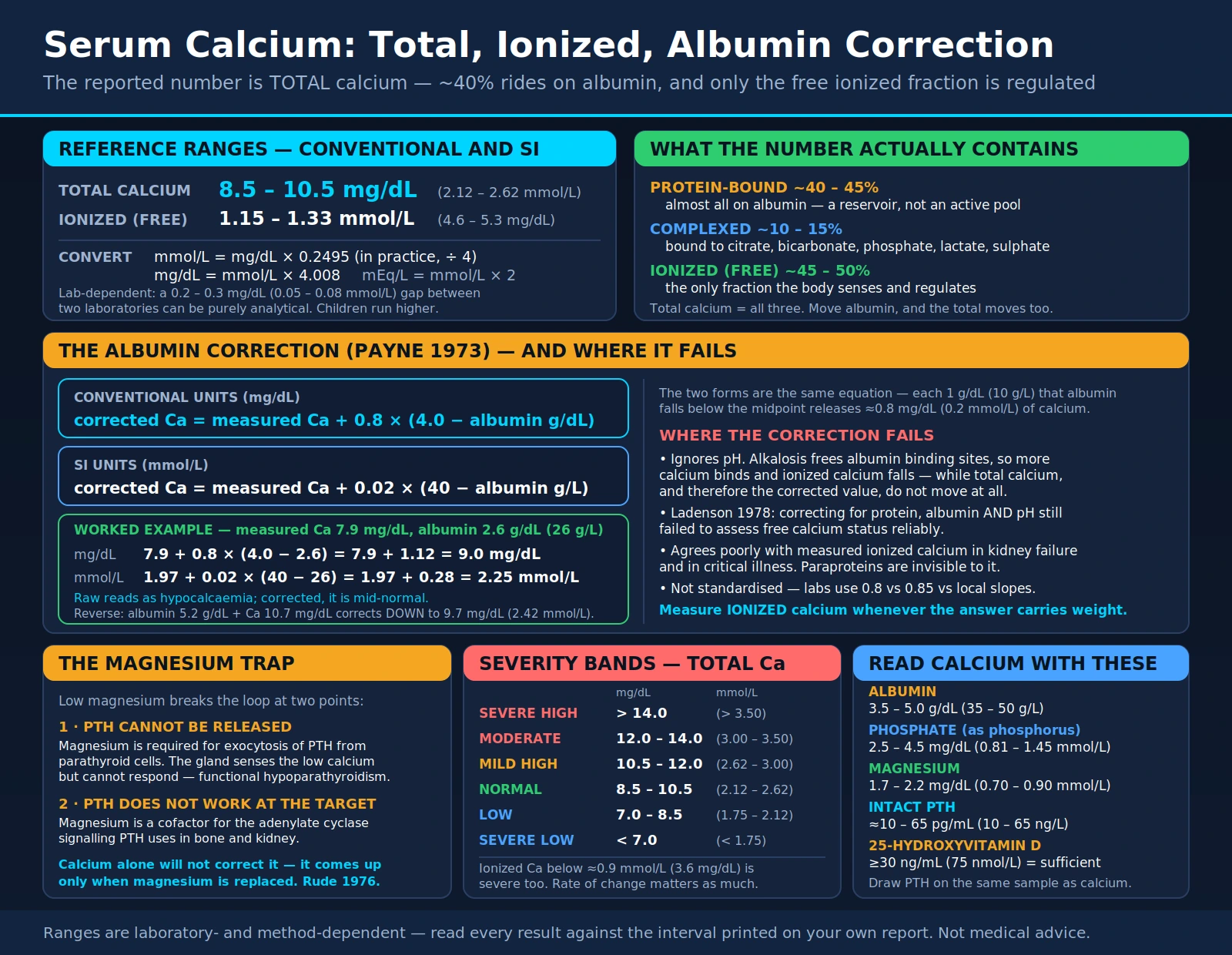
Serum calcium is one of the most frequently measured analytes in medicine — it is part of both the basic and the comprehensive metabolic panel (MedlinePlus) — and it is also one of the most frequently misread. The number on the report is total calcium, which sums three physically different pools: calcium bound to proteins (mainly albumin), calcium complexed to small anions, and the free ionized calcium that is the biologically active fraction. MedlinePlus states that about one half of the calcium in the blood is attached to proteins, mainly albumin, so anything that moves albumin moves total calcium without changing the active fraction. This page covers what the two calcium measurements mean, when each is used, how the albumin-adjustment formula was derived and how it has performed in studies, what raises and lowers calcium, and why calcium is read alongside parathyroid hormone, phosphate, vitamin D, magnesium, and albumin.
Table of Contents
- What Serum Calcium Measures
- Total Calcium vs Ionized Calcium
- Reference Ranges and Units
- The Albumin Correction and Its Limits
- When the Test Is Ordered
- Causes of Hypercalcaemia
- Symptoms and Severity of Hypercalcaemia
- Causes of Hypocalcaemia
- Symptoms and Signs of Hypocalcaemia
- Magnesium Depletion: The Calcium That Will Not Correct
- Reading Calcium With PTH, Phosphate, Vitamin D and Albumin
- Pre-Analytical and Sample-Handling Pitfalls
- Key Research Papers
- Connections
- Featured Videos
What Serum Calcium Measures
Approximately 99 percent of total body calcium is held in the skeleton as hydroxyapatite (StatPearls, Physiology, Calcium). Everything a blood test can see lives in the small remainder dissolved in extracellular fluid. That circulating pool is tightly controlled, because free calcium ions take part in nerve signalling, muscle contraction, heart function and blood clotting (MedlinePlus).
Circulating calcium exists in three forms, and a standard "serum calcium" result is the sum of all three. Sources give slightly different splits: the StatPearls chapter on hypercalcaemia gives about 45 percent protein-bound, 45 percent free (ionized) and 10 percent bound to anions; the StatPearls physiology chapter gives roughly 50 percent ionized, about 40 percent protein-bound and 9 percent complexed; MedlinePlus describes the bound and free forms as normally present in about equal amounts.
- Protein-bound calcium. Predominantly bound to albumin, and to a lesser extent globulins (StatPearls). It is physiologically inactive, but it changes the total when albumin is low or high.
- Complexed calcium. Calcium paired with small diffusible anions such as phosphate, bicarbonate, citrate and oxalate; StatPearls describes this fraction as diffusible but not biologically active.
- Ionized (free) calcium. The biologically active fraction, regulated by parathyroid hormone (PTH), vitamin D metabolites and, to a lesser extent, calcitonin (StatPearls). It is the fraction read by the calcium-sensing receptor (CaSR) on the parathyroid glands (Hannan 2018).
The control loop runs through the parathyroid glands. Their calcium-sensing receptor regulates PTH secretion (Hannan 2018); a fall in ionized calcium increases PTH release, and PTH raises calcium by stimulating bone resorption and, with activated vitamin D, increasing calcium reabsorption in the distal renal tubules (StatPearls, Hypocalcemia); activated vitamin D (calcitriol) also drives intestinal calcium absorption. PTH also lowers serum phosphorus by causing phosphate loss in the urine (StatPearls), which is why phosphate so often moves in the opposite direction to calcium and is a useful interpretive clue. Calcium and activated vitamin D suppress PTH secretion.
Because the body tightly controls blood calcium (MedlinePlus), a persistently abnormal calcium usually points to a problem in the regulating system itself — a gland, a kidney, a receptor, a tumour, or a drug. Diet is not irrelevant, though: MedlinePlus lists drinking 2 or more quarts (2 liters) of milk a day, a large amount of other dairy products, or too much supplemental vitamin D among things that can raise blood calcium, and lists too little calcium in the diet among possible reasons for a low result.
Total Calcium vs Ionized Calcium
These are two different tests measuring two different things, and they are not interchangeable.
Total calcium is measured colorimetrically on a general chemistry analyser, most often with an o-cresolphthalein complexone or Arsenazo III dye method. MedlinePlus describes it as the most common blood calcium test, often part of the basic metabolic panel (BMP) and comprehensive metabolic panel (CMP), and notes that because the body normally holds bound and ionized calcium in balance, total calcium usually gives a good estimate of ionized calcium.
Ionized calcium (also written iCa or free calcium) is measured directly with an ion-selective electrode, usually on a blood-gas analyser, and reports the active fraction independent of albumin. It is more demanding pre-analytically — the sample must be handled anaerobically and analysed promptly, because pH affects the result — and MedlinePlus notes that it is more difficult to do, so it is usually ordered when total calcium is abnormal, when a condition disturbs the balance between bound and free calcium, or in people who are seriously ill or having surgery.
Situations in which total calcium is a less reliable guide to ionized calcium include:
- Abnormal albumin or immunoglobulin levels. MedlinePlus names these as reasons for a separate ionized calcium test. Cirrhosis, nephrotic syndrome, protein-losing enteropathy, malnutrition and prolonged critical illness all lower albumin.
- Hospitalised and critically ill patients. In 5,553 hospitalised patients, albumin-adjusted calcium agreed with ionized calcium in only 56.9 percent of paired results (Smith 2018); StatPearls notes that assessing ionized calcium is particularly important in critical illness and low albumin.
- Massive transfusion or apheresis. Citrate anticoagulant chelates calcium; total calcium can look normal while ionized calcium falls, because the chelated calcium is still counted in the total.
- Cardiopulmonary bypass, liver transplantation, and large-volume plasma exchange. Same citrate mechanism.
- Chronic kidney disease and dialysis. In 34 haemodialysis patients, albumin-corrected calcium classified 26 percent as hypercalcaemic, against 9 percent by measured ionized calcium (Gøransson 2005); renal failure was also associated with worse agreement in the Smith 2018 study.
- Acid–base disorders. Hydrogen ions also bind albumin, so acidosis and alkalosis change free calcium (StatPearls): alkalosis increases calcium binding to albumin and lowers ionized calcium without changing total calcium — the mechanism behind tetany during hyperventilation.
- Multiple myeloma and other paraprotein disorders. Abnormal immunoglobulins can bind calcium and produce a raised total calcium with a normal ionized calcium (pseudohypercalcaemia).
- Borderline results. Alhenc-Gelas and colleagues (2022) concluded that ionized calcium measurement should be performed to confirm a calcium disorder when total calcium is abnormal, and to rule out hypocalcaemia when total calcium is in the lower part of the normal range.
In ambulatory people with a normal albumin, total calcium is the usual measurement; the albumin result is a central piece of information in deciding whether total calcium reflects the active fraction.
Reference Ranges and Units
Reference ranges are laboratory-dependent. The ranges below are the example normal ranges given in the MedlinePlus Medical Encyclopedia (U.S. National Library of Medicine). MedlinePlus notes that normal value ranges may vary slightly among laboratories and that some labs use different measurements or test different specimens; a result is read against the range printed on the report from the laboratory that ran it. Other sources print other intervals: the MedlinePlus comprehensive metabolic panel article gives 8.5 to 10.2 mg/dL (2.13 to 2.55 mmol/L) for total calcium, and the StatPearls chapters give approximately 8.9 to 10.1 mg/dL (Hypercalcemia), 8.8 to 10.4 mg/dL (Physiology, Calcium) and 8.5 to 10.5 mg/dL (Hypocalcemia).
Total Serum Calcium — MedlinePlus example range (mg/dL)
Total Serum Calcium — MedlinePlus example range (mmol/L, SI)
Ionized (Free) Calcium — Adults, MedlinePlus example range (mmol/L)
Ionized (Free) Calcium — Adults, MedlinePlus example range (mg/dL)
Unit conversion. Calcium has an atomic mass of 40.08, so:
- mmol/L = mg/dL × 0.2495 (in practice, divide mg/dL by 4)
- mg/dL = mmol/L × 4.008
- Some laboratories, particularly in the United States, also report mEq/L: mEq/L = mmol/L × 2, because calcium is divalent.
Severity of hypercalcaemia. A 2022 JAMA review defines mild hypercalcaemia as a total calcium of less than 12 mg/dL (<3 mmol/L) or an ionized calcium of 5.6 to 8.0 mg/dL (1.4–2 mmol/L), and severe hypercalcaemia as a total calcium of 14 mg/dL or greater (>3.5 mmol/L) or an ionized calcium of 10 mg/dL or greater (≥2.5 mmol/L) (Walker 2022). The same review notes that hypercalcaemia that develops rapidly, over days to weeks, can cause severe symptoms too.
Age matters. Children have their own reference intervals. MedlinePlus, for example, gives a separate ionized calcium range for children of 4.8 to 5.3 mg/dL (1.20 to 1.32 mmol/L), and its phosphorus range for children (4.0 to 7.0 mg/dL) is higher than the adult range. Neonates have their own intervals and their own physiology, and paediatric results are read against age-specific reference data.
Companion analytes and their MedlinePlus example ranges, given here because calcium is never read alone:
- Albumin: 3.4–5.4 g/dL (34–54 g/L) (MedlinePlus comprehensive metabolic panel article)
- Phosphorus: 2.8–4.5 mg/dL in adults
- Magnesium: 1.7–2.2 mg/dL (0.70–0.91 mmol/L)
- PTH: 10–65 pg/mL; MedlinePlus notes that a PTH value in the normal range can still be inappropriate when serum calcium is high
- 25-hydroxyvitamin D: MedlinePlus states that many experts recommend a level of 30 to 80 ng/mL (75 to 200 nmol/L); health bodies differ on the cutoffs used to define deficiency and sufficiency
The Albumin Correction and Its Limits
Because much of total calcium is carried on albumin, a low albumin lowers total calcium without touching the ionized fraction. Albumin-adjustment formulas are an attempt to estimate what the total calcium would have been if albumin were normal.
Payne 1973. Payne and colleagues measured 200 consecutive specimens sent for liver function tests, found that calcium correlated closely with albumin (r = 0.867), and derived a simple formula from the regression of calcium on albumin (Payne 1973):
Adjusted calcium = calcium − albumin + 4.0 (calcium in mg/100 mL, albumin in g/100 mL)
That is, Payne's formula adds back 1.0 mg/dL of calcium for each 1 g/dL that albumin is below 4.0 g/dL — a factor of 1.0, not the 0.8 often attributed to it.
The 0.8 version. Other versions of the formula use different factors. The StatPearls chapter on hypocalcaemia states that total calcium decreases by approximately 0.8 mg/dL for every 1 g/dL reduction in serum albumin below a reference value of 4 g/dL, which gives the widely quoted form:
Corrected calcium = measured calcium + 0.8 × (4.0 − albumin in g/dL)
How the formulas have performed
An adjusted calcium is a population-derived regression, not a measurement, and studies comparing it with directly measured ionized calcium have repeatedly found it unreliable in individual patients:
- Thirteen published algorithms, 1978. Ladenson and colleagues tested 13 published algorithms for correcting total calcium for protein, albumin and pH in 55 samples from healthy controls and 404 from patients with disorders of calcium metabolism; none produced substantially better agreement with measured free calcium than uncorrected total calcium did (Ladenson 1978).
- pH is not in the formula. Hydrogen ions compete with calcium for albumin binding sites (StatPearls). Alkalosis increases calcium binding and lowers ionized calcium with no change in total calcium, and therefore no change in the adjusted value; acidosis does the reverse.
- Globulins and paraproteins are not in the formula. Calcium bound to a paraprotein is invisible to an albumin-based adjustment. Payne 1973 did report that, unlike adjustments based on total protein, the albumin adjustment gave normal calcium values in 39 specimens with raised gamma globulins.
- Kidney disease. In 34 haemodialysis patients, albumin-corrected calcium classified 10 (26 percent) as hypercalcaemic, against three (9 percent) by ionized calcium, and five (15 percent) were hypocalcaemic by ionized calcium against one (3 percent) by corrected calcium (Gøransson 2005).
- Hospital patients. In 13,604 paired results from 5,553 hospitalised patients, adjusted calcium (from a modified Payne formula) agreed with ionized calcium status in 56.9 percent; renal failure and albumin below 3.0 g/dL were associated with worse agreement, adjusted calcium overestimated calcium in those groups, and unadjusted total calcium performed better than adjusted calcium when albumin was below 3.0 g/dL (Smith 2018).
- General internal medicine. In 210 hospitalised patients, adjusting total calcium for protein or albumin did not increase, and sometimes decreased, diagnostic accuracy; total calcium had 50 percent sensitivity for hypocalcaemia and 93 percent sensitivity for hypercalcaemia, and the authors concluded that their data argue against using albumin- or protein-adjusted calcium (Alhenc-Gelas 2022).
- It is not standardised. Laboratories use different factors and different albumin assays (bromocresol green and bromocresol purple methods give systematically different albumin values), which carries straight into the adjusted calcium.
Payne’s own group took the opposite view in 1979: in 1,693 patients, they concluded that adjustment for albumin was essential to detect abnormal values and to assess changes in a value (Payne 1979). The later studies above, which compared adjusted calcium with measured ionized calcium rather than with total calcium, found that adjustment did not reliably improve classification.
A "corrected calcium" on a report is a calculated field, and the number can differ from a hand calculation if the laboratory uses a different factor; serial results are most comparable when raw is compared with raw, adjusted with adjusted, and both come from the same laboratory.
When the Test Is Ordered
Serum calcium is ordered so routinely that most abnormal results are found by accident rather than by suspicion. Before the routine measurement of serum calcium in the 1970s, primary hyperparathyroidism was a rare and symptomatic disorder; once routine calcium testing became widespread, unrecognised asymptomatic cases were identified and the incidence initially rose fivefold (Walker 2018).
MedlinePlus lists signs or symptoms of the following among reasons for the test: certain bone diseases; certain cancers, such as multiple myeloma or cancer of the breast, lung, neck and kidney; chronic kidney disease; chronic liver disease; disorders of the parathyroid glands; intestinal malabsorption; a high vitamin D level; an overactive thyroid gland or too much thyroid hormone medicine; and a long period of bed rest. Other settings in which calcium is measured include:
- Any routine metabolic panel — calcium is a standard component of both the BMP and the CMP.
- Kidney disease of any stage — calcium, phosphate, PTH and vitamin D together define the CKD–mineral and bone disorder picture covered by the KDIGO guideline (Ketteler 2017).
- Kidney stones or nephrocalcinosis — MedlinePlus states that people with kidney stones should have tests to check for hypercalcaemia.
- Osteoporosis, unexplained fractures, or bone pain — part of the search for secondary causes of bone loss.
- Known or suspected malignancy — hypercalcaemia has been reported in up to 30 percent of people with a malignancy, is most common in later-stage disease, and predicts a poor prognosis (Goldner 2016).
- Neck surgery — checked after thyroidectomy or parathyroidectomy; in adults, chronic hypoparathyroidism most often follows thyroid surgery (Bollerslev 2015).
- Tingling, cramps, or tetany — symptoms listed by MedlinePlus for low calcium.
- Constipation, nausea, thirst, frequent urination, fatigue, or confusion — symptoms listed by MedlinePlus for high calcium.
- An unexplained short or long QT interval on the ECG, or new-onset seizures.
- Pancreatitis — listed by MedlinePlus both as a complication of hypercalcaemia and as a cause of a low calcium result.
- Malabsorption — coeliac disease, Crohn's disease, chronic pancreatitis, post-bariatric anatomy.
- Drug monitoring — lithium, thiazides, high-dose vitamin D, teriparatide, denosumab, bisphosphonates, cinacalcet.
- Critical illness — ionized calcium specifically, often repeatedly during resuscitation or massive transfusion.
- Sarcoidosis and other granulomatous disease — listed by MedlinePlus among causes of a high calcium.
The test is an ordinary venous blood draw. MedlinePlus states that no special preparation is usually needed for a calcium test or a metabolic panel, that fasting for several hours may be needed if other tests are run on the same sample, and that a provider may ask a person to stop certain medicines or supplements for a time beforehand — it lists calcium salts (in supplements or antacids), lithium, thiazide diuretics, levothyroxine and vitamin D. Ionized calcium requires the specific handling described in the pre-analytical section below.
Causes of Hypercalcaemia
A 2022 JAMA review states that approximately 90 percent of people with hypercalcaemia have primary hyperparathyroidism or malignancy, and that serum intact PTH is the most important initial test: an elevated or normal PTH in a person with hypercalcaemia is consistent with primary hyperparathyroidism, while a suppressed PTH (below about 20 pg/mL, depending on the assay) indicates another cause (Walker 2022). Primary hyperparathyroidism is the leading cause of hypercalcaemia in outpatients (StatPearls, Primary Hyperparathyroidism); hypercalcaemia of malignancy is associated with poor survival (Walker 2022).
PTH-dependent hypercalcaemia (PTH high or inappropriately normal)
- Primary hyperparathyroidism — caused by a solitary parathyroid adenoma in 80 percent of cases, four-gland hyperplasia in 10–15 percent, multiple adenomas in 5 percent and parathyroid cancer in under 1 percent (Walker 2018). Incidence increases with age and is higher in women; half of all patients are postmenopausal women (Walker 2018). Typical biochemistry: high calcium, high or inappropriately normal PTH (Bilezikian 2018), often with low or low-normal phosphate. The Fifth International Workshop guidelines (Bilezikian 2022) cover its evaluation and management.
- Tertiary hyperparathyroidism — prolonged, severe secondary hyperparathyroidism (as in end-stage kidney disease) leads to autonomous functioning of hyperplastic parathyroid glands; it can occur on dialysis and after kidney transplantation (Walker 2018).
- Familial hypocalciuric hypercalcaemia (FHH) — caused by loss-of-function mutations in the calcium-sensing receptor (Hannan 2018). Serum biochemistry resembles primary hyperparathyroidism, but urinary calcium excretion is low. FHH has to be distinguished from primary hyperparathyroidism because prognosis and treatment differ (Christensen 2008). Walker and Silverberg (2018) note that a fractional calcium excretion below 1 percent is consistent with FHH but that values overlap; in a study of 54 genetically confirmed FHH patients and 97 with surgically confirmed primary hyperparathyroidism, the optimal calcium/creatinine clearance ratio cutoff was below 0.0115, and the authors proposed a screening cutoff below 0.020 followed by gene testing (Christensen 2008).
- Lithium therapy — chronic lithium use decreases the sensitivity of the parathyroid glands to calcium and is associated with primary hyperparathyroidism (Walker 2018); MedlinePlus lists lithium among medicines that can raise calcium.
- Inherited syndromes — MEN1, MEN2A, MEN4, hyperparathyroidism–jaw tumour syndrome and familial isolated hyperparathyroidism (Walker 2018). Up to 10 percent of primary hyperparathyroidism presenting under the age of 45 is estimated to have an underlying genetic predisposition (Turner 2017).
PTH-independent hypercalcaemia (PTH suppressed)
- Humoral hypercalcaemia of malignancy — mediated by tumour secretion of parathyroid hormone-related peptide (PTHrP), one of the most common causes of cancer-related hypercalcaemia (Goldner 2016). PTH suppressed, PTHrP measurable.
- Osteolytic bone involvement — osteolytic cytokine production (Goldner 2016); MedlinePlus lists multiple myeloma, metastatic bone tumours, and T-cell lymphoma and certain other cancers.
- Excess 1,25-dihydroxyvitamin D production — by some lymphomas (Goldner 2016; Walker 2022).
- Granulomatous disease — sarcoidosis, tuberculosis, and certain fungal and mycobacterial infections (MedlinePlus). Granulomas make calcitriol outside the kidney's control loop.
- Vitamin D excess — MedlinePlus lists taking too much vitamin D over a long period; 25-hydroxyvitamin D is markedly elevated and PTH suppressed.
- Vitamin A excess — listed by MedlinePlus among causes of high ionized calcium, and by Walker 2022 among supplements that can cause hypercalcaemia.
- Milk-alkali (calcium-alkali) syndrome — MedlinePlus describes it as occurring most often with large calcium supplement intake together with high doses of vitamin D.
- Overactive thyroid gland — listed by MedlinePlus; Walker 2022 names thyroid disease among endocrine causes.
- Adrenal insufficiency and phaeochromocytoma (directly, and as part of MEN2A).
- Immobilisation — prolonged bed rest (MedlinePlus; Walker 2022).
- Paget disease of bone — listed by MedlinePlus.
- Medicines — MedlinePlus lists lithium, tamoxifen and thiazide diuretics; thiazides reduce urinary calcium excretion. Walker 2022 also lists calcium and vitamin D supplements and notes that hypercalcaemia has been associated with sodium-glucose cotransporter 2 inhibitors, immune checkpoint inhibitors and stopping denosumab, which together with several other rare associations account for less than 1 percent of causes.
- Recovery phase of rhabdomyolysis or acute kidney injury — calcium deposited in damaged muscle is remobilised.
Pseudohypercalcaemia is a separate category: a raised total calcium with a normal ionized calcium, caused by high albumin (dehydration, prolonged tourniquet) or by high globulins or a calcium-binding paraprotein in myeloma. A directly measured ionized calcium distinguishes it from true hypercalcaemia.
Symptoms and Severity of Hypercalcaemia
The classical teaching mnemonic is "stones, bones, abdominal groans, and psychiatric moans," to which "thrones" is often added for the polyuria. The mnemonic is memorable but misleading about frequency. Mild hypercalcaemia is usually asymptomatic but may be associated with constitutional symptoms such as fatigue and constipation in approximately 20 percent of people; severe hypercalcaemia, or hypercalcaemia that develops rapidly over days to weeks, can cause nausea, vomiting, dehydration, confusion, somnolence and coma (Walker 2022). MedlinePlus notes that the condition is most often diagnosed at an early stage on routine blood tests and that most people have no symptoms.
- Renal — "stones and thrones": increased thirst and more frequent urination (MedlinePlus); dehydration, which can raise calcium further; kidney stones; nephrocalcinosis; reduced kidney function.
- Skeletal — "bones": bone pain and fragile bones (MedlinePlus); in primary hyperparathyroidism, bone density is lost preferentially at cortical sites such as the distal one-third of the forearm, while osteitis fibrosa cystica is rare (Walker 2018).
- Gastrointestinal — "groans": constipation, nausea, vomiting and poor appetite (MedlinePlus); pancreatitis; peptic ulcer disease, which MedlinePlus describes as seldom occurring except in rare cases of MEN type 1.
- Neuropsychiatric — "moans": tiredness, confusion, difficulty concentrating and depression (MedlinePlus); confusion, somnolence and coma in severe hypercalcaemia (Walker 2022). Walker and Silverberg (2018) note that the extent to which neuropsychological features remain part of modern primary hyperparathyroidism, and the mechanisms behind them, are unclear.
- Cardiovascular: a shortened QT interval — the electrocardiographic signature of hypercalcaemia; high blood pressure (MedlinePlus); arrhythmias at high levels.
What clinicians monitor. For people with primary hyperparathyroidism who do not have surgery, reviews describe monitoring serum calcium and bone density (Bilezikian 2018); in severe hypercalcaemia, calcium is monitored frequently alongside kidney function, volume status, electrolytes and the ECG. Management decisions belong with the treating clinician.
Causes of Hypocalcaemia
The first question in any low calcium is whether it is real. A low total calcium with a normal ionized calcium is a protein artefact, not hypocalcaemia, and MedlinePlus lists a low blood albumin level among the causes of a low total calcium. StatPearls names disorders of the parathyroid glands, vitamin D deficiency, chronic kidney disease and disturbances of magnesium balance as common causes of true hypocalcaemia, and notes that most causes are acquired, though inherited forms occur (StatPearls, Hypocalcemia). Once genuine hypocalcaemia is established, the PTH level splits the causes into two groups.
Low or inappropriately normal PTH (the parathyroid axis has failed)
- Post-surgical hypoparathyroidism — in adults, chronic hypoparathyroidism is most often secondary to thyroid surgery (Bollerslev 2015). It follows total thyroidectomy, extensive neck dissection, or parathyroid surgery when the glands are removed, devascularised or bruised, and may be transient or permanent.
- Autoimmune hypoparathyroidism — isolated, or part of autoimmune polyglandular syndrome type 1 alongside adrenal insufficiency and mucocutaneous candidiasis.
- Genetic and developmental causes — DiGeorge (22q11.2 deletion) syndrome, and gain-of-function calcium-sensing receptor mutations causing autosomal dominant hypocalcaemia (Hannan 2018): the mirror image of FHH.
- Infiltrative and destructive causes — haemochromatosis (iron), Wilson disease (copper), metastatic infiltration, neck radiation.
- Magnesium depletion — causes both impaired PTH secretion and PTH resistance (Rude 1976). Covered in its own section below.
- Severe hypermagnesaemia — typically from intravenous magnesium; very high magnesium activates the calcium-sensing receptor and suppresses PTH.
- Hungry bone syndrome — a recognised complication of parathyroid surgery (StatPearls, Primary Hyperparathyroidism), in which previously resorbing bone takes up calcium, phosphate and magnesium rapidly.
High PTH (the parathyroid axis is working but losing)
- Vitamin D deficiency or insufficiency — without enough calcitriol, intestinal absorption falls and PTH rises to compensate (secondary hyperparathyroidism). Walker and Silverberg (2018) list vitamin D deficiency, malabsorption, kidney disease and hypercalciuria as the most common causes of secondary hyperparathyroidism, in which calcium may be frankly low or still normal. MedlinePlus lists lack of sun exposure, darkly pigmented skin, consistent use of high-SPF sunscreen, low dietary vitamin D, liver and kidney disease, poor absorption (including after weight-loss surgery) and medicines including phenytoin, phenobarbital and rifampin as causes of a low vitamin D level.
- Chronic kidney disease — the failing kidney makes less calcitriol and excretes less phosphate; both push calcium down and PTH up. This is the CKD–mineral and bone disorder complex addressed by the KDIGO guideline (Ketteler 2017).
- Pseudohypoparathyroidism — failure of the body to respond to PTH (MedlinePlus): calcium low, phosphate high, PTH elevated. Albright hereditary osteodystrophy is the classic phenotype.
- Acute pancreatitis — listed by MedlinePlus; free fatty acids released around the inflamed pancreas bind calcium.
- Tumour lysis syndrome — massive cell breakdown after chemotherapy releases intracellular phosphate; calcium phosphate precipitates and calcium falls, alongside high potassium, high uric acid and acute kidney injury (Coiffier 2008).
- Rhabdomyolysis — calcium deposits into damaged muscle acutely, and is released again during recovery, sometimes causing rebound hypercalcaemia.
- Massive transfusion, apheresis and citrate anticoagulation — citrate chelates ionized calcium. Total calcium may look normal because the chelated fraction is still counted; only ionized calcium reveals it.
- Osteoblastic metastases — prostate and some breast cancers take calcium into rapidly forming bone.
- Drugs — bisphosphonates and denosumab (especially with unrecognised vitamin D deficiency or kidney impairment), cinacalcet, foscarnet, phosphate preparations, loop diuretics, proton pump inhibitors (via magnesium depletion), aminoglycosides, cisplatin, calcineurin inhibitors.
- Sepsis and critical illness — ionized hypocalcaemia in intensive care is multifactorial: magnesium depletion, citrate load and altered protein binding all contribute.
- Acute respiratory alkalosis — hyperventilation raises pH, increases calcium binding to albumin, and lowers ionized calcium without lowering total calcium; in this setting only ionized calcium shows the change.
Symptoms and Signs of Hypocalcaemia
Hypocalcaemia is fundamentally a disorder of nerve and muscle excitability. StatPearls describes manifestations ranging from mild paraesthesias to life-threatening tetany, seizures and cardiac arrhythmias (StatPearls, Hypocalcemia). As with hypercalcaemia, the rate of change matters as well as the absolute number: chronic hypocalcaemia can be well tolerated, while a rapid fall produces more marked symptoms.
- Neuromuscular — the dominant picture: tingling in the lips, tongue, fingers and feet; muscle cramps, spasms or stiffness (MedlinePlus); carpopedal spasm (involuntary flexion of the wrist and knuckle joints with extended fingers), the hallmark of tetany; laryngospasm and bronchospasm with stridor, one of the dangerous manifestations; seizures if calcium is extremely low (MedlinePlus); hyperreflexia.
- Chvostek sign — tapping over the facial nerve just in front of the ear twitches the facial muscles on that side. Easy to elicit but not specific; some people with normal calcium show it.
- Trousseau sign — inflating a blood pressure cuff above systolic pressure for a few minutes provokes carpal spasm; it is considered more specific than Chvostek sign.
- Cardiac: irregular heartbeat (MedlinePlus); a prolonged QT interval — the mirror image of hypercalcaemia's short QT — with the associated risk of torsades de pointes; reduced heart muscle contractility and, in severe or prolonged cases, a reversible cardiomyopathy; low blood pressure that responds poorly to fluids and pressors in critical illness.
- Psychiatric and cognitive: anxiety, irritability, emotional lability, depression and confusion are described.
Chronic hypocalcaemia:
- Cataracts and basal ganglia calcification on CT are described as long-term consequences of hypoparathyroidism.
- Dry skin, coarse hair, and nails that break easily after a long period of low levels (MedlinePlus).
- Dental abnormalities when hypocalcaemia occurs during tooth development in childhood — enamel hypoplasia, delayed eruption, root defects.
- Rickets in children and osteomalacia in adults when the driver is vitamin D deficiency; MedlinePlus lists both among causes of low ionized calcium.
What clinicians monitor. In established hypoparathyroidism, the monitored quantities typically include calcium (often albumin-adjusted or ionized), phosphate, magnesium, creatinine and eGFR, and 24-hour urinary calcium. The European Society of Endocrinology 2015 guideline gives guidance on treating and monitoring chronic hypoparathyroidism in adults; it notes that little evidence is available on how best to treat the condition and that no studies relate target calcium levels to clinically relevant endpoints (Bollerslev 2015).
Magnesium Depletion: The Calcium That Will Not Correct
Magnesium depletion gets a section of its own because the hypocalcaemia it causes behaves differently from other kinds: calcium does not come back up until the magnesium deficit is addressed. MedlinePlus lists magnesium deficiency among causes of a low calcium result, and StatPearls names disturbances of magnesium balance among the common causes of hypocalcaemia.
In their 1976 study of 17 patients with hypocalcaemia from magnesium deficiency, Rude, Oldham and Singer found evidence that low magnesium breaks the calcium control loop at two points (Rude 1976):
- Impaired PTH secretion. Despite severe hypocalcaemia (mean initial calcium 5.6 mg/dL), PTH was undetectable or normal in all but two of the patients measured. Within one minute of intravenous magnesium, PTH rose sharply, which the authors interpreted as a defect in PTH secretion rather than in its synthesis. Because PTH is low or inappropriately normal, the picture resembles hypoparathyroidism, which is why it is called functional hypoparathyroidism.
- End-organ PTH resistance. Calcium did not rise during the first days of magnesium repletion, even when PTH was normal or high, and the kidney's cyclic-AMP response to PTH was minimal. The authors suggested that magnesium deficiency impairs cyclic-AMP generation both in the parathyroid glands and in PTH's target organs.
In that study, PTH rose within 24 hours of starting magnesium therapy, while calcium took approximately four days to reach normal values (Rude 1976). This is why magnesium is measured in hypocalcaemia that does not respond as expected, and is often measured at the outset alongside calcium, phosphate and PTH.
Who is magnesium-depleted
- Proton pump inhibitor use — drug-induced hypomagnesaemia, particularly related to proton pump inhibitor therapy, is being increasingly recognised (Ayuk 2014).
- Loop and thiazide diuretics — renal magnesium wasting.
- Alcohol use disorder — listed by MedlinePlus among causes of a low blood magnesium; poor intake, renal wasting and gastrointestinal losses together.
- Chronic diarrhoea, malabsorption, and short bowel — coeliac disease, Crohn's disease, and post-bariatric anatomy.
- Uncontrolled diabetes — listed by MedlinePlus; osmotic diuresis increases magnesium loss.
- Nephrotoxic and magnesium-wasting drugs — cisplatin, aminoglycosides, amphotericin B, calcineurin inhibitors (ciclosporin, tacrolimus), and cetuximab.
- Refeeding syndrome — magnesium, phosphate and potassium all shift into cells as nutrition is restored.
- Genetic renal magnesium-wasting disorders such as Gitelman syndrome.
Hypomagnesaemia is relatively common, with an estimated prevalence in the general population of 2.5 to 15 percent (Ayuk 2014).
A caveat about the magnesium test itself
Serum magnesium is an imperfect marker of magnesium status. Only a small fraction of total body magnesium is in the blood, so serum magnesium can sit within the reference interval while body stores are depleted. A normal serum magnesium therefore does not fully exclude magnesium deficiency as a contributor to hypocalcaemia that does not respond as expected. Red-cell magnesium and magnesium loading tests exist but are not routinely available. The Magnesium Test page in this section covers the detail.
The mirror situation: very high magnesium — uncommon, and generally occurring only with kidney insufficiency and excessive magnesium intake (Ayuk 2014), or with intravenous magnesium therapy — also suppresses PTH, by activating the calcium-sensing receptor. So both ends of the magnesium range can lower calcium, for opposite reasons.
Reading Calcium With PTH, Phosphate, Vitamin D and Albumin
A calcium result in isolation answers little. The same mildly raised calcium can mean a parathyroid adenoma, an occult malignancy, sarcoidosis, a benign inherited receptor variant, or a tight tourniquet, and the tests that distinguish them are widely available. The reverse is also true: PTH cannot be interpreted without a simultaneous calcium. MedlinePlus notes that a PTH value in the normal range can still be inappropriate when serum calcium is high. Ideally both are drawn from the same blood sample, because both move.
The core panel
- Albumin — bears on whether total calcium reflects ionized calcium, and is the input to adjustment formulas. The CMP includes it automatically.
- Intact PTH — described as the most important initial test in hypercalcaemia (Walker 2022); splits calcium abnormalities into PTH-dependent and PTH-independent.
- Phosphate — PTH increases phosphate loss in the urine, so phosphate usually moves opposite to PTH activity. Low or low-normal phosphate with high calcium supports hyperparathyroidism; MedlinePlus lists hypoparathyroidism and kidney failure among causes of a high phosphorus, and hypercalcaemia among causes of a low one.
- Magnesium — in unexplained or persistent hypocalcaemia, for the reasons above.
- 25-hydroxyvitamin D — the Endocrine Society's 2011 guideline names serum 25-hydroxyvitamin D, measured by a reliable assay, as the initial diagnostic test for vitamin D deficiency in people at risk (Holick 2011). Low values help explain hypocalcaemia with high PTH. Walker and Silverberg (2018) note that a coexisting hypocalcaemic stimulus such as vitamin D deficiency can mask the hypercalcaemia of primary hyperparathyroidism.
- 1,25-dihydroxyvitamin D (calcitriol) — not a general status test. Ordered when granulomatous disease or a calcitriol-producing lymphoma is suspected, where it is high while PTH is suppressed.
- Creatinine and eGFR — kidney function shapes the whole calcium–phosphate–PTH axis, and the kidney is also a target organ for hypercalcaemic damage.
- 24-hour urine calcium, or calcium/creatinine clearance ratio — helps separate FHH (low urinary calcium) from primary hyperparathyroidism (Christensen 2008); MedlinePlus lists urine calcium among tests used in hypercalcaemia.
- PTHrP — when hypercalcaemia has a suppressed PTH and malignancy is suspected; MedlinePlus lists serum PTHrP among tests used in hypercalcaemia.
- Alkaline phosphatase — a rough index of bone turnover; raised in osteitis fibrosa cystica, osteomalacia, bone metastases and Paget disease.
- Serum and urine protein electrophoresis — when myeloma is possible, both as a cause of true hypercalcaemia and of paraprotein pseudohypercalcaemia.
The interpretive patterns
High calcium:
- High calcium + high or inappropriately normal PTH + low/low-normal phosphate + normal-to-high urine calcium → primary hyperparathyroidism.
- High calcium + high or normal PTH + low urine calcium (low calcium/creatinine clearance ratio) → familial hypocalciuric hypercalcaemia is in the differential; lithium produces a similar biochemistry.
- High calcium + suppressed PTH + raised PTHrP → humoral hypercalcaemia of malignancy.
- High calcium + suppressed PTH + raised 1,25-dihydroxyvitamin D with normal 25-hydroxyvitamin D → granulomatous disease or lymphoma.
- High calcium + suppressed PTH + markedly raised 25-hydroxyvitamin D → vitamin D toxicity.
- High calcium + suppressed PTH + normal vitamin D metabolites → thyrotoxicosis, immobilisation, vitamin A excess, adrenal insufficiency, milk-alkali syndrome are in the differential.
- High total calcium + normal ionized calcium → pseudohypercalcaemia from high albumin, paraprotein, or a pre-analytical artefact.
Low calcium:
- Low calcium + low or inappropriately normal PTH + high phosphate + normal kidney function → hypoparathyroidism (in adults, most often after thyroid surgery).
- Low calcium + low or inappropriately normal PTH + low magnesium → magnesium-dependent functional hypoparathyroidism (Rude 1976).
- Low calcium + high PTH + low phosphate + low 25-hydroxyvitamin D → vitamin D deficiency with secondary hyperparathyroidism.
- Low calcium + high PTH + high phosphate + reduced eGFR → chronic kidney disease–mineral and bone disorder.
- Low calcium + high PTH + high phosphate + normal kidney function and normal vitamin D → pseudohypoparathyroidism (PTH resistance).
- Low total calcium + normal ionized calcium + low albumin → not hypocalcaemia; a protein artefact.
- Normal total calcium + low ionized calcium + alkalosis → binding shift, as in hyperventilation or citrate load.
A last note on normal-calcium states. Normocalcaemic primary hyperparathyroidism is the term used for people with normal albumin-corrected and ionized calcium and an elevated PTH in whom all known causes of secondary hyperparathyroidism have been excluded (Walker 2018; Bilezikian 2018). It is a diagnosis of exclusion, and it depends on having the whole panel rather than a single number.
Pre-Analytical and Sample-Handling Pitfalls
Some abnormal calcium results are created between the needle and the analyser. Because the physiological range is narrow, small artefacts can produce results that cross decision thresholds.
At the draw
- Prolonged tourniquet time. Venous stasis drives fluid out of the vessel while proteins stay behind, so albumin and protein-bound calcium rise. In a study using standardised 60 mm Hg pressure, calcium rose by 1.6 percent after 1 minute of stasis and 3.6 percent after 3 minutes, and albumin by 3.5 and 8.6 percent; the calcium change reached clinical significance after 1 minute (Lippi 2005).
- Fist clenching and pumping — repeated contraction lowers local pH, shifting calcium off albumin and spuriously raising ionized calcium.
- Posture — standing shifts fluid out of the vascular space and concentrates albumin, raising total calcium. A patient sampled seated in clinic and later supine on a ward has changed two things at once.
- Hydration status — dehydration concentrates albumin and raises total calcium; intravenous fluids dilute it.
- Drawing above a running intravenous line — dilution, or contamination with calcium- or citrate-containing fluid; this is why samples are usually taken from the other arm.
- Tube contamination and order of draw — EDTA carry-over from a lavender-top tube chelates calcium and produces a spuriously very low calcium, classically alongside a spuriously high potassium. Citrate from a blue-top tube does the same less dramatically.
Handling, analysis, and patient-side factors
- Ionized calcium is handled anaerobically. Air exposure lets carbon dioxide escape, pH rises, more calcium binds to albumin, and measured ionized calcium falls falsely. Samples are drawn into an appropriate syringe or tube, bubbles expelled, capped, and analysed promptly.
- Heparin choice matters for ionized calcium — liquid heparin both dilutes the sample and binds calcium; electrolyte-balanced (calcium-titrated) heparin syringes exist specifically to avoid this.
- Delay and temperature — prolonged storage at room temperature allows glycolysis, lowering pH and shifting ionized calcium upward; laboratories specify transport conditions for this reason.
- Gadolinium-based MRI contrast. Some gadolinium agents interfere with colorimetric calcium methods: in a laboratory spiking study, gadodiamide and gadoversetamide produced strong interference while gadopentetate dimeglumine and gadobenate dimeglumine did not (Löwe 2005). In 896 inpatients, calcium spuriously fell by more than 2 mg/dL after 42 gadodiamide-enhanced examinations, producing reports of "critical" hypocalcaemia after 25; the effect was larger with higher doses and kidney insufficiency, lasted up to 4½ days in patients with kidney insufficiency, and seven patients were inappropriately treated with intravenous calcium and eleven with oral calcium (Prince 2003).
- Haemolysis, icterus, lipaemia and paraproteins all interfere with colorimetric assays to varying degrees.
- Method differences between laboratories. Total calcium is not perfectly harmonised across platforms; serial monitoring is most comparable on the same analyser.
- Medicines and supplements. MedlinePlus lists calcium salts (in supplements or antacids), lithium, thiazide diuretics, levothyroxine and vitamin D among medicines that a provider may ask a person to stop temporarily before the test.
- High-dose biotin does not affect the colorimetric calcium measurement itself but interferes with many immunoassays, including some PTH and 25-hydroxyvitamin D assays — exactly the results calcium is read against. Recent neck surgery, transfusion, contrast imaging and current intravenous fluids are all relevant information for the laboratory.
Because of these artefacts, an isolated unexpected calcium result is commonly repeated with careful technique, ideally with albumin measured on the same sample; Alhenc-Gelas and colleagues (2022) concluded that ionized calcium should be measured to confirm a calcium disorder when total calcium is abnormal.
Key Research Papers
Key Research Papers and Guidelines
- Payne RB, Little AJ, Williams RB, Milner JR. Interpretation of serum calcium in patients with abnormal serum proteins. British Medical Journal. 1973;4(5893):643–646. — doi:10.1136/bmj.4.5893.643 · PubMed PMID: 4758544
- Payne RB, Carver ME, Morgan DB. Interpretation of serum total calcium: effects of adjustment for albumin concentration on frequency of abnormal values and on detection of change in the individual. Journal of Clinical Pathology. 1979;32(1):56–60. — doi:10.1136/jcp.32.1.56 · PubMed PMID: 429580
- Ladenson JH, Lewis JW, Boyd JC. Failure of total calcium corrected for protein, albumin, and pH to correctly assess free calcium status. Journal of Clinical Endocrinology and Metabolism. 1978;46(6):986–993. — doi:10.1210/jcem-46-6-986 · PubMed PMID: 45478
- Smith JD, Wilson S, Schneider HG. Misclassification of calcium status based on albumin-adjusted calcium: studies in a tertiary hospital setting. Clinical Chemistry. 2018;64(12):1713–1722. — doi:10.1373/clinchem.2018.291377 · PubMed PMID: 30352866
- Baird GS. Ionized calcium. Clinica Chimica Acta. 2011;412(9–10):696–701. — doi:10.1016/j.cca.2011.01.004 · PubMed PMID: 21238441
- Gøransson LG, Skadberg Ø, Bergrem H. Albumin-corrected or ionized calcium in renal failure? What to measure? Nephrology Dialysis Transplantation. 2005;20(10):2126–2129. — doi:10.1093/ndt/gfh988 · PubMed PMID: 16030044
- Bushinsky DA, Monk RD. Electrolyte quintet: calcium. The Lancet. 1998;352(9124):306–311. — doi:10.1016/S0140-6736(97)12331-5 · PubMed PMID: 9690425
- Minisola S, Pepe J, Piemonte S, Cipriani C. The diagnosis and management of hypercalcaemia. BMJ. 2015;350:h2723. — doi:10.1136/bmj.h2723 · PubMed PMID: 26037642
- Turner JJO. Hypercalcaemia — presentation and management. Clinical Medicine (London). 2017;17(3):270–273. — doi:10.7861/clinmedicine.17-3-270 · PubMed PMID: 28572230
- Stewart AF. Hypercalcemia associated with cancer. New England Journal of Medicine. 2005;352(4):373–379. — doi:10.1056/NEJMcp042806 · PubMed PMID: 15673803
- Goldner W. Cancer-related hypercalcemia. Journal of Oncology Practice. 2016;12(5):426–432. — doi:10.1200/JOP.2016.011155 · PubMed PMID: 27170690
- Bilezikian JP, Bandeira L, Khan A, Cusano NE. Hyperparathyroidism. The Lancet. 2018;391(10116):168–178. — doi:10.1016/S0140-6736(17)31430-7 · PubMed PMID: 28923463
- Walker MD, Silverberg SJ. Primary hyperparathyroidism. Nature Reviews Endocrinology. 2018;14(2):115–125. — doi:10.1038/nrendo.2017.104 · PubMed PMID: 28885621
- Bilezikian JP, Khan AA, Silverberg SJ, El-Hajj Fuleihan G, et al. Evaluation and management of primary hyperparathyroidism: summary statement and guidelines from the Fifth International Workshop. Journal of Bone and Mineral Research. 2022;37(11):2293–2314. — doi:10.1002/jbmr.4677 · PubMed PMID: 36245251
- Christensen SE, Nissen PH, Vestergaard P, Heickendorff L, et al. Discriminative power of three indices of renal calcium excretion for the distinction between familial hypocalciuric hypercalcaemia and primary hyperparathyroidism. Clinical Endocrinology (Oxford). 2008;69(5):713–720. — doi:10.1111/j.1365-2265.2008.03259.x · PubMed PMID: 18410554
- Hannan FM, Kallay E, Chang W, Brandi ML, Thakker RV. The calcium-sensing receptor in physiology and in calcitropic and noncalcitropic diseases. Nature Reviews Endocrinology. 2018;15(1):33–51. — doi:10.1038/s41574-018-0115-0 · PubMed PMID: 30443043
- Cooper MS, Gittoes NJL. Diagnosis and management of hypocalcaemia. BMJ. 2008;336(7656):1298–1302. — doi:10.1136/bmj.39582.589433.BE · PubMed PMID: 18535072
- Shoback D. Hypoparathyroidism. New England Journal of Medicine. 2008;359(4):391–403. — doi:10.1056/NEJMcp0803050 · PubMed PMID: 18650515
- Brandi ML, Bilezikian JP, Shoback D, Bouillon R, et al. Management of hypoparathyroidism: summary statement and guidelines. Journal of Clinical Endocrinology and Metabolism. 2016;101(6):2273–2283. — doi:10.1210/jc.2015-3907 · PubMed PMID: 26943719
- Bollerslev J, Rejnmark L, Marcocci C, Shoback DM, et al. European Society of Endocrinology clinical guideline: treatment of chronic hypoparathyroidism in adults. European Journal of Endocrinology. 2015;173(2):G1–G20. — doi:10.1530/EJE-15-0628 · PubMed PMID: 26160136
- Rude RK, Oldham SB, Singer FR. Functional hypoparathyroidism and parathyroid hormone end-organ resistance in human magnesium deficiency. Clinical Endocrinology (Oxford). 1976;5(3):209–224. — doi:10.1111/j.1365-2265.1976.tb01947.x · PubMed PMID: 182417
- Ayuk J, Gittoes NJL. Contemporary view of the clinical relevance of magnesium homeostasis. Annals of Clinical Biochemistry. 2014;51(Pt 2):179–188. — doi:10.1177/0004563213517628 · PubMed PMID: 24402002
- Ketteler M, Block GA, Evenepoel P, Fukagawa M, et al. Executive summary of the 2017 KDIGO Chronic Kidney Disease–Mineral and Bone Disorder (CKD-MBD) guideline update: what's changed and why it matters. Kidney International. 2017;92(1):26–36. — doi:10.1016/j.kint.2017.04.006 · PubMed PMID: 28646995
- Holick MF, Binkley NC, Bischoff-Ferrari HA, Gordon CM, et al. Evaluation, treatment, and prevention of vitamin D deficiency: an Endocrine Society clinical practice guideline. Journal of Clinical Endocrinology and Metabolism. 2011;96(7):1911–1930. — doi:10.1210/jc.2011-0385 · PubMed PMID: 21646368
- Lippi G, Salvagno GL, Montagnana M, Brocco G, et al. Influence of short-term venous stasis on clinical chemistry testing. Clinical Chemistry and Laboratory Medicine. 2005;43(8):869–875. — doi:10.1515/CCLM.2005.146 · PubMed PMID: 16201899
- Coiffier B, Altman A, Pui CH, Younes A, Cairo MS. Guidelines for the management of pediatric and adult tumor lysis syndrome: an evidence-based review. Journal of Clinical Oncology. 2008;26(16):2767–2778. — doi:10.1200/JCO.2007.15.0177 · PubMed PMID: 18509186
- Alhenc-Gelas M, Lefevre G, Bachmeyer C, M'Bappe P, et al. Poor performance of albumin or protein-adjusted plasma calcium to diagnose dyscalcemia in hospitalized patients: a confirmatory study in a general internal medicine department. La Revue de Médecine Interne. 2022;43(4):206–211. — doi:10.1016/j.revmed.2021.11.006 · PubMed PMID: 34953621
- Walker MD, Shane E. Hypercalcemia: a review. JAMA. 2022;328(16):1624–1636. — doi:10.1001/jama.2022.18331 · PubMed PMID: 36282253
- Prince MR, Erel HE, Lent RW, Blumenfeld J, et al. Gadodiamide administration causes spurious hypocalcemia. Radiology. 2003;227(3):639–646. — doi:10.1148/radiol.2273012007 · PubMed PMID: 12773671
- Löwe A, Balzer T, Hirt U. Interference of gadolinium-containing contrast-enhancing agents with colorimetric calcium laboratory testing. Investigative Radiology. 2005;40(8):521–525. — doi:10.1097/01.rli.0000170447.45577.5e · PubMed PMID: 16024990
- Sadiq NM, Anastasopoulou C, Patel G, Badireddy M. Hypercalcemia. In: StatPearls. Treasure Island (FL): StatPearls Publishing; 2024 (updated 2024 May 7). — PubMed PMID: 28613465
- Anastasopoulou C, Goyal A. Hypocalcemia. In: StatPearls. Treasure Island (FL): StatPearls Publishing; 2026 (updated 2026 Jun 8). — PubMed PMID: 28613662
- Sahota RJ, Leslie SW, Sharma S. Physiology, Calcium. In: StatPearls. Treasure Island (FL): StatPearls Publishing; 2026 (updated 2026 Sep 14). — PubMed PMID: 29489276
- Helbing A, Leslie SW, Levine SN. Primary Hyperparathyroidism. In: StatPearls. Treasure Island (FL): StatPearls Publishing; 2026 (updated 2026 Jan 31). — PubMed PMID: 28722924
PubMed Topic Searches
- Ionized calcium versus albumin-adjusted calcium — PubMed literature search
- Albumin-corrected calcium formula accuracy — PubMed literature search
- Hypercalcaemia differential diagnosis and PTH — PubMed literature search
- Humoral hypercalcaemia of malignancy and PTHrP — PubMed literature search
- Refractory hypocalcaemia and magnesium depletion — PubMed literature search
- Post-thyroidectomy hypoparathyroidism and calcium — PubMed literature search
- Familial hypocalciuric hypercalcaemia and calcium-creatinine clearance ratio — PubMed literature search
- Sarcoidosis, hypercalcaemia and calcitriol — PubMed literature search
- CKD–mineral and bone disorder: calcium, phosphate and PTH — PubMed literature search
- Pre-analytical variables in serum calcium: tourniquet and posture — PubMed literature search
- EDTA contamination and spurious hypocalcaemia — PubMed literature search
- Gadolinium contrast interference with calcium assays — PubMed literature search
- Normocalcaemic primary hyperparathyroidism — PubMed literature search
- Citrate anticoagulation and ionized calcium in massive transfusion — PubMed literature search
External Authoritative Resources
- MedlinePlus — Calcium Blood Test
- Testing.com (formerly Lab Tests Online, ADLM/AACC) — Calcium
- Endotext (NCBI Bookshelf) — Hypocalcemia: Diagnosis and Treatment
- KDIGO — Chronic Kidney Disease–Mineral and Bone Disorder Guideline
- NIH Office of Dietary Supplements — Calcium Fact Sheet for Health Professionals
- MedlinePlus Medical Encyclopedia — Calcium blood test (normal values)
- MedlinePlus Medical Encyclopedia — Calcium, ionized
- MedlinePlus Medical Encyclopedia — Comprehensive metabolic panel
- MedlinePlus Medical Encyclopedia — Hypercalcemia
- MedlinePlus Medical Encyclopedia — Phosphorus blood test
- MedlinePlus Medical Encyclopedia — Magnesium blood test
- MedlinePlus Medical Encyclopedia — Parathyroid hormone (PTH) blood test
- MedlinePlus Medical Encyclopedia — 25-hydroxy vitamin D test
- StatPearls (NCBI Bookshelf) — Hypercalcemia
- StatPearls (NCBI Bookshelf) — Hypocalcemia
- StatPearls (NCBI Bookshelf) — Physiology, Calcium
- StatPearls (NCBI Bookshelf) — Primary Hyperparathyroidism
- StatPearls (NCBI Bookshelf) — Familial Hypocalciuric Hypercalcemia
Connections
- All Lab Tests
- Parathyroid Hormone (PTH) Test
- Albumin Test
- Vitamin D Test (25-Hydroxyvitamin D)
- Magnesium Test
- Comprehensive Metabolic Panel
- Basic Metabolic Panel
- Kidney Function Tests
- Calcium — Essential Mineral
- Hypocalcemia (Low Calcium)
- Hypercalcemia (High Calcium)
- Magnesium
- Magnesium Deficiency
- Phosphorus
- Vitamin D3 (Cholecalciferol)
- Hyperparathyroidism
- Primary Hyperparathyroidism
- Secondary Hyperparathyroidism
- Hypoparathyroidism
- Chronic Kidney Disease
- Kidney Stones
- Osteoporosis
- Sarcoidosis
- Bone Loss and Osteoporosis
- Calcium and Kidney Stones
- Calcium & PTH Homeostasis — interactive animation
- Bone Remodeling & Calcium Balance — interactive animation