Basic Metabolic Panel (BMP)
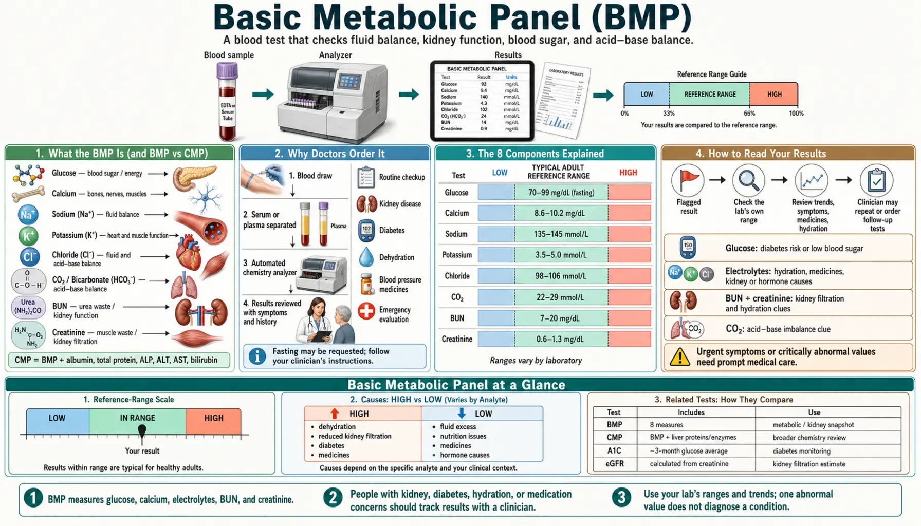
The Basic Metabolic Panel (BMP) is a blood test that measures eight substances in one blood sample: glucose, calcium, four electrolytes, and two waste products cleared by the kidneys. MedlinePlus describes it as often done as part of a regular checkup, and as a test used to screen for or diagnose health conditions and to monitor treatment. MedlinePlus also notes that it is common for healthy people to sometimes have results outside the reference range, and that results are interpreted together with symptoms, medical history, and other tests. The sections below cover what the panel measures, why it is ordered, the example reference ranges published by MedlinePlus (which vary between laboratories), what high or low values can point to, and which results the cited sources describe as urgent.
Interactive Visualization Kidney Nephron — watch blood become urine Follow blood into the glomerulus and see filtration and reabsorption turn 180 litres of filtrate into urine — then toggle dehydration or diabetes. Launch → Interactive Visualization The Sodium–Potassium Pump — why you have a voltage Run the pump that gives every cell its charge — three sodium out, two potassium in, on Mg-ATP — then drop the magnesium and see exactly why low potassium refuses to correct. Launch →
Table of Contents
What the BMP Is (and BMP vs CMP)
A Basic Metabolic Panel is a blood test that measures eight substances in one blood sample. MedlinePlus describes it as giving information about the body’s fluid balance, metabolism, and how well the kidneys are working. Other names for it include chem 7, CHEM-7, SMA7, chemistry panel, chemistry screen, and electrolyte panel (MedlinePlus: BMP test; MedlinePlus Medical Encyclopedia: Basic metabolic panel). The eight measurements are glucose (blood sugar), calcium, four electrolytes (sodium, potassium, chloride, and carbon dioxide/bicarbonate), and two waste products cleared by the kidneys (blood urea nitrogen and creatinine). Creatinine results are often used to calculate an estimated glomerular filtration rate (eGFR), and sodium is often reported with the other electrolytes as an anion gap (MedlinePlus), so a printout can list more lines than the eight tests.
The Comprehensive Metabolic Panel (CMP) includes the same eight tests plus six more: albumin, total protein, the liver enzymes ALP, ALT, and AST, and bilirubin — fourteen in all. MedlinePlus notes that a CMP may be ordered instead of a BMP to get a more complete picture of the organs or to check for liver disease or other specific conditions. A report that lists albumin and liver enzymes is therefore from a CMP. The companion Comprehensive Metabolic Panel page covers the extra six tests.
Why the BMP Is Ordered
MedlinePlus lists these common reasons for a BMP:
- Routine check-ups — a BMP is often done as part of a regular checkup, which may find health problems before there are symptoms.
- Screening, diagnosis, and monitoring treatment — information on kidney function, fluid and electrolyte balance, blood glucose, and acid–base balance.
- Monitoring a long-term condition — such as high blood pressure or kidney disease (see Kidney Function).
- Medicines that affect these values — MedlinePlus lists taking medicines that can affect potassium or fluid balance as a reason for these tests; diuretics (“water pills”) are among the medicines it names as possible causes of both high and low potassium.
- Diabetes screening — a high glucose may be a sign of prediabetes or diabetes (see Diabetes).
- Symptoms and emergency care — such as fatigue, confusion, breathing problems, or vomiting that lasts a long time, and treatment in an emergency room.
The 8 Components Explained
Each analyte in plain language, as MedlinePlus describes it unless another source is named.
Glucose (blood sugar)
Glucose is a type of sugar and the body’s main source of energy; the hormone insulin helps move it from the bloodstream into cells. High blood glucose may be a sign of prediabetes or diabetes. Symptoms MedlinePlus lists for low blood glucose include feeling shaky or jittery, hunger, dizziness, confusion, a fast heartbeat, and, when severe, fainting or seizures. Because food raises blood sugar, a fasting glucose is taken after at least 8 hours without eating or drinking anything except water.
Calcium
About 1% of the body’s calcium is in the blood; the rest is stored in bones and teeth. Calcium in the blood is needed for nerves, muscles, and the heart to work properly, and it helps with blood clotting. Total calcium (the BMP measurement) includes calcium bound to proteins in the blood and “free” (ionized) calcium, normally present in about equal amounts; Cooper and Gittoes write that serum calcium must be interpreted in relation to serum albumin (Cooper 2008). Abnormal levels may be a sign of bone, thyroid, parathyroid, or kidney disease, among other conditions. See the mineral page on Calcium.
The Electrolytes: Sodium, Potassium, Chloride, and CO2/Bicarbonate
Electrolytes are electrically charged minerals that help control the amount of fluid and the balance of acids and bases (pH) in the body, and they help nerves and muscles work.
- Sodium — the body normally keeps blood sodium in a very narrow range, and the kidneys remove extra sodium in urine. Low sodium (hyponatremia) most commonly results from water retention (Adrogué 2022). See Sodium.
- Potassium — almost all of the body’s potassium is inside cells, with a small amount in the blood; potassium is the most abundant cation in the fluid inside cells (Palmer 2015). The kidneys remove potassium in urine to keep the blood level steady. MedlinePlus lists arrhythmia among the symptoms of high potassium and irregular heartbeat among the symptoms of low potassium. See Potassium.
- Chloride — comes mainly from salt in food, helps control the amount of fluid in the body, and helps maintain blood volume and blood pressure. It is often measured with the other electrolytes. See Chloride.
- CO2 (carbon dioxide / bicarbonate) — most of the carbon dioxide in the body is in the form of bicarbonate (HCO3), an electrolyte. Abnormal results may be a sign that the body is having a hard time keeping the right acid–base balance: metabolic acidosis is characterized by a primary fall in serum bicarbonate (Kraut 2010), and MedlinePlus lists metabolic alkalosis among the causes of a high value.
The Kidney Markers: BUN and Creatinine
- BUN (blood urea nitrogen) is a waste product that forms as the body breaks down proteins and is removed by the kidneys. A high BUN is generally a sign that the kidneys are not working well, but MedlinePlus also lists dehydration, burns, certain medicines, a high-protein diet, and a recent heart attack as other reasons for a higher level, and a lack of protein in the diet, malnutrition, and liver disease as reasons for a lower one. BUN levels can vary with sex and usually increase with age.
- Creatinine is a waste product made when muscles are used and some muscle tissue breaks down; the kidneys filter it into the urine. MedlinePlus notes that creatinine alone is not the best measure of kidney function, because people make different amounts depending on muscle mass, diet, age, and activity, and that blood creatinine can stay in the normal range during the early stages of kidney disease. The 2021 CKD-EPI creatinine equation estimates GFR from creatinine, age, and sex without the race term used in earlier equations (Inker 2021); MedlinePlus describes eGFR as a more accurate measure of kidney health than creatinine alone.
Creatinine and BUN measured in the same panel may be compared with each other to help find the cause of a kidney problem (MedlinePlus). A high BUN-to-creatinine ratio (above 20) has traditionally been read as suggesting pre-renal azotemia; in a 2012 study of two cohorts of critically ill patients (1,010 and 10,228 patients), a ratio above 20 was associated with higher mortality, and the authors concluded that it should not be used to classify acute kidney injury in critically ill patients (Rachoin 2012).
Reading the Results and Reference Ranges
Ranges vary by laboratory. A reference range is based on the test results of large groups of healthy people, and because laboratories use different testing methods, they often use different reference ranges. MedlinePlus therefore says results are compared with the reference range printed on the lab report itself, that results from different labs cannot be compared directly, and that tracking results over time is easier at the same lab (MedlinePlus: How to Understand Your Lab Results). Results are also reported in different units, such as mg/dL or mmol/L.
The example ranges below are those given in the MedlinePlus Medical Encyclopedia article on the basic metabolic panel, which notes that normal value ranges may vary slightly among laboratories and that some laboratories use different measurements or test different specimens. Where MedlinePlus’s comprehensive metabolic panel article gives a different figure, it is shown too; the companion CMP page uses that article’s ranges, so the two pages differ only where the two MedlinePlus articles do.
- Glucose: 64 to 100 mg/dL (3.55 to 5.55 mmol/L); the CMP article gives 70 to 100 mg/dL (3.9 to 5.6 mmol/L). Diagnostic thresholds for fasting plasma glucose from the American Diabetes Association’s Standards of Care: 100 to 125 mg/dL (5.6 to 6.9 mmol/L) is prediabetes (impaired fasting glucose) and 126 mg/dL (7.0 mmol/L) or higher is diabetes, with fasting defined as no caloric intake for at least 8 hours; in the absence of unequivocal hyperglycemia, diagnosis requires two abnormal results, from the same sample or from two separate samples (ADA 2024).
- Calcium (total): 8.5 to 10.2 mg/dL (2.13 to 2.55 mmol/L), the same in both articles.
- Sodium: 136 to 144 mEq/L (136 to 144 mmol/L); the CMP article gives 135 to 145 mEq/L. A 2022 review defines hyponatremia as a serum sodium below 135 mEq/L (Adrogué 2022).
- Potassium: 3.7 to 5.2 mEq/L (3.7 to 5.2 mmol/L), the same in both articles.
- Chloride: 96 to 106 mmol/L, the same in both articles.
- CO2 (bicarbonate): 23 to 29 mmol/L, the same in both articles.
- BUN: 6 to 20 mg/dL (2.14 to 7.14 mmol/L), the same in both articles.
- Creatinine: 0.8 to 1.2 mg/dL (70.72 to 106.08 µmol/L); the CMP article gives 0.6 to 1.3 mg/dL, and the MedlinePlus creatinine blood test article gives 0.7 to 1.3 mg/dL (62 to 115 µmol/L) for men and 0.5 to 0.95 mg/dL (44 to 84 µmol/L) for women, noting that women often have less muscle mass (MedlinePlus: Creatinine blood test).
Calculated values that may also appear:
- eGFR: the KDIGO 2024 guideline defines chronic kidney disease as abnormalities of kidney structure or function present for more than 3 months; an eGFR below 60 mL/min/1.73 m² (categories G3a to G5) for more than 3 months meets that definition. An eGFR of 60 to 89 is category G2, “mildly decreased” relative to the young-adult level, and 90 or above is G1; without other evidence of kidney damage, such as albumin in the urine, neither G1 nor G2 meets the criteria for chronic kidney disease (KDIGO 2024).
- Anion gap: the normal value can vary widely, reflecting both differences in the methods used to measure its parts and substantial differences between individuals (Kraut 2007), so it is read against the reporting laboratory’s own range.
- BUN:creatinine ratio: see above; laboratories that report it use their own reference values.
MedlinePlus notes that it is common for healthy people to sometimes have results outside the reference range, that a result outside the range may or may not be a sign of a health problem, and that a result inside it is not a guarantee of good health. It also notes that in a healthy person creatinine levels are usually fairly stable over time, which is one reason earlier results are part of reading a new one.
What High or Low Values Can Mean
The possible causes below are those listed by the cited sources. A value outside the range does not always mean a condition that needs treatment, and each value is read together with the others, with symptoms, and with medical history (MedlinePlus).
Kidney function (BUN, creatinine, eGFR)
MedlinePlus lists kidney disease or injury (including infection, poor blood flow to the kidneys, a blockage in the urinary system, or kidney failure) and conditions that affect the kidneys, such as heart failure or diabetes, as possible causes of a high creatinine. A high creatinine can also be caused by dehydration, muscle disorders and injuries, intense exercise, or a diet high in meat; a low creatinine may reflect malnutrition, muscle loss, or serious liver disease. Causes of a high or low BUN are listed in the components section above. A single high creatinine result cannot diagnose a specific condition, and retesting or other tests are usually needed.
Electrolyte and fluid balance
Low sodium (hyponatremia) is the most common electrolyte disorder, affecting about 5% of adults and 35% of hospitalized patients, and most commonly results from water retention; even mild hyponatremia is associated with longer hospital stays and higher mortality (Adrogué 2022). MedlinePlus lists sodium loss from diarrhea or vomiting, conditions that make the body hold extra fluid (kidney disease, cirrhosis, heart failure), certain brain and lung diseases, certain cancers, certain medicines, Addison disease, and malnutrition among the causes. High sodium (hypernatremia) may be a sign of dehydration (from not drinking enough, diarrhea, or diuretic medicines), an adrenal disorder, kidney disease, or, uncommonly, diabetes insipidus.
Potassium: hyperkalemia is one of the few potentially lethal electrolyte disturbances, and prompt recognition and treatment of severe hyperkalemia are expected to save lives (Weisberg 2008). MedlinePlus lists kidney disease, Addison disease, injuries, burns or surgery, poorly controlled type 1 diabetes, and side effects of certain medicines among the causes of high potassium, and diuretics, fluid loss from diarrhea, vomiting or heavy sweating, laxative overuse, adrenal disorders, kidney disease, and alcohol use disorder among the causes of low potassium. A high result is not always real: pseudohyperkalemia is induced by hemolysis and by excessive leakage of potassium from cells during or after blood collection, and is increasingly seen in blood disorders with high white-cell or platelet counts (Meng 2015). MedlinePlus adds that repeatedly clenching and relaxing the fist just before or during the draw may temporarily raise blood potassium.
Chloride and CO2/bicarbonate point to acid–base disturbances. MedlinePlus lists dehydration, kidney disease, and metabolic acidosis as possible causes of high chloride, and heart failure, lung disease, Addison disease, and metabolic alkalosis as possible causes of low chloride. For CO2, it lists metabolic acidosis (which can follow kidney or liver disease or long-lasting diarrhea), diabetic ketoacidosis, Addison disease, shock, and respiratory alkalosis as possible causes of a low value, and lung diseases, Cushing’s syndrome, kidney failure, and metabolic alkalosis (for example from vomiting or dehydration) as possible causes of a high value.
Blood sugar
Prediabetes and diabetes are the most common causes of high glucose; other causes MedlinePlus lists include an overactive thyroid, pancreatic problems, stress from surgery, serious illness or trauma, adrenal disorders, and certain medicines. The ADA thresholds quoted above are for fasting plasma glucose (ADA 2024). A related test is the Hemoglobin A1C. In people without diabetes, low blood glucose is not common; MedlinePlus lists liver or kidney disease, underactive adrenal or thyroid glands, certain pituitary disorders, malnutrition, certain medicines, and alcohol use disorder among its causes.
Calcium
Primary hyperparathyroidism, hypercalcemia associated with cancer, and chronic kidney failure (with calcium and vitamin D metabolite treatment, or tertiary hyperparathyroidism) are the most common causes of high calcium (hypercalcemia); less common causes include vitamin D intoxication and other vitamin D–related conditions, an overactive thyroid, and medicines such as thiazides and lithium, as well as milk-alkali syndrome (Endres 2012). For low calcium (hypocalcemia), MedlinePlus lists low blood protein levels (from liver disease or malnutrition), underactive parathyroid glands, too little calcium in the diet, too little vitamin D or magnesium, pancreatitis, and kidney disease; a clinical review gives vitamin D deficiency as the most common cause of hypocalcemia in primary care (Cooper 2008). Because part of total calcium is bound to albumin, a low albumin can make total calcium read low; Cooper and Gittoes describe an albumin adjustment of 0.02 mmol/L of calcium for every 1 g/L of albumin above or below a reference value (Cooper 2008).
Fasting & Preparation
MedlinePlus says a person may need to fast (not eat or drink) for eight hours before a BMP, and that fasting before a blood test usually lasts 8 to 12 hours, with the ordering clinician giving the exact time. For the potassium, calcium, and BUN tests on their own, MedlinePlus says no special preparation is usually needed; fasting is asked for when other tests, such as a fasting glucose, are drawn from the same sample. A lipid panel is one of the tests MedlinePlus lists as usually requiring fasting (MedlinePlus: Fasting for a Blood Test).
Other points from the MedlinePlus pages:
- Water: plain water is allowed during a fast; juice, coffee, soda, and other drinks can affect results. Staying well hydrated can make it easier to draw blood.
- Timing: fasting tests can usually be scheduled early in the morning, so that most of the fasting time falls during sleep.
- Medicines and supplements: some medicines and supplements can affect results, and MedlinePlus advises telling the provider about everything taken and not stopping any medicine unless the provider says to.
- Creatinine: meat can temporarily raise creatinine, and a person may be told not to eat meat for 24 hours before a creatinine test.
- Potassium: natural black licorice may lower potassium, and MedlinePlus says not to eat it for at least two weeks before the test.
The Anion Gap
Sodium is often reported with the other electrolytes in a calculated value called the anion gap (MedlinePlus). It is not separately measured: it is calculated from the electrolytes as the serum sodium minus the sum of the serum chloride and bicarbonate. It is used to detect and analyze acid–base disorders, for quality control in the laboratory, and to help detect conditions such as multiple myeloma and bromide or lithium intoxication (Kraut 2007).
Elevated values most commonly indicate metabolic acidosis, but can also reflect laboratory error, metabolic alkalosis, high phosphate, or an abnormal protein (paraproteinemia); low values most commonly indicate laboratory error or low albumin. Metabolic acidosis is divided into high–anion gap and normal–anion gap (hyperchloremic) types, which can occur alone or together (Kraut 2007; Kraut 2010). Acute metabolic acidosis most often results from overproduction of organic acids such as ketoacids or lactic acid, while chronic metabolic acidosis often reflects bicarbonate wasting or impaired acid excretion by the kidneys (Kraut 2010). Kraut and Madias call the anion gap an inexpensive and effective tool, with the caveat that its normal value varies widely between methods and between individuals (Kraut 2007).
Related Tests
Tests often discussed alongside the BMP, each covered on its own page:
- Comprehensive Metabolic Panel (CMP) — the BMP plus six protein and liver tests.
- Complete Blood Count (CBC) — red cells, white cells, and platelets.
- Hemoglobin A1C and Fasting Insulin — blood sugar over time and insulin.
- Kidney Function testing, including Microalbumin / Urine ACR — KDIGO 2024 classifies chronic kidney disease by GFR together with urine albumin.
- Lipid Panel — cholesterol and triglycerides.
- Uric Acid — another blood chemistry test.
Browse the full menu on the Lab Tests landing page.
Results Treated as Urgent
MedlinePlus notes that an abnormal result does not always mean a condition that needs treatment, and that results are interpreted together with symptoms, medical history, and other tests. The sources cited on this page describe these situations as serious:
- Potassium: hyperkalemia is one of the few potentially lethal electrolyte disturbances (Weisberg 2008).
- Sodium: without treatment, extremely high or extremely low sodium may lead to coma and become life-threatening (MedlinePlus); severely symptomatic hyponatremia, with signs such as somnolence, coma, seizures, or cardiorespiratory distress, is a medical emergency (Adrogué 2022).
- Calcium: extremely low calcium can cause seizures (MedlinePlus).
- Glucose: without treatment, severe low blood glucose can lead to seizures and brain damage (MedlinePlus).
- Kidney function: KDIGO 2024 classifies a low eGFR by category, but a single low reading does not by itself establish chronic kidney disease, because the definition requires the abnormality to be present for more than 3 months.
This page is general health information, not medical advice; decisions about abnormal results belong with a clinician.
Key Research Papers
- Stevens PE, Ahmed SB, Carrero JJ, et al. KDIGO 2024 Clinical Practice Guideline for the Evaluation and Management of Chronic Kidney Disease. Kidney International. 2024;105(4S):S117–S314. — the current global guideline defining and staging chronic kidney disease from eGFR and albuminuria. — doi:10.1016/j.kint.2023.10.018
- Inker LA, Eneanya ND, Coresh J, et al. New Creatinine- and Cystatin C–Based Equations to Estimate GFR without Race. New England Journal of Medicine. 2021;385(19):1737–1749. — the race-free CKD-EPI equations for estimating GFR from creatinine and cystatin C. — doi:10.1056/NEJMoa2102953
- Levey AS, Stevens LA, Schmid CH, et al. A New Equation to Estimate Glomerular Filtration Rate. Annals of Internal Medicine. 2009;150(9):604–612. — the original CKD-EPI creatinine equation, which performed better than the MDRD Study equation in validation. — doi:10.7326/0003-4819-150-9-200905050-00006
- Adrogué HJ, Madias NE. Hyponatremia. New England Journal of Medicine. 2000;342(21):1581–1589. — a review of low blood sodium. — doi:10.1056/NEJM200005253422107
- Sterns RH. Disorders of Plasma Sodium — Causes, Consequences, and Correction. New England Journal of Medicine. 2015;372(1):55–65. — an accessible modern overview of both high and low sodium. — doi:10.1056/NEJMra1404489
- Palmer BF. Regulation of Potassium Homeostasis. Clinical Journal of the American Society of Nephrology. 2015;10(6):1050–1060. — a review of how the body regulates potassium distribution and its excretion by the kidneys. — doi:10.2215/CJN.08580813
- Weisberg LS. Management of severe hyperkalemia. Critical Care Medicine. 2008;36(12):3246–3251. — a review of the pathophysiology and management of severe hyperkalemia. — doi:10.1097/CCM.0b013e31818f222b
- Kraut JA, Madias NE. Serum Anion Gap: Its Uses and Limitations in Clinical Medicine. Clinical Journal of the American Society of Nephrology. 2007;2(1):162–174. — a clear explanation of the anion gap calculated from BMP electrolytes. — doi:10.2215/CJN.03020906
- Kraut JA, Madias NE. Metabolic acidosis: pathophysiology, diagnosis and management. Nature Reviews Nephrology. 2010;6(5):274–285. — how a low bicarbonate (CO2) on a panel is interpreted. — doi:10.1038/nrneph.2010.33
- ElSayed NA, Aleppo G, et al.; American Diabetes Association. Diagnosis and Classification of Diabetes: Standards of Care in Diabetes—2024. Diabetes Care. 2024;47(Suppl 1):S20–S42. — the fasting-glucose thresholds used to read the BMP sugar value. — doi:10.2337/dc24-S002
- Cooper MS, Gittoes NJL. Diagnosis and management of hypocalcaemia. BMJ. 2008;336(7656):1298–1302. — a clinical review of low blood calcium, including interpretation of calcium in relation to albumin. — doi:10.1136/bmj.39582.589433.BE
- Minisola S, Pepe J, Piemonte S, et al. The diagnosis and management of hypercalcaemia. BMJ. 2015;350:h2723. — a clinical review of the diagnosis and management of high blood calcium. — doi:10.1136/bmj.h2723
- Adrogué HJ, Tucker BM, Madias NE. Diagnosis and Management of Hyponatremia: A Review. JAMA. 2022;328(3):280–291. — hyponatremia as the most common electrolyte disorder, its definition and usual cause. — doi:10.1001/jama.2022.11176
- Meng QH, Wagar EA. Pseudohyperkalemia: A new twist on an old phenomenon. Critical Reviews in Clinical Laboratory Sciences. 2015;52(2):45–55. — falsely high potassium from hemolysis and cell leakage during or after blood collection. — doi:10.3109/10408363.2014.966898
- Endres DB. Investigation of hypercalcemia. Clinical Biochemistry. 2012;45(12):954–963. — the most common and less common causes of high blood calcium. — doi:10.1016/j.clinbiochem.2012.04.025
- Rachoin JS, Daher R, Moussallem C, et al. The fallacy of the BUN:creatinine ratio in critically ill patients. Nephrology Dialysis Transplantation. 2012;27(6):2248–2254. — a ratio above 20 and outcomes in two cohorts of critically ill patients. — doi:10.1093/ndt/gfr705
Connections
- All Lab Tests
- The Sodium–Potassium Pump — interactive animation
- Kidney Nephron — interactive animation
- Comprehensive Metabolic Panel (CMP)
- Complete Blood Count (CBC)
- Kidney Function
- Hemoglobin A1C
- Fasting Insulin
- Microalbumin / Urine ACR
- Lipid Panel
- Uric Acid
- Potassium
- Sodium
- Calcium
- Chloride
- Diabetes