Parathyroid Hormone (PTH) Test
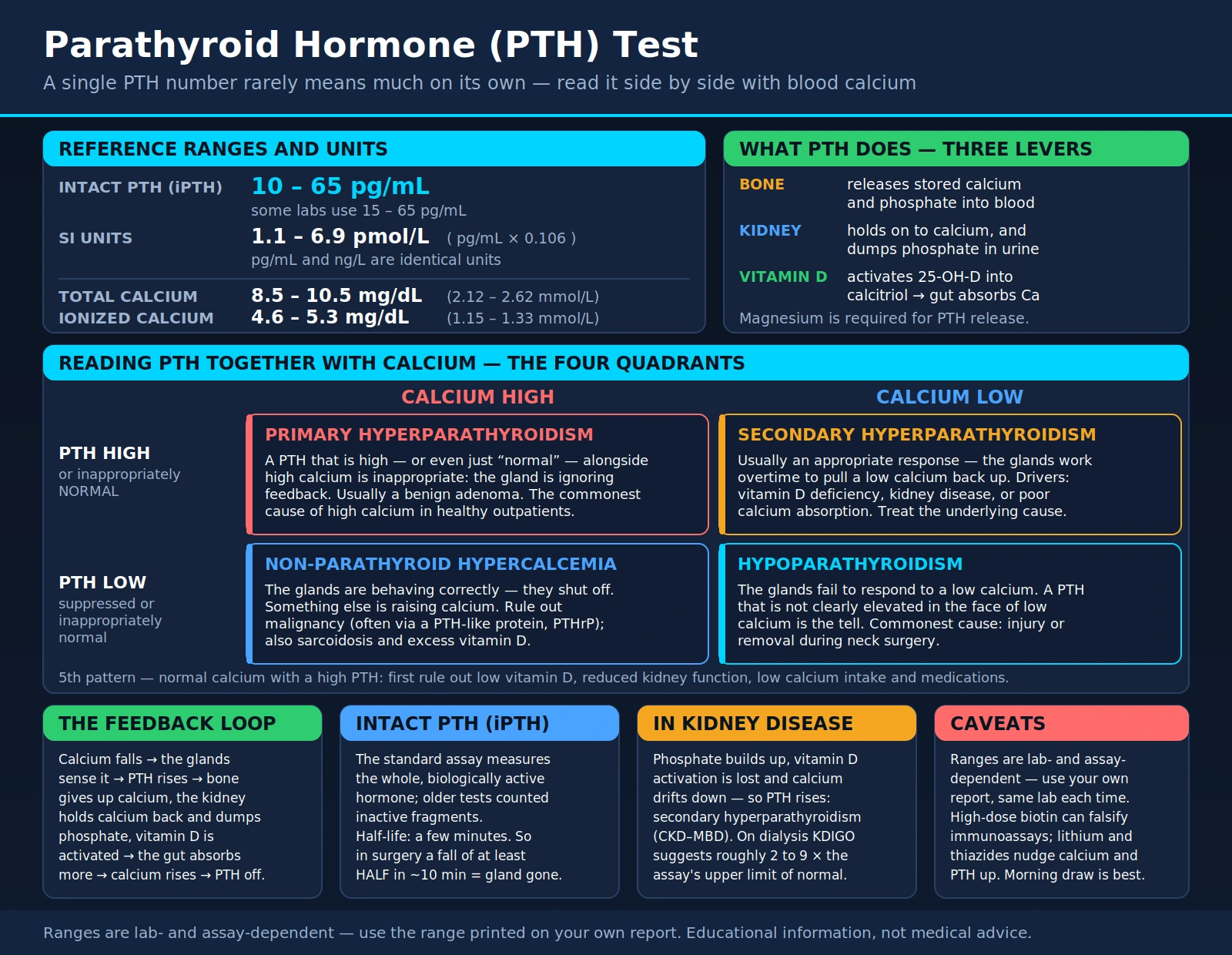
The parathyroid hormone (PTH) test measures the level of parathyroid hormone in the blood. PTH is a protein hormone released by four small parathyroid glands in the neck, near or attached to the back of the thyroid gland, and it controls calcium, phosphorus and vitamin D levels in the blood (MedlinePlus). Calcium matters for heart function, muscle contraction, nerve signaling, blood clotting and bone (MedlinePlus). A single PTH number rarely means much on its own: MedlinePlus notes that blood calcium is measured at the same time to judge whether a PTH result is normal, and that a PTH value in the normal range can still be inappropriate when calcium is high. This page covers what PTH does, why the test is ordered, how it is read side by side with blood calcium, and what the common patterns — the different kinds of hyperparathyroidism and hypoparathyroidism — mean.
Interactive Visualization Bone Remodeling & Calcium Balance Watch osteoclasts tunnel and osteoblasts rebuild — then drop vitamin D and see PTH quietly strip calcium out of your skeleton. Launch →
Table of Contents
- What PTH Is and What It Does
- Why the Test Is Ordered
- Reference Ranges and Units
- Reading PTH Together With Calcium
- Hyperparathyroidism: Primary, Secondary, Tertiary
- Hypoparathyroidism (Too Little PTH)
- PTH in Kidney Disease (CKD–MBD)
- Intact PTH and Intraoperative PTH
- How to Prepare and What to Expect
- Key Research Papers
- Connections
- Featured Videos
What PTH Is and What It Does
Parathyroid hormone is made by the parathyroid glands, which MedlinePlus describes as four small glands in the neck, near or attached to the back side of the thyroid gland. Despite the similar name, they are separate from the thyroid: PTH controls calcium, phosphorus and vitamin D levels in the blood and is important for regulating bone growth (MedlinePlus).
The glands track calcium through a receptor. On the chief cells of the parathyroid glands, the calcium-sensing receptor senses the calcium concentration outside the cell and alters the release of PTH; the same receptor is also found in bone, kidney and gut, the three main organs that move calcium in and out of the blood (Tfelt-Hansen 2005). Mutations that make the receptor more active cause low calcium, and mutations that inactivate it cause high calcium (Tfelt-Hansen 2005).
When calcium is low, PTH can raise it through three routes (Goltzman 2018):
- Bone. PTH mobilizes calcium from bone by increasing bone turnover, mainly but not only in cortical bone. PTH may also help build bone, particularly trabecular bone, and that action is used as an anabolic osteoporosis therapy (Goltzman 2018). In primary hyperparathyroidism, a fracture or a low bone density is among the 2022 international guideline criteria for surgery (Bilezikian 2022).
- Kidneys. PTH increases how much calcium the kidneys reabsorb (Goltzman 2018). PTH also affects phosphorus: MedlinePlus lists primary hyperparathyroidism among the causes of a low blood phosphorus, and a low phosphorus as one reason the PTH test is ordered.
- Vitamin D activation. PTH increases production of active vitamin D, which in turn increases calcium absorption from the gut (Goltzman 2018). The vitamin D level usually measured in blood is a different form, 25-hydroxyvitamin D (MedlinePlus).
Magnesium also belongs in the picture. Reversible hypoparathyroidism can occur with magnesium deficiency due to malabsorption, alcoholism and other states of poor nutrition, and magnesium excess (as from magnesium given for preterm labor) can also inhibit PTH secretion (Bilezikian 2011). MedlinePlus lists low blood magnesium among the causes of a low PTH. A 2025 literature review describes impaired PTH secretion as one of the most important factors behind the low calcium seen with low magnesium, and reports that this low calcium cannot be adequately corrected with calcium, vitamin D, or both (Albert 2025).
Why the Test Is Ordered
PTH is usually a second-step test ordered to explain something already noticed, most often an abnormal calcium. MedlinePlus lists a high calcium or low phosphorus level, severe osteoporosis that cannot be explained or does not respond to treatment, and kidney disease as reasons for the test. Situations described in the guidelines and research include:
- High blood calcium (hypercalcemia). PTH separates parathyroid-driven high calcium (PTH high or inappropriately normal) from high calcium with a suppressed PTH, which points to a cause outside the glands (MedlinePlus; Goldner 2016).
- Low blood calcium (hypocalcemia). Low calcium with a frankly low or inappropriately low-normal intact PTH is the initial biochemical profile that leads clinicians to suspect hypoparathyroidism (Bilezikian 2011).
- Bone disease. MedlinePlus lists severe osteoporosis that cannot be explained or does not respond to treatment.
- Kidney stones. Kidney stones are a classical feature of primary hyperparathyroidism, and stones or kidney calcification on imaging are among the 2022 guideline criteria for surgery (Bilezikian 2022).
- Chronic kidney disease. The KDIGO 2017 guideline recommends monitoring calcium, phosphate, PTH and alkaline phosphatase beginning in CKD stage G3a (see CKD–MBD).
- Inherited endocrine syndromes. MedlinePlus lists multiple endocrine neoplasia (MEN) type I and type II among other conditions for which the test may be ordered.
- Parathyroid surgery. The surgical guideline recommends PTH monitoring during minimally invasive parathyroidectomy (Wilhelm 2016) (see Intraoperative PTH).
MedlinePlus states that blood calcium is measured at the same time as PTH to help understand whether the PTH is normal. The two numbers are read as a pair.
Reference Ranges and Units
MedlinePlus gives normal values for intact PTH of 10 to 65 picograms per milliliter (pg/mL), and notes that normal ranges may vary slightly among laboratories and that some labs use different measurements or test different specimens. The Fourth International Workshop concluded that reference ranges for PTH should be established in vitamin D-replete healthy people (Eastell 2014).
The companion tests have their own MedlinePlus example ranges, which also vary by laboratory:
- Total calcium: 8.8 to 10.3 mg/dL (2.20 to 2.58 mmol/L).
- Ionized calcium (the free, biologically active fraction): adults 4.8 to 5.6 mg/dL (1.20 to 1.40 mmol/L); children 4.8 to 5.3 mg/dL (1.20 to 1.32 mmol/L).
- Phosphorus: adults 2.8 to 4.5 mg/dL; children 4.0 to 7.0 mg/dL.
- 25-hydroxyvitamin D: MedlinePlus reports that many experts recommend a level from 30 to 80 ng/mL (75 to 200 nmol/L).
Other sources print slightly different calcium intervals; the Calcium Test page compares several. Three points about ranges recur in these sources:
- The laboratory's own range is the reference. Different assays and labs produce different ranges, so a result is read against the range printed by the laboratory that ran it (MedlinePlus).
- Trends compare best on one lab. MedlinePlus describes going back to the same lab each time so PTH results can be compared reliably over time.
- "Normal" is not automatically reassuring. MedlinePlus notes that a PTH in the normal range can still be inappropriate when calcium is high — the subject of the next section.
Reading PTH Together With Calcium
In a healthy person, calcium and PTH move in opposite directions: the calcium-sensing receptor on the parathyroid glands alters PTH release as calcium changes (Tfelt-Hansen 2005), and PTH works to raise calcium when it is low (Goltzman 2018). So the question is not simply "is PTH high or low?" but "does this PTH fit the calcium drawn with it?" The patterns are often pictured as quadrants:
- High calcium + high (or normal-range) PTH. This is the pattern of primary hyperparathyroidism: high calcium with a PTH that is high or inappropriately normal. MedlinePlus notes that a PTH in the normal range can still be inappropriate when calcium is high.
- High calcium + low (suppressed) PTH. The glands have switched off, so the high calcium comes from elsewhere. Cancer-related high calcium is most often caused by a PTH-related peptide (PTHrP) made by the tumor, by bone-dissolving cytokines, or by excess production of active vitamin D, and it is most common in later-stage cancers (Goldner 2016). MedlinePlus also lists cancers that spread to bone, sarcoidosis and tuberculosis, excess vitamin D intake, and long-term excess calcium intake among causes of a low PTH.
- Low calcium + high PTH. This is an appropriate response. Secondary hyperparathyroidism is an increase in PTH in response to a stimulus, most commonly a low serum calcium; Jamal and Miller describe the serum calcium in secondary hyperparathyroidism as normal, with the PTH elevated (Jamal 2013). Causes include vitamin D deficiency, chronic kidney disease (stages 3–5) and calcium malabsorption (Bilezikian 2022).
- Low calcium + low (or normal-range) PTH. This is the pattern of hypoparathyroidism: low calcium with a frankly low or inappropriately low-normal intact PTH (Bilezikian 2011).
A fifth situation is normal calcium with a high PTH. The 2022 international guidelines define normocalcemic primary hyperparathyroidism as a normal albumin-adjusted total calcium and a normal ionized calcium with an elevated intact PTH on at least two occasions over 3–6 months, after all alternative causes of secondary hyperparathyroidism have been ruled out; the causes they list include stage 3–5 chronic kidney disease, vitamin D deficiency, calcium malabsorption, and bisphosphonate or denosumab use (Bilezikian 2022). The Fourth Workshop described normocalcemic disease as a variant of the more common presentation with high calcium (Eastell 2014).
The ionized-calcium requirement matters because about one half of the calcium in the blood is attached to proteins, mainly albumin (MedlinePlus). In 13,604 paired results from 5,553 hospitalized patients, albumin-adjusted calcium agreed with measured ionized calcium status in only 56.9% of cases, and it overestimated calcium in patients with kidney impairment and albumin below 3.0 g/dL (Smith 2018). The Albumin page covers the adjustment formulas and these studies.
Hyperparathyroidism: Primary, Secondary, Tertiary
"Hyperparathyroidism" means increased activity of the parathyroid glands. Fraser (2009) describes two routes: an intrinsic change in the glands themselves (primary or tertiary hyperparathyroidism), or an outside change in calcium balance that stimulates PTH production (secondary hyperparathyroidism).
Primary hyperparathyroidism
Sporadic primary hyperparathyroidism is most often caused by a single benign parathyroid adenoma (85%) (Bilezikian 2022). It is the third most common endocrine disorder, with the highest incidence in postmenopausal women (Fraser 2009); the 2022 guidelines cite an age-adjusted United States prevalence of 233 per 100,000 women and 85 per 100,000 men (Bilezikian 2022).
Asymptomatic disease is common, and severe disease with kidney stones and metabolic bone disease arises less often than it did 20–30 years ago (Fraser 2009). Before standardized blood chemistry panels arrived in the 1970s, the disease typically came to attention through bone pain and fractures, chronic kidney disease, and renal colic from kidney stones (Bilezikian 2022). The diagnostic pattern is high calcium with a PTH that is high or inappropriately normal (MedlinePlus). The surgical guideline lists 25-hydroxyvitamin D, 24-hour urine calcium and bone density (DXA) in the initial evaluation (Wilhelm 2016). Urine calcium also bears on an inherited look-alike, familial hypocalciuric hypercalcemia (FHH): the 2022 guidelines state that FHH may be suspected in younger people with a urinary calcium/creatinine clearance ratio below 0.01 and/or a family history of high calcium (Bilezikian 2022).
Surgery. In the systematic review of randomized trials behind the 2022 guidelines, parathyroidectomy achieved a biochemical cure in 97.8% of patients with asymptomatic disease, and the guidelines state that all symptomatic patients should be offered surgery unless it is medically contraindicated (Bilezikian 2022). For people without symptoms, the 2022 guidelines list these indications for surgery:
- serum calcium more than 1 mg/dL (0.25 mmol/L) above the upper limit of normal;
- a vertebral fracture on vertebral fracture assessment or X-ray, or a bone density T-score of −2.5 or lower at any site;
- eGFR or creatinine clearance below 60 mL/min;
- kidney stones or kidney calcification (nephrolithiasis or nephrocalcinosis) on X-ray, ultrasound or other imaging;
- high urine calcium (for example, more than 250 mg/day in women or more than 300 mg/day in men);
- age under 50 years, which the guidelines describe as a sufficient indication on its own.
For people who do not have surgery, the guidelines state that they can be followed without drug treatment, and list serum calcium and 25-hydroxyvitamin D yearly, creatinine clearance yearly, and three-site DXA every 1 or 2 years unless bone density is normal (Bilezikian 2022). The surgeons' guideline states that parathyroidectomy is indicated for all symptomatic patients and should be considered for most asymptomatic patients (Wilhelm 2016).
Secondary hyperparathyroidism
Secondary hyperparathyroidism is an increase in PTH that is appropriate and in response to a stimulus, most commonly a low serum calcium; Jamal and Miller describe the serum calcium as normal and the PTH as elevated (Jamal 2013). Vitamin D deficiency is a common cause, particularly in elderly people, although the biochemical definition of vitamin D deficiency and its treatment are debated (Fraser 2009). Chronic kidney disease and calcium malabsorption are other causes listed in the 2022 guidelines (Bilezikian 2022), and MedlinePlus adds low calcium intake, calcium loss in the urine and, uncommonly, pregnancy or breastfeeding among causes of a high PTH. Jamal and Miller describe the cause and treatment of secondary hyperparathyroidism as relatively straightforward (Jamal 2013), and the Fourth Workshop concluded that vitamin D insufficiency found during evaluation for primary hyperparathyroidism should be treated as part of management (Eastell 2014).
Tertiary hyperparathyroidism
Tertiary hyperparathyroidism is excessive PTH secretion after longstanding secondary hyperparathyroidism, in which high calcium has developed. It typically occurs in people with chronic kidney disease, usually after a kidney transplant, and data on its management are limited to a few small trials with short follow-up (Jamal 2013). For severe hyperparathyroidism in chronic kidney disease that does not respond to medical or drug therapy, the KDIGO 2017 guideline suggests parathyroidectomy (KDIGO 2017).
Hypoparathyroidism (Too Little PTH)
Hypoparathyroidism is a rare disorder characterized by low calcium and absent or deficient PTH (Brandi 2016), with blood phosphorus typically elevated (Bilezikian 2011). The most common acquired cause in adults is surgery: operations on the thyroid or parathyroid glands, nearby neck structures, or neck dissection for cancer can lead to acute or chronic hypoparathyroidism. Surgical centers with experienced endocrine surgeons and a high case volume report permanent hypoparathyroidism after thyroid surgery in 0.9–1.6% of cases, while earlier reports gave figures as high as 6.6% (Bilezikian 2011). Autoimmune hypoparathyroidism, alone or as part of an autoimmune polyglandular syndrome, is the next most common form in adults. Genetic causes include mutations in the genes for PTH, GCMB or the calcium-sensing receptor, and DiGeorge syndrome, which affects 1 in 4,000–5,000 live births and includes hypocalcemia from hypoparathyroidism in 60% of cases (Bilezikian 2011). Reversible hypoparathyroidism can occur with magnesium deficiency (Bilezikian 2011).
Patients most often present with tingling (paresthesia), cramps or tetany, and the disorder can also appear acutely with seizures, bronchospasm, laryngospasm or heart rhythm disturbances; patients also describe numbness around the mouth and twitching of the facial muscles (Bilezikian 2011). On the lab report, the pattern is low calcium with a frankly low or inappropriately low-normal intact PTH (Bilezikian 2011). For chronic management, treatment options include oral calcium, vitamin D (including its active metabolites and analogs) and thiazide diuretics, and PTH itself is used as replacement therapy (Bilezikian 2011; Goltzman 2018). High urine calcium is a common complication of therapy and can lead to kidney stones, kidney calcification and reduced kidney function; the management targets described include a 24-hour urine calcium below 7.5 mmol/day (Bilezikian 2011).
PTH in Kidney Disease (CKD–MBD)
Chronic kidney disease (CKD) gets its own section because PTH is tracked routinely there. A KDIGO position statement defines CKD–Mineral and Bone Disorder (CKD–MBD) as a systemic disorder of mineral and bone metabolism due to CKD, shown by abnormalities in bone and mineral metabolism and/or calcification outside the skeleton, and reserves the term renal osteodystrophy for the changes in bone structure itself (Moe 2006). These disturbances are an important cause of illness, reduced quality of life and extraskeletal calcification, which has been associated with higher cardiovascular mortality (Moe 2006). Secondary hyperparathyroidism from CKD is important in the development of renal bone disease (Fraser 2009).
The KDIGO 2017 guideline update recommends monitoring calcium, phosphate, PTH and alkaline phosphatase from CKD stage G3a in adults (from G2 in children). For people with CKD G3a–G5 not on dialysis, it states that the optimal PTH level is not known, and suggests that a PTH that is progressively rising or persistently above the assay's upper limit be evaluated for modifiable factors: high blood phosphate, low calcium, high phosphate intake and vitamin D deficiency (KDIGO 2017).
In people on dialysis (CKD G5D), the guideline does not use the ordinary normal range as the target. It suggests keeping intact PTH in the range of approximately 2 to 9 times the upper normal limit for the assay, and suggests that marked changes in PTH in either direction within this range prompt starting or changing therapy (KDIGO 2017). Its review of a bone-biopsy study in 492 dialysis patients found that no marker, alone or combined, was robust enough to classify bone turnover in an individual patient, which supported using trends in PTH rather than absolute target values when deciding to start or stop PTH-lowering treatment (KDIGO 2017). For dialysis patients who need PTH-lowering therapy, it suggests calcimimetics, calcitriol or vitamin D analogs, or a calcimimetic combined with calcitriol or a vitamin D analog (KDIGO 2017).
Intact PTH and Intraoperative PTH
PTH circulates alongside PTH fragments, one of six main types of interference known to affect PTH immunoassays; the others are heterophilic antibodies, biotin, oxidized PTH, phosphorylated PTH and some sample-handling factors (Cheng 2023). The authors of that review describe misdiagnosis and inappropriate parathyroidectomy among the reported consequences. MedlinePlus lists "intact PTH" among the test's alternative names, and the Fourth International Workshop concluded that second- and third-generation PTH assays are both helpful in diagnosing primary hyperparathyroidism (Eastell 2014).
PTH has a short half-life: in 306 patients having minimally invasive parathyroidectomy, the median intraoperative half-life was 3 minutes 9 seconds (Leiker 2013). Surgeons use this in intraoperative PTH monitoring, measuring PTH before and after the suspected gland is removed. Under the Miami criterion — a PTH drop of at least 50% from the highest of the pre-incision or pre-excision level, 10 minutes after the gland is removed — postoperative calcium was correctly predicted in 329 of 341 patients (3 false positives, 9 false negatives); other published criteria raised the false-negative rate from 3% to between 6% and 24%, which would have led to unnecessary neck explorations in search of more abnormal glands (Carneiro 2003). The surgical guideline recommends intraoperative PTH monitoring with a reliable protocol for minimally invasive parathyroidectomy, and states that both focused (minimally invasive) surgery and bilateral neck exploration achieve high cure rates (Wilhelm 2016).
How to Prepare and What to Expect
The PTH test is an ordinary blood sample, and MedlinePlus states that most often no fasting is needed. Factors that affect the result:
- Timing. PTH follows a daily rhythm. In 11 healthy men, PTH had a main peak around 3 a.m., a second peak in the late afternoon and a late-morning low point, with an average amplitude of 4.2 pg/mL (el-Hajj Fuleihan 1997); a daytime study in 12 healthy young men found a low point at 9:30 a.m. and a peak in the afternoon (Herfarth 1992).
- The companion tests. Calcium is measured at the same time (MedlinePlus); the surgical guideline's initial evaluation for primary hyperparathyroidism also includes 25-hydroxyvitamin D, 24-hour urine calcium and bone density (Wilhelm 2016), and the CKD guideline monitors calcium, phosphate, PTH and alkaline phosphatase together (KDIGO 2017).
- Medications and supplements. Lithium treatment is associated with higher calcium and PTH and more hyperparathyroidism; a 2024 meta-analysis put the pooled prevalence of lithium-associated hyperparathyroidism at around 4%, compared with 0.5% in the healthy population (Vandermeulen 2024). Thiazide diuretics are commonly associated with high calcium, and in a Minnesota population study 24% of people with thiazide-associated high calcium were diagnosed with primary hyperparathyroidism (Griebeler 2016). MedlinePlus lists long-term excess calcium intake (from supplements or certain antacids) and excess vitamin D intake among causes of a low PTH. Biotin supplements may interfere with some PTH assays and give a falsely low result, and the 2022 guidelines note that stopping biotin for at least 48 hours before PTH measurement has been suggested (Bilezikian 2022). In a small study of 6 healthy adults taking 10 mg/day of biotin for 7 days, 9 of 23 biotin-based immunoassays showed interference (Li 2017).
- Consistency. MedlinePlus notes that going back to the same lab each time lets PTH results be compared reliably over time.
A PTH value is read together with calcium and the rest of the panel; MedlinePlus notes that a PTH in the normal range can still be inappropriate when calcium is high. Questions about a result belong with a clinician.
Key Research Papers
- Goltzman D. Physiology of Parathyroid Hormone. Endocrinology and Metabolism Clinics of North America. 2018;47(4):743–758. — A clear review of how PTH regulates calcium and phosphate through bone, kidney, and vitamin D — the biology underlying every result on this page. — doi:10.1016/j.ecl.2018.07.003 · PubMed PMID: 30390810
- Insogna KL. Primary Hyperparathyroidism. New England Journal of Medicine. 2018;379(11):1050–1059. — A practical clinical-practice overview of the most common reason PTH is tested: high calcium with an inappropriately high or normal PTH. — doi:10.1056/NEJMcp1714213 · PubMed PMID: 30207907
- Bilezikian JP, Khan AA, Silverberg SJ, El-Hajj Fuleihan G, et al. Evaluation and Management of Primary Hyperparathyroidism: Summary Statement and Guidelines from the Fifth International Workshop. Journal of Bone and Mineral Research. 2022;37(11):2293–2314. — The Fifth International Workshop guidelines on evaluating and managing primary hyperparathyroidism, including the indications for surgery and the definition of normocalcemic disease. — doi:10.1002/jbmr.4677 · PubMed PMID: 36245251
- Eastell R, Brandi ML, Costa AG, D'Amour P, et al. Diagnosis of Asymptomatic Primary Hyperparathyroidism: Proceedings of the Fourth International Workshop. The Journal of Clinical Endocrinology & Metabolism. 2014;99(10):3570–3579. — Concluded that PTH reference ranges should come from vitamin D-replete healthy people, that second- and third-generation assays are both helpful, and that normocalcemic disease is a variant of primary hyperparathyroidism. — doi:10.1210/jc.2014-1414 · PubMed PMID: 25162666
- Wilhelm SM, Wang TS, Ruan DT, Lee JA, et al. The American Association of Endocrine Surgeons Guidelines for Definitive Management of Primary Hyperparathyroidism. JAMA Surgery. 2016;151(10):959–968. — Surgical guidelines covering diagnosis, imaging, and the role of parathyroidectomy. — doi:10.1001/jamasurg.2016.2310 · PubMed PMID: 27532368
- Carneiro DM, Solorzano CC, Nader MC, Ramirez M, Irvin GL 3rd. Comparison of intraoperative iPTH assay (QPTH) criteria in guiding parathyroidectomy: which criterion is the most accurate? Surgery. 2003;134(6):973–979. — Compared the Miami criterion (a PTH drop of at least 50% at 10 minutes) with five other published criteria in 341 patients. — doi:10.1016/j.surg.2003.06.001 · PubMed PMID: 14668730
- Fraser WD. Hyperparathyroidism. The Lancet. 2009;374(9684):145–158. — A broad review spanning primary and secondary hyperparathyroidism and how the PTH–calcium relationship guides diagnosis. — doi:10.1016/S0140-6736(09)60507-9 · PubMed PMID: 19595349
- Jamal SA, Miller PD. Secondary and Tertiary Hyperparathyroidism. Journal of Clinical Densitometry. 2013;16(1):64–68. — Explains how appropriate (secondary) overactivity can progress to autonomous (tertiary) disease. — doi:10.1016/j.jocd.2012.11.012 · PubMed PMID: 23267748
- Ketteler M, Block GA, Evenepoel P, Fukagawa M, et al. Executive summary of the 2017 KDIGO Chronic Kidney Disease–Mineral and Bone Disorder (CKD–MBD) Guideline Update. Kidney International. 2017;92(1):26–36. — The guideline behind the "trend, not a single value" and the higher PTH targets used in dialysis patients. — doi:10.1016/j.kint.2017.04.006 · PubMed PMID: 28646995 (erratum: Kidney Int 2017;92(6):1558)
- Bilezikian JP, Khan A, Potts JT, Brandi ML, et al. Hypoparathyroidism in the adult: epidemiology, diagnosis, pathophysiology, target-organ involvement, treatment, and challenges for future research. Journal of Bone and Mineral Research. 2011;26(10):2317–2337. — A comprehensive reference on the low-PTH state, including its most common cause — neck surgery. — doi:10.1002/jbmr.483 · PubMed PMID: 21812031
- Kidney Disease: Improving Global Outcomes (KDIGO) CKD-MBD Update Work Group. KDIGO 2017 Clinical Practice Guideline Update for the Diagnosis, Evaluation, Prevention, and Treatment of Chronic Kidney Disease–Mineral and Bone Disorder (CKD-MBD). Kidney International Supplements. 2017;7(1):1–59. — The full guideline text, source of the monitoring, dialysis PTH-range, treatment and parathyroidectomy statements quoted above (erratum: Kidney Int Suppl 2017;7(3):e1). — doi:10.1016/j.kisu.2017.04.001 · PubMed PMID: 30675420
- Moe S, Drüeke T, Cunningham J, Goodman W, et al. Definition, evaluation, and classification of renal osteodystrophy: a position statement from Kidney Disease: Improving Global Outcomes (KDIGO). Kidney International. 2006;69(11):1945–1953. — doi:10.1038/sj.ki.5000414 · PubMed PMID: 16641930
- Brandi ML, Bilezikian JP, Shoback D, Bouillon R, et al. Management of Hypoparathyroidism: Summary Statement and Guidelines. The Journal of Clinical Endocrinology & Metabolism. 2016;101(6):2273–2283. — doi:10.1210/jc.2015-3907 · PubMed PMID: 26943719
- Tfelt-Hansen J, Brown EM. The calcium-sensing receptor in normal physiology and pathophysiology: a review. Critical Reviews in Clinical Laboratory Sciences. 2005;42(1):35–70. — doi:10.1080/10408360590886606 · PubMed PMID: 15697170
- Albert A, Hinkel UP, Bohlender T, Stieger P, et al. Paradoxical Inadequate Parathyroid Hormone Secretion Secondary to Severe Hypomagnesemia: A Review of the Literature. Kidney Medicine. 2025;7(8):101046. — doi:10.1016/j.xkme.2025.101046 · PubMed PMID: 40740723
- Goldner W. Cancer-Related Hypercalcemia. Journal of Oncology Practice. 2016;12(5):426–432. — doi:10.1200/JOP.2016.011155 · PubMed PMID: 27170690
- Smith JD, Wilson S, Schneider HG. Misclassification of Calcium Status Based on Albumin-Adjusted Calcium: Studies in a Tertiary Hospital Setting. Clinical Chemistry. 2018;64(12):1713–1722. — doi:10.1373/clinchem.2018.291377 · PubMed PMID: 30352866
- Leiker AJ, Yen TW, Eastwood DC, Doffek KM, et al. Factors that influence parathyroid hormone half-life: determining if new intraoperative criteria are needed. JAMA Surgery. 2013;148(7):602–606. — doi:10.1001/jamasurg.2013.104 · PubMed PMID: 23677330
- Cheng J, Mu D, Wang D, Qiu L, Cheng X. Preanalytical considerations in parathyroid hormone measurement. Clinica Chimica Acta. 2023;539:259–265. — doi:10.1016/j.cca.2022.12.022 · PubMed PMID: 36566956
- el-Hajj Fuleihan G, Klerman EB, Brown EN, Choe Y, et al. The parathyroid hormone circadian rhythm is truly endogenous — a general clinical research center study. The Journal of Clinical Endocrinology & Metabolism. 1997;82(1):281–286. — doi:10.1210/jcem.82.1.3683 · PubMed PMID: 8989274
- Herfarth K, Schmidt-Gayk H, Graf S, Maier A. Circadian rhythm and pulsatility of parathyroid hormone secretion in man. Clinical Endocrinology. 1992;37(6):511–519. — doi:10.1111/j.1365-2265.1992.tb01482.x · PubMed PMID: 1286521
- Vandermeulen L, Van Melkebeke L, Sienaert P. Lithium-associated hypercalcemia and hyperparathyroidism: a systematic review and meta-analysis. The World Journal of Biological Psychiatry. 2024;25(8):417–429. — doi:10.1080/15622975.2024.2393373 · PubMed PMID: 39192574
- Griebeler ML, Kearns AE, Ryu E, Thapa P, et al. Thiazide-Associated Hypercalcemia: Incidence and Association With Primary Hyperparathyroidism Over Two Decades. The Journal of Clinical Endocrinology & Metabolism. 2016;101(3):1166–1173. — doi:10.1210/jc.2015-3964 · PubMed PMID: 26751196
- Li D, Radulescu A, Shrestha RT, Root M, et al. Association of Biotin Ingestion With Performance of Hormone and Nonhormone Assays in Healthy Adults. JAMA. 2017;318(12):1150–1160. (erratum: JAMA 2017;318(19):1937) — doi:10.1001/jama.2017.13705 · PubMed PMID: 28973622