Cardiac Troponin Test (Troponin I & T)
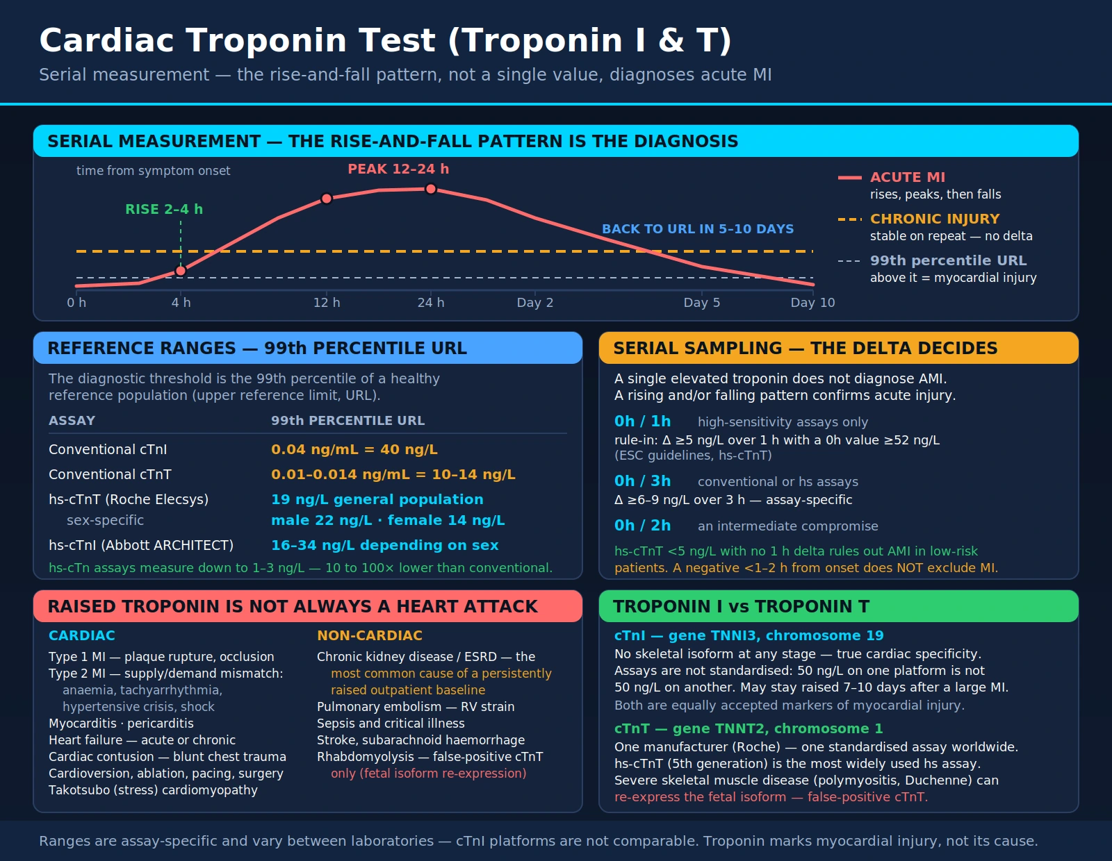
Cardiac troponin (cTnI and cTnT) is the gold-standard biomarker for diagnosing acute myocardial infarction. Released from cardiomyocytes when the cell membrane is disrupted, it combines high sensitivity — it detects even minor myocardial injury — with high cardiac specificity, because the cardiac isoforms are structurally distinct from skeletal muscle troponin. The arrival of high-sensitivity troponin (hs-cTn) assays transformed the emergency evaluation of chest pain, making 0h/1h and 0h/2h rapid rule-in / rule-out algorithms possible where earlier assays needed six hours or more.
Interactive Visualization The Heart & Circulation — watch a blood cell make the loop Trace a blood cell through all four chambers and both circuits, with a live ECG, chamber pressures, and an exercise mode. Launch →Table of Contents
- What the Cardiac Troponin Test Measures
- Biochemistry of the Troponin Complex
- Why Troponin Testing Is Ordered
- How the Test Is Performed
- Troponin I vs. Troponin T: Differences
- Reference Ranges and Result Interpretation
- The Troponin Rise-and-Fall Pattern
- Rapid Rule-In and Rule-Out Algorithms
- The HEART Score
- The Five Types of Myocardial Infarction
- Conditions That Elevate Troponin
- Non-Coronary Causes in Depth
- High-Sensitivity Troponin (hs-cTn)
- What Trials Report on Lowering Cardiac Risk
- Limitations of Troponin Testing
- Key Research Papers
- Connections
- Featured Videos
What the Cardiac Troponin Test Measures
Cardiac troponin is a protein complex found exclusively in heart muscle cells (cardiomyocytes). It plays a central role in regulating the contraction of the heart by controlling the interaction between actin and myosin, the two proteins that generate the mechanical force of heartbeat. The troponin complex has three subunits: troponin C (which binds calcium), troponin I (which inhibits contraction in the resting state), and troponin T (which anchors the complex to tropomyosin on the actin filament).
In a healthy heart, troponin remains inside cardiomyocytes. When heart muscle cells are damaged or die — as occurs during a myocardial infarction, myocarditis, or significant cardiac stress — the cell membranes break down and troponin leaks into the bloodstream. Because cardiac troponin I (cTnI) and cardiac troponin T (cTnT) are structurally distinct from their skeletal muscle counterparts, laboratory assays can detect them with high cardiac specificity. Even small amounts of myocardial injury can produce a measurable rise in troponin.
Troponin testing has become the gold-standard biomarker for diagnosing acute myocardial infarction (heart attack) and for risk-stratifying patients presenting with chest pain. The advent of high-sensitivity troponin (hs-cTn) assays in the 2010s further transformed cardiac emergency medicine by measuring far lower concentrations: in the Dallas Heart Study, troponin T was detectable in 25.0% of adults aged 30 to 65 with a highly sensitive assay, against 0.7% with the standard assay (de Lemos 2010).
How Troponin Became the Standard
Troponin entered routine clinical use as a cardiac biomarker in the 1990s and rapidly displaced CK-MB (the MB isoform of creatine kinase), which had been the previous standard but was also present in skeletal muscle and therefore far less cardiac-specific. The 2000 ESC/ACC consensus document redefined myocardial infarction around the new marker: any troponin elevation above the 99th percentile of a healthy reference population, accompanied by a rise and/or fall pattern and evidence of ischemia. That document became the first "Universal Definition of Myocardial Infarction," and it has been revised three times since — in 2007, 2012, and 2018.
The two cardiac-specific isoforms measured in clinical practice are distinguished from their skeletal-muscle counterparts by structural features that the assay antibodies target directly:
- Cardiac troponin I (cTnI): the inhibitory subunit, carrying a cardiac-specific N-terminal extension that skeletal-muscle troponin I lacks.
- Cardiac troponin T (cTnT): the tropomyosin-binding subunit, with a cardiac-specific region in its N-terminal portion that the assay antibodies target.
Both are measured with highly specific monoclonal antibodies that do not cross-react with skeletal muscle isoforms under normal conditions — which makes the test cardiac-specific, not merely cardiac-sensitive. It is worth being precise about what that specificity does and does not buy: troponin identifies myocardial cell injury or death, but it does not identify the cause of that injury. Coronary atherosclerosis, cardiomyopathy, myocarditis, pulmonary embolism, and sepsis all produce the same signal. Sorting out which one is present relies on the clinical history, the ECG, echocardiography, and where appropriate coronary angiography.
Biochemistry of the Troponin Complex
The troponin complex is a trimeric regulatory protein embedded in the thin filament of striated muscle, sitting at the core of the calcium-regulated actin-myosin contraction cycle. Each of the three subunits has a distinct structural and functional role:
- Troponin T (TnT) — the tropomyosin-binding subunit. It anchors the entire complex to tropomyosin along the thin filament. Its elongated N-terminal tail extends along tropomyosin, while its globular C-terminal domain interacts with TnI and TnC.
- Troponin I (TnI) — the inhibitory subunit. At low intracellular calcium, TnI's inhibitory region binds actin, holding tropomyosin over the myosin-binding sites and preventing contraction. The cardiac-specific N-terminal extension is phosphorylated by protein kinase A in response to beta-adrenergic stimulation, which modulates the calcium sensitivity of contraction — part of how adrenaline makes the heart contract harder and relax faster.
- Troponin C (TnC) — the calcium-sensing subunit. It contains four EF-hand calcium-binding motifs. Sites III and IV at the C-terminal end are high-affinity structural sites that are permanently occupied; site II at the N-terminal end is the low-affinity regulatory site that binds calcium during depolarization and triggers the conformational cascade.
The Contraction Cycle
When intracellular calcium rises during an action potential, calcium binds TnC's regulatory site II. The resulting conformational change causes TnC to grab TnI's inhibitory domain, releasing TnI from actin. Tropomyosin then shifts and exposes the myosin-binding sites on actin, permitting cross-bridge cycling and contraction. When calcium is pumped back into the sarcoplasmic reticulum by SERCA, TnC releases its calcium, TnI rebinds actin, tropomyosin slides back over the myosin-binding sites, and the muscle relaxes.
Why Troponin Leaks in Two Phases
Ischemia that lasts long enough produces irreversible cardiomyocyte injury and disruption of the sarcolemmal membrane. Troponin then escapes from two pools, which Katus and colleagues described in 1991 as an unbound cytosolic pool and the structurally bound troponin of the myofibrils (Katus 1991, Am J Cardiol):
- The free cytosolic pool — a small unbound reservoir — washes out first, producing the early rise in serum troponin within hours.
- The structurally bound complexes — the great majority of the protein, tethered to the thin filament — are degraded and released far more slowly over the following days, which is why troponin stays elevated long after the event.
That slow second phase is diagnostically valuable: a patient who presents days after an episode of chest pain may still have measurable troponin from the original event, allowing a subacute MI to be identified retrospectively.
Why Troponin Testing Is Ordered
Cardiac troponin testing is ordered in a wide range of clinical situations:
- Suspected acute myocardial infarction (AMI): Any patient presenting to an emergency department with chest pain, shortness of breath, arm or jaw pain, or other symptoms suggestive of a heart attack receives serial troponin measurements as part of the standard workup. Under the Fourth Universal Definition of Myocardial Infarction, a rising and/or falling pattern with at least one value above the 99th percentile indicates acute myocardial injury, and it is called an acute MI when the injury is caused by myocardial ischaemia.
- Risk stratification in acute coronary syndromes (ACS): Patients with unstable angina or non-ST-elevation myocardial infarction (NSTEMI) are stratified by troponin level to guide urgency of intervention — those with elevated troponin face higher short-term risk (in Keller 2009, a raised sensitive troponin I was independently associated with adverse outcome at 30 days, hazard ratio 1.96), and guidelines use troponin in deciding how soon invasive evaluation takes place.
- Post-cardiac surgery monitoring: Troponin is released during bypass surgery and other cardiac procedures; serial measurements help identify procedure-related myocardial injury.
- Suspected myocarditis or pericarditis: Inflammatory conditions of the heart muscle or pericardium can elevate troponin to varying degrees.
- Heart failure assessment: Chronically elevated low-level troponin in heart failure patients correlates with ongoing myocardial stress and adverse prognosis.
- Cardiotoxicity monitoring: Cancer patients receiving cardiotoxic chemotherapy agents (such as anthracyclines or trastuzumab) are monitored with serial troponin to detect early myocardial injury.
- Evaluation of other cardiac conditions: Pulmonary embolism, hypertensive emergencies, and sepsis can all cause secondary cardiac injury detectable by troponin elevation.
How the Test Is Performed
Cardiac troponin is measured from a standard venous blood draw. No fasting is required. The sample is sent to the clinical laboratory immediately — troponin testing in emergency settings is typically processed as a STAT (urgent) order.
Because troponin rises progressively after cardiac injury rather than instantly, diagnosis rests on serial sampling. Rapid rule-in/rule-out protocols use one of these schedules:
- 0h/3h protocol: Blood drawn at presentation and repeated 3 hours later. A significant rise or fall between the two samples confirms acute myocardial injury.
- 0h/1h protocol (high-sensitivity assays only): The faster kinetics of high-sensitivity assays allow meaningful risk stratification within one hour of the first draw. The 2020 ESC guideline ranked it first among the high-sensitivity algorithms, and the 2023 ESC guideline again sets out the 0h/1h and 0h/2h algorithms.
- 0h/2h protocol: the ESC guidelines' second option, used where a precise one-hour re-draw is hard to organise.
Point-of-care (POC) troponin devices are also available for bedside testing in pre-hospital settings or resource-limited environments, though laboratory-based assays remain the gold standard for sensitivity and precision.
Troponin I vs. Troponin T: Differences
Both cardiac troponin I (cTnI) and cardiac troponin T (cTnT) are equally accepted as diagnostic markers for myocardial injury. They are structurally distinct proteins encoded by different genes, and their clinical performance characteristics differ slightly:
Cardiac Troponin I (cTnI):
- Encoded by the TNNI3 gene on chromosome 19.
- No skeletal muscle isoform at any developmental stage — true cardiac specificity.
- Multiple manufacturers produce cTnI assays, but without standardization across platforms, reference ranges vary significantly between laboratories and cannot be directly compared.
- Not raised by diseased skeletal muscle: in a Mayo Clinic series, 16 patients with myopathies had raised cTnT but normal cTnI (Jaffe 2011).
Cardiac Troponin T (cTnT):
- Encoded by the TNNT2 gene on chromosome 1.
- Healthy adult skeletal muscle does not express it, but diseased skeletal muscle can: in Jaffe 2011, the antibodies of both the fourth-generation and the high-sensitivity cTnT assays recognised a protein in biopsies of diseased skeletal muscle, a possible source of false-positive cTnT results; the authors suggest re-expressed isoforms as the explanation. The Fourth Universal Definition likewise notes that injured skeletal muscle expresses proteins detected by the cTnT assay.
- Made by a single manufacturer, so cTnT results come from one assay design rather than from several unstandardized ones.
- In kidney disease, the Fourth Universal Definition notes that hs-cTnT is more often elevated than hs-cTnI.
Both are internationally endorsed as the standard biomarkers for myocardial injury and MI diagnosis (Apple 2012); the practical differences show up mainly in skeletal muscle disease and kidney disease.
Reference Ranges and Result Interpretation
Troponin reference ranges are assay-specific and vary between laboratories. The universal diagnostic threshold for acute myocardial injury is the 99th percentile of a healthy reference population (also called the upper reference limit or URL). Any troponin elevation above the 99th percentile URL is considered abnormal and indicates myocardial injury.
Typical conventional troponin ranges (exact values differ by assay platform):
- Sensitive (contemporary) cTnI: the sensitive troponin I assay studied by Keller and colleagues in 2009 used a cut-off of 0.04 ng/mL; values vary widely by platform.
- High-sensitivity cTnT: the 99th percentile in the assay's 2010 analytical validation was 13.5 ng/L (Giannitsis 2010), and 14 ng/L is the cut point commonly used (Gore 2014). A single presenting value below the assay's limit of detection (<0.005 µg/L) together with a non-ischaemic ECG classified 30.6% of 9,241 patients as low risk in a meta-analysis, with a pooled sensitivity for AMI of 98.7% (Pickering 2017).
- High-sensitivity cTnI (varies by manufacturer): for one widely studied hs-cTnI assay, sex-specific 99th percentiles of 16 ng/L for women and 34 ng/L for men were used in a Scottish cohort and in the High-STEACS trial (Shah 2015; Lee 2019). Other hs-cTnI assays have their own thresholds.
A note on the hs-cTnT numbers above. The 99th percentile of one assay depends on the reference population it is measured in. For hs-cTnT, three US population cohorts gave 99th percentiles of 18, 22 and 36 ng/L (the Dallas Heart Study, ARIC and the Cardiovascular Health Study, after excluding people with recent hospitalisation, overt cardiovascular disease or kidney disease), differences that paralleled the cohorts' ages (Gore 2014). A Swedish random population sample gave 24 ng/L (Hammarsten 2012). The limits that apply to any one result are therefore those of the laboratory and assay that produced it. The 99th percentile itself, despite arguments for higher cutoffs, is described in a 2020 review as remaining the best-established criterion for diagnosing acute MI (Sandoval 2020).
Results are read in clinical context:
- A single elevated troponin does not diagnose AMI — serial measurements showing a rising and/or falling pattern (delta change) confirm acute myocardial injury.
- A stable chronically elevated troponin (same level on serial measurements hours apart) points to a non-acute cause such as chronic kidney disease, heart failure, or a stable cardiomyopathy.
- Troponin below the URL does not rule out all cardiac pathology, and a very early sample may precede a measurable rise: in Hammarsten 2012, troponin T stayed below the 99th percentile in 1% of patients with NSTEMI until 8.5 hours after symptom onset.
Why the Reference Range Differs by Sex
Women consistently have lower 99th percentile troponin concentrations than men, for both hs-cTnT and hs-cTnI. Applying a sex-specific URL rather than one combined number does two things at once:
- It identifies more injury in women: in a Scottish cohort, an hs-cTnI assay with sex-specific thresholds (women 16 ng/L, men 34 ng/L) raised the diagnosis of MI in women from 11% to 22%, against 19% to 21% in men, compared with a contemporary assay with a single 50 ng/L threshold (Shah 2015).
- It avoids over-calling men: in three US cohorts the hs-cTnT 99th percentile was higher in men, and the authors concluded that a uniform 14 ng/L cutoff may over-diagnose MI, particularly in men and older people (Gore 2014).
Finding more injury has not, so far, been shown to improve outcomes. In the High-STEACS stepped-wedge trial of 48,282 patients across 10 hospitals, sex-specific thresholds increased the identification of myocardial injury by 42% in women and 6% in men, but women with injury still received roughly half as many coronary treatments as men, and the one-year rate of reinfarction or cardiovascular death in women was not improved (Lee 2019).
Why the Reference Range Rises With Age
The 99th percentile climbs with age even among people considered healthy. In emergency-room patients without obvious conditions that raise troponin, the hs-cTnT 99th percentile was 12 ng/L under age 65 but 82 ng/L over 65 (Hammarsten 2012), and in the three US cohorts more than 10% of men aged 65 to 74 with no cardiovascular disease had values above the MI threshold (Gore 2014). In the Dallas Heart Study, detectable troponin T was linked to left ventricular hypertrophy, systolic dysfunction and kidney disease (de Lemos 2010), so the rise is not simply an artifact. For an older patient with a raised value, the rise-and-fall pattern across serial draws is what separates a stable personal baseline from an acute event.
The Rise/Fall (Delta) Criterion
A single value above the 99th percentile establishes myocardial injury. It does not establish acute myocardial infarction, which under the Fourth Universal Definition additionally requires a rising and/or falling pattern and evidence that ischaemia caused the injury. Delta criteria are assay-specific; the published examples for hs-cTnT include:
- 0h/1h algorithm (TRAPID-AMI): a 1-hour rise of at least 5 ng/L, or a 0h value of at least 52 ng/L, was a rule-in criterion; a 0h value below 12 ng/L with a 1-hour change below 3 ng/L was a rule-out criterion (Mueller 2016).
- Relative change: a change of more than 20% has often been used alongside a raised value, but in hospitalised patients without MI, serial hs-cTnT changes of more than 20% were common — the 97.5th percentile of change was 51–67% (Hammarsten 2012).
- Chronic stable elevation: no meaningful delta across serial draws, indicating ongoing but non-acute myocardial injury — heart failure, chronic kidney disease, or a stable cardiomyopathy.
The Troponin Rise-and-Fall Pattern
The characteristic biokinetic pattern of troponin in acute MI is a rise-and-fall curve that reflects the release and gradual clearance of troponin from damaged cardiomyocytes. Understanding this pattern is essential for interpreting serial results:
- Onset: with a first-generation assay, troponin T appeared in serum as early as 3 hours after the onset of pain in 50% of patients with MI (Katus 1991, Circulation). High-sensitivity assays detect the rise earlier: their accuracy at presentation stayed high even in patients arriving within 3 hours of chest-pain onset (Reichlin 2009).
- Peak: in thrombolysis-treated patients whose artery reopened early, troponin T showed a marked early peak at about 14 hours after the onset of pain — a "washout" of the cytosolic pool. That early peak was absent when reperfusion came more than 5.5 hours after onset or not at all (Katus 1991, Am J Cardiol).
- How long it stays raised: in the same group's 1991 study, troponin T remained elevated in every patient with MI for more than 130 hours (over five days), reflecting release from both the free cytosolic and the structurally bound pools, and its diagnostic efficiency stayed at 98% until 5.5 days after admission (Katus 1991, Circulation). This long window allows diagnosis of patients who present days after their event (subacute MI).
The delta change (absolute or relative change between serial measurements) is the central diagnostic concept. The ESC algorithms set assay-specific delta cut-offs; for one hs-cTnT assay, the 0h/1h algorithm validated in TRAPID-AMI treated a 1-hour rise of at least 5 ng/L, or a 0h value of at least 52 ng/L, as a rule-in criterion (Mueller 2016).
Rapid Rule-In and Rule-Out Algorithms
The 2020 ESC guideline for acute coronary syndromes without persistent ST-segment elevation recommended the 0h/1h and 0h/2h high-sensitivity troponin algorithms as the first and second choices, ahead of the 0h/3h algorithm (as summarised by Chiang 2022). The 2023 ESC guideline for acute coronary syndromes sets out the ESC 0h/1h and 0h/2h rule-out and rule-in algorithms in its Figure 6, with cut-offs that are specific to each assay (Byrne 2023). These algorithms replaced the older practice of serial draws over six hours or more before a decision.
The 0h/1h Algorithm (Preferred)
Two hs-cTn measurements are taken: one at presentation and one exactly one hour later. Patients then fall into one of three zones. The figures below are for one hs-cTnT assay, as validated in the TRAPID-AMI multicenter study of 1,282 patients, 16.6% of whom had a final diagnosis of AMI (Mueller 2016):
- Rule out — 0h hs-cTnT below 12 ng/L and a 1-hour change below 3 ng/L. In TRAPID-AMI this captured 63.4% of all patients, with a negative predictive value of 99.1% (95% CI 98.2–99.7%) and sensitivity of 96.7%. The ESC algorithms also allow rule-out on a single very low presenting value in patients whose chest pain began more than three hours earlier; in a meta-analysis, a single hs-cTnT below the limit of detection with a non-ischaemic ECG had a pooled sensitivity for AMI of 98.7% (Pickering 2017).
- Rule in — 0h hs-cTnT of 52 ng/L or above, or a 1-hour rise of 5 ng/L or more. This captured 14.4% of patients, with a positive predictive value of 77.2% and specificity of 96.1%.
- Observation zone — neither criterion met, covering the remaining 22.2%. The MI prevalence in this group was 22.5%, so it is genuinely indeterminate rather than reassuring; under the ESC algorithm these patients have a further troponin measurement and clinical reassessment.
The specific ng/L values are assay-dependent and are not transferable. The thresholds above apply to one hs-cTnT assay; each hs-cTnI assay has its own separately validated numbers, which the ESC guideline lists assay by assay, and applying one assay's cut-points to another assay's results is a recognised source of error.
The 0h/2h Algorithm
An alternative for departments where a precise one-hour re-draw is logistically difficult. Two samples are taken at presentation and two hours later. Rule-out requires a low presenting value with a small 2-hour change; rule-in requires a markedly elevated presenting value or a large 2-hour change, with assay-specific cut-offs. In a 2022 meta-analysis of 32 studies (30,066 patients), the 0h/2h algorithm had a pooled sensitivity of 98.6% and negative predictive value of 99.6% for ruling out AMI, against 99.1% and 99.8% for the 0h/1h algorithm (Chiang 2022).
The 0h/3h Algorithm
The older rapid algorithm, which uses the 99th percentile threshold plus a significant change across three hours. In the same meta-analysis it had a pooled sensitivity of 93.7% and negative predictive value of 98.7%, and its sensitivity was lower in studies that did not add clinical criteria (a GRACE score below 140 and being pain-free) than in studies that did (90.2% vs 98.4%). The authors concluded that the 0h/1h and 0h/2h algorithms have higher sensitivities and negative predictive values than the 0h/3h algorithm (Chiang 2022).
The HEART Score
Troponin is one input, not the whole decision. The HEART score is a validated five-component clinical decision tool — History, ECG, Age, Risk factors, Troponin — that scores each element 0, 1, or 2 for a total of 0 to 10. In its prospective validation in 2,440 patients at ten Dutch hospitals, the rate of major adverse cardiac events (MACE) within six weeks fell into three bands (Backus 2013):
- Low (0–3): 36.4% of patients; MACE in 1.7%.
- Intermediate (4–6): MACE in 16.6%.
- High (7–10): MACE in 50.1%.
The HEART score's c-statistic (0.83) was significantly higher than those of the TIMI (0.75) and GRACE (0.70) scores. The authors wrote that low scores excluded short-term MACE with more than 98% certainty, so that "reserved policies" might be considered, while high scores might indicate "more aggressive policies" (Backus 2013).
The Five Types of Myocardial Infarction
The Fourth Universal Definition of Myocardial Infarction classifies MI into five types by pathophysiology, not by troponin magnitude. Two patients can have identical troponin values and very different underlying problems, so the classification shapes how each is managed.
Type 1 — Atherosclerotic Plaque Event
Spontaneous MI caused by atherosclerotic plaque rupture, erosion, or fissuring with intraluminal thrombus formation, leading to acute coronary occlusion or sharply reduced coronary flow. This is the classic heart attack, presenting as either STEMI or NSTEMI. Guideline management covers revascularization and antithrombotic and secondary-prevention drug therapy (Byrne 2023).
Type 2 — Supply-Demand Mismatch
Myocardial injury caused by something other than plaque disruption, in which oxygen demand outstrips supply. Troponin rises and falls as it does in Type 1, but coronary angiography reveals no culprit lesion. Common triggers include:
- Tachyarrhythmia — atrial fibrillation with rapid ventricular response, producing rate-related subendocardial ischemia.
- Hypotension and shock — hemorrhagic, distributive (sepsis), or cardiogenic, all reducing global coronary supply.
- Severe anemia — reduced oxygen-carrying capacity lets demand outrun supply, especially in a territory served by a fixed coronary stenosis.
- Hypertensive emergency — extreme afterload driving subendocardial ischemia.
- Coronary vasospasm — Prinzmetal angina, and cocaine-induced spasm.
- Pulmonary embolism — acute right-heart pressure overload causing right ventricular free-wall ischemia.
Management of Type 2 MI centers on the underlying condition. Evidence for antiplatelet and anticoagulant therapy in Type 2 MI is much more limited than in Type 1, and these drugs carry bleeding risk — a particular concern in a bleeding or septic patient.
Types 3, 4, and 5
- Type 3: sudden cardiac death with ischemic symptoms and new ECG changes, where the patient died before biomarkers could be drawn or before results returned.
- Type 4a: MI associated with percutaneous coronary intervention — defined, in patients with normal baseline values, as troponin elevation greater than 5× the URL within 48 hours of the procedure, together with new ischaemic ECG changes, new pathological Q waves, imaging evidence of new loss of viable myocardium, or an angiographic procedural complication. When baseline troponin is already raised and stable (≤20% variation) or falling, it must rise by more than 20% to above 5× the URL.
- Type 4b: MI caused by stent thrombosis.
- Type 4c: MI related to in-stent restenosis.
- Type 5: MI associated with coronary artery bypass grafting — defined, in patients with normal baseline values, as troponin elevation greater than 10× the URL within 48 hours, together with new pathological Q waves, angiographic evidence of new graft or native coronary occlusion, or imaging evidence of new loss of viable myocardium.
The higher multiples required for Types 4a and 5 exist because both procedures cause some myocardial injury by design; the threshold has to clear that expected background before a procedural infarction can be called.
Conditions That Elevate Troponin
While troponin elevation is most strongly associated with acute MI, it can also be elevated in a broad range of other conditions. These alternatives — type 2 MI and non-ischaemic myocardial injury — are part of the differential for a positive result:
Primary cardiac causes:
- Acute myocardial infarction (Type 1 MI): Plaque rupture causing coronary artery occlusion — the classic heart attack.
- Type 2 MI: Myocardial ischemia due to supply-demand mismatch without plaque rupture (e.g., severe anemia, tachyarrhythmias, hypertensive crisis, shock).
- Myocarditis: Viral or autoimmune inflammation of the heart muscle can cause significant troponin elevation mimicking AMI.
- Heart failure (acute decompensated or chronic): Low-level troponin release is common in heart failure due to ongoing myocardial stretch and ischemia.
- Cardiac contusion: Blunt chest trauma can cause myocardial injury detectable by troponin elevation.
- Cardiac procedures: Cardioversion, ablation, pacemaker implantation, and cardiac surgery all cause procedural troponin release.
- Takotsubo (stress) cardiomyopathy: Transient apical ballooning syndrome caused by catecholamine surge, often after emotional or physical stress.
Non-cardiac causes (secondary myocardial injury):
- Pulmonary embolism: Right ventricular strain causes troponin elevation, and a raised troponin is associated with higher short-term mortality (Becattini 2007).
- Chronic kidney disease (CKD) and end-stage renal disease (ESRD): Reduced troponin clearance and chronic low-level myocardial injury commonly cause persistently elevated baseline troponin in CKD patients.
- Sepsis and critical illness: troponin was elevated in 61% of patients with sepsis across 13 studies, and elevation was associated with death (Bessière 2013).
- Stroke and subarachnoid hemorrhage: Neurogenic cardiac injury from catecholamine surges caused by intracranial events elevates troponin.
- Skeletal muscle disease: diseased skeletal muscle can raise cTnT (not cTnI) without cardiac injury (Jaffe 2011).
- Strenuous endurance exercise: troponin T rose above the assay's detection limit after endurance events in almost half of athletes studied (Shave 2007).
Non-Coronary Causes in Depth
Any condition that injures cardiomyocytes will raise troponin, whether or not a coronary artery is involved. The list above is the quick reference; what follows is the detail that matters when a troponin comes back positive and the coronary arteries turn out to be clean.
Myocarditis
Inflammation of the myocardium arising from viral infection (coxsackievirus B, adenovirus, parvovirus B19, influenza, SARS-CoV-2), autoimmune processes (giant cell myocarditis, eosinophilic myocarditis), or toxic exposures (anthracyclines, immune checkpoint inhibitors). Troponin can be markedly elevated, and the ECG may show diffuse ST elevation that mimics a STEMI closely enough to send a patient to the catheterization laboratory. The angiogram shows normal or non-obstructive arteries. Cardiac MRI with late gadolinium enhancement is the key diagnostic tool, showing a patchy, often subepicardial pattern that does not follow a coronary territory.
Pulmonary Embolism
Acute massive or submassive pulmonary embolism causes abrupt right ventricular pressure overload. The RV free wall, which is normally perfused at low pressure and has little reserve, sustains subendocardial ischemia when intracavitary pressure rises suddenly. A meta-analysis of 20 studies (1,985 patients) found that 19.7% of patients with raised troponin died in the short term, against 3.7% of those with normal troponin (odds ratio 5.24), and the association held in hemodynamically stable patients (odds ratio 5.90) and for both troponin I and T (Becattini 2007). Troponin is therefore used, alongside clinical scores and imaging of the right ventricle, in grading the severity of a pulmonary embolism.
Acute Decompensated Heart Failure
Both acute and chronic heart failure are associated with troponin elevation. During acute decompensation, cardiomyocyte injury comes from several directions at once: a raised left ventricular end-diastolic pressure compresses subendocardial perfusion, catecholamine excess drives demand up, and neurohormonal activation adds further stress. In 5,284 patients with chronic heart failure from the Val-HeFT and GISSI-HF trials, increases in hs-cTnT over three to four months were associated with all-cause mortality (hazard ratios 1.59 and 1.88) after adjustment for risk factors and for baseline hs-cTnT and NT-proBNP, although the change added only modest prognostic discrimination beyond the baseline value, and only for fatal outcomes (Masson 2012).
Takotsubo (Stress) Cardiomyopathy
Also called apical ballooning syndrome or broken heart syndrome. A massive catecholamine surge — emotional shock, severe physical stress, the post-surgical state — produces a transient wall-motion abnormality with ballooning of the left ventricular apex and preserved or hyperdynamic basal contraction, giving the characteristic "octopus pot" shape on ventriculography that named the syndrome. Troponin rises, though usually to a lower peak than an equivalent-territory STEMI would produce, and the disproportion between a modest troponin and a large wall-motion abnormality is itself a diagnostic clue. Coronary angiography shows no obstructive disease, and ejection fraction typically recovers.
Sepsis-Induced Cardiomyopathy
Direct cardiomyocyte injury from circulating cytokines (TNF-alpha, IL-1beta, IL-6), endotoxin, reactive oxygen species, and nitric oxide produces a reversible myocardial depression in some septic patients. In a meta-analysis of 13 studies (1,227 patients), troponin was elevated in 61%, and elevation was associated with all-cause mortality (relative risk 1.91), including after adjustment for prognostic scores in four studies (odds ratio 1.92); the authors note that the precise role of troponin and its optimal cut-offs in sepsis are still undefined (Bessière 2013).
Chronic Kidney Disease and Dialysis
Both cTnI and cTnT run chronically elevated in chronic kidney disease. The cause is a combination of reduced clearance, uremia-mediated cardiomyocyte injury, left ventricular hypertrophy, and a genuinely high prevalence of subclinical coronary disease — it is not simply a laboratory artifact, and the elevation carries real prognostic weight. The Fourth Universal Definition notes that hs-cTnT is more often elevated than hs-cTnI in this population. In dialysis patients a single number says little on its own; serial measurement is used to look for a dynamic change against the patient's own elevated baseline.
Skeletal Muscle Disease and Rhabdomyolysis
This is the one situation where the choice of assay changes the answer, and it is easy to get backwards. Diseased skeletal muscle can contain a protein that cTnT assays detect, producing an elevated cTnT without cardiac injury. In the Mayo Clinic series that documented this, 16 patients with myopathies had increased cTnT but not cTnI, and biopsies of their diseased muscle reacted with the cTnT assays' antibodies; the authors proposed re-expressed isoforms as the source (Jaffe 2011). Measuring cTnI was how that study separated the two.
Strenuous Endurance Exercise
Troponin can rise after prolonged hard exercise in people with no known heart disease. A meta-analysis of 26 studies (1,120 cases) found post-exercise troponin T above the assay's detection limit in 47% of participants; detection after cycling events was about half that after running events (27% vs 52%), and heavier individuals in shorter endurance events, primarily marathon runners, were slightly more likely to show it (Shave 2007).
Other Causes
- Cardiac contusion: blunt chest trauma from a motor vehicle collision or a direct precordial blow can injure the myocardium and raise troponin.
- Ablation and cardioversion: iatrogenic myocardial injury from radiofrequency or electrical energy, expected rather than alarming.
- Chemotherapy: anthracyclines (doxorubicin, daunorubicin) cause dose-dependent cardiomyocyte injury; trastuzumab causes a distinct, more often reversible cardiomyopathy; immune checkpoint inhibitors can trigger myocarditis: in a multicenter registry, its prevalence was 1.14%, 46% of 35 affected patients had a major adverse cardiac event over a median of 102 days, troponin T of 1.5 ng/mL or more carried a fourfold higher risk, and lower steroid doses were associated with higher event rates (Mahmood 2018). Serial troponin is part of cardio-oncology monitoring for these reasons.
- Stroke and subarachnoid hemorrhage: neurogenic cardiac injury from a massive catecholamine release, presenting with troponin elevation, wall-motion abnormalities, and QTc prolongation that together can mimic an MI.
- Severe hypothyroidism: myxedema can raise troponin through a metabolic cardiomyopathy.
High-Sensitivity Troponin (hs-cTn)
High-sensitivity cardiac troponin (hs-cTn) assays represent a major technological advance over conventional troponin assays. The key differences:
- Lower detection limit: hs-cTn assays measure far lower concentrations than conventional assays; the first hs-cTnT assay had an analytical range starting at 3 ng/L (Giannitsis 2010).
- Measurability in healthy individuals: A defining characteristic of hs-cTn assays is that they can measure troponin in at least 50% of a healthy reference population, in contrast to conventional assays, with which most healthy individuals have undetectable levels.
- Faster rule-out: The superior sensitivity enables rapid 0h/1h rule-out protocols — in TRAPID-AMI, a very low hs-cTnT at presentation with a minimal 1-hour change had a negative predictive value of 99.1% for AMI (Mueller 2016).
- Sex-specific thresholds: Women have lower baseline troponin levels than men. hs-cTn assays have sex-specific 99th percentile URLs (for one hs-cTnI assay, 16 ng/L in women vs. 34 ng/L in men), which increased the diagnosis of MI in women in a Scottish cohort (Shah 2015).
- Population-based chronic disease monitoring: Because hs-cTn detects subclinical myocardial injury, it is increasingly studied as a chronic disease risk marker — in the Dallas Heart Study, troponin T detected with a highly sensitive assay was independently associated with all-cause mortality in the general population (de Lemos 2010).
The adoption of hs-cTn has increased the diagnosis of MI, particularly in women (Shah 2015), while the rapid algorithms allow many patients with chest pain to be ruled out sooner than with extended observation (Mueller 2016; Chiang 2022).
What Officially Makes an Assay "High-Sensitivity"
The generation of a troponin assay is not defined by a particular technology but by two analytical performance criteria discussed by the International Federation of Clinical Chemistry (IFCC) task force (Apple 2012):
- Conventional (contemporary) assays: a coefficient of variation greater than 10% at the 99th percentile, and measurable troponin in fewer than 50% of healthy individuals. Older troponin I and T assays fall here.
- High-sensitivity assays: a coefficient of variation of 10% or less at the 99th percentile and measurable troponin in more than 50% of healthy individuals — ideally more than 95%.
The imprecision criterion matters as much as the detection criterion. An assay that can technically report very low numbers but scatters them widely from run to run cannot support a rule-out decision built on a 1-hour change of less than 3 ng/L; the whole 0h/1h approach depends on the assay being able to distinguish a small real change from its own noise. The second criterion produces the conceptual shift: because troponin is now measurable at physiological background levels in nearly everyone, clinicians can track a dynamic change from an individual's own baseline instead of waiting for a threshold to be crossed from "undetectable."
Major Validated Platforms
- hs-cTnT: made by a single manufacturer; the assay used in the TRAPID-AMI validation of the 0h/1h algorithm (Mueller 2016).
- hs-cTnI: made by several manufacturers, each assay with its own 99th percentile and its own validated algorithm cut-offs.
- Performance across assays: in the 2022 meta-analysis, the ESC algorithms performed similarly across the three high-sensitivity assays studied (one hs-cTnT and two hs-cTnI) (Chiang 2022).
- Point-of-care hs-cTn devices are now available for settings where central laboratory turnaround exceeds 60 minutes, though laboratory analyzers remain the reference for precision.
Before high-sensitivity assays, confirming a rise commonly took serial draws on admission and at 3 and 6 hours (as in Keller 2009). In TRAPID-AMI, the 0h/1h algorithm assigned 63.4% of patients to rule-out after the one-hour sample (Mueller 2016). The corresponding hazard is that assay thresholds are not interchangeable: a hospital switching platforms needs a new set of cut-points, and published guidance exists specifically for institutions making that transition.
What Trials Report on Lowering Cardiac Risk
For people who have had a troponin-positive event or who are at elevated cardiovascular risk, this is what the research reports on lifestyle, nutrition and medicines. Decisions about treatment belong with a clinician.
Lifestyle Foundations
- Mediterranean diet: a diet built on vegetables, fruits, legumes, nuts, olive oil and fish. In the PREDIMED trial (7,447 people at high cardiovascular risk without cardiovascular disease, median follow-up 4.8 years), a Mediterranean diet supplemented with extra-virgin olive oil or with nuts was associated with fewer major cardiovascular events than a control diet in which participants were advised to reduce dietary fat (hazard ratios 0.69 and 0.72). These are the figures of the 2018 republication, which replaced the withdrawn 2013 report after protocol deviations in randomization were found (Estruch 2018).
- Physical activity: moderate-intensity aerobic exercise is associated with lower cardiovascular risk through effects on endothelial function, blood pressure, inflammation and lipid profiles, and cardiac rehabilitation is a standard part of care after MI.
- Smoking: smoking is a powerful independent risk factor for coronary artery disease and thrombosis, and MI risk falls after stopping.
- Body weight: obesity, particularly visceral adiposity, drives systemic inflammation, insulin resistance, hypertension, and dyslipidemia — all contributors to coronary artery disease.
- Psychological stress: acute psychological stress triggers catecholamine surges that can precipitate takotsubo cardiomyopathy and coronary spasm. Chronic stress is linked to inflammation and unhealthy behaviors, and meditation, yoga and stress-reduction programs are being studied for cardiovascular effects.
Nutrition Research
- Omega-3 fatty acids: EPA and DHA lower triglycerides. In the REDUCE-IT trial (8,179 statin-treated patients with raised triglycerides and cardiovascular disease or diabetes plus risk factors), a purified prescription EPA ethyl ester (icosapent ethyl, 2 g twice daily) was followed by fewer primary end-point events than placebo over a median of 4.9 years (17.2% vs 22.0%; hazard ratio 0.75). Hospitalization for atrial fibrillation or flutter was more common with the drug (3.1% vs 2.1%) (Bhatt 2019).
- Magnesium: magnesium deficiency is associated with arrhythmias, hypertension, and endothelial dysfunction.
- Coenzyme Q10: CoQ10 takes part in mitochondrial energy production in cardiomyocytes, and supplementation has been studied in heart failure and in statin-associated muscle symptoms.
Medical Management After AMI
For patients who have had a confirmed MI with troponin elevation, guideline-directed medical therapy typically includes dual antiplatelet therapy, high-intensity statin therapy, ACE inhibitors or ARBs (especially with reduced ejection fraction), beta-blockers (in the acute setting and for reduced ejection fraction), and aldosterone antagonists when indicated. These medications have robust evidence for reducing recurrent MI, heart failure, and death.
Limitations of Troponin Testing
- Not synonymous with heart attack: Troponin elevation means myocardial injury, not necessarily coronary artery disease. The clinical context, ECG, symptoms, and imaging findings are all required for proper diagnosis.
- Raised baselines in kidney disease: CKD commonly causes a persistently elevated baseline troponin, so the acute change (delta) rather than the absolute value carries the diagnosis of AMI in these patients.
- Early false negatives: If a patient presents within 1-2 hours of symptom onset, troponin may not yet have risen to detectable levels. A negative initial troponin in this early window does not exclude MI, which is why the algorithms rely on serial testing.
- Assay non-standardization for cTnI: Unlike cTnT (single manufacturer), cTnI assays from different companies cannot be directly compared. A result of 50 ng/L on one cTnI platform does not equal 50 ng/L on another platform.
- Does not localize injury: Troponin indicates that cardiac injury occurred but does not identify which coronary territory is affected. ECG, echocardiography, and coronary angiography are required to localize and characterize the injury.
- Does not measure cardiac function: Troponin is a biomarker of injury, not of systolic or diastolic function. BNP/NT-proBNP are the preferred biomarkers for assessing heart failure severity and ventricular wall stress.
- Analytical interference: rarely, a result is falsely raised with no myocardial injury. A 2026 review of reported cases lists heterophile antibodies, rheumatoid factor, macrotroponin complexes, fibrin clots, hemolysis and analyzer malfunction among the causes (Lai 2026).
- Biotin: high-dose biotin, often found in dietary supplements, can interfere with some troponin immunoassays. After its 2017 safety communication and a later update, the FDA reported continuing adverse-event reports of falsely low troponin results caused by biotin, and it publishes a list of troponin assays that have not addressed this interference (FDA).
Key Research Papers
Every citation below was checked against the PubMed record (most recently on 14 October 2026); the title, journal, volume, and pages match the linked PMID.
Guidelines and Definitions
- Thygesen K, Alpert JS, Jaffe AS, et al. Fourth Universal Definition of Myocardial Infarction (2018). J Am Coll Cardiol. 2018;72(18):2231–2264. — PubMed PMID: 30153967
- Collet JP, Thiele H, Barbato E, et al. 2020 ESC Guidelines for the management of acute coronary syndromes in patients presenting without persistent ST-segment elevation. Eur Heart J. 2021;42(14):1289–1367. — PubMed PMID: 32860058
- Januzzi JL Jr, Mahler SA, Christenson RH, et al. Recommendations for Institutions Transitioning to High-Sensitivity Troponin Testing: JACC Scientific Expert Panel. J Am Coll Cardiol. 2019;73(9):1059–1077. — PubMed PMID: 30798981
- Byrne RA, Rossello X, Coughlan JJ, et al. 2023 ESC Guidelines for the management of acute coronary syndromes. Eur Heart J. 2023;44(38):3720–3826. — PubMed PMID: 37622654
Rapid Rule-Out Algorithms and Early Diagnosis
- Mueller C, Giannitsis E, Christ M, et al. Multicenter Evaluation of a 0-Hour/1-Hour Algorithm in the Diagnosis of Myocardial Infarction With High-Sensitivity Cardiac Troponin T (TRAPID-AMI). Ann Emerg Med. 2016;68(1):76–87. — PubMed PMID: 26794254
- Reichlin T, Hochholzer W, Bassetti S, et al. Early diagnosis of myocardial infarction with sensitive cardiac troponin assays. N Engl J Med. 2009;361(9):858–867. — PubMed PMID: 19710484
- Keller T, Zeller T, Peetz D, et al. Sensitive troponin I assay in early diagnosis of acute myocardial infarction. N Engl J Med. 2009;361(9):868–877. — PubMed PMID: 19710485
- Body R, Carley S, McDowell G, et al. Rapid exclusion of acute myocardial infarction in patients with undetectable troponin using a high-sensitivity assay. J Am Coll Cardiol. 2011;58(13):1332–1339. — PubMed PMID: 21920261
- Shah AS, Anand A, Sandoval Y, et al. High-sensitivity cardiac troponin I at presentation in patients with suspected acute coronary syndrome: a cohort study. Lancet. 2015;386(10012):2481–2488. — PubMed PMID: 26454362
- Backus BE, Six AJ, Kelder JC, et al. A prospective validation of the HEART score for chest pain patients at the emergency department. Int J Cardiol. 2013;168(3):2153–2158. — PubMed PMID: 23465250
- Chiang CH, Chiang CH, Pickering JW, et al. Performance of the European Society of Cardiology 0/1-Hour, 0/2-Hour, and 0/3-Hour Algorithms for Rapid Triage of Acute Myocardial Infarction: An International Collaborative Meta-analysis. Ann Intern Med. 2022;175(1):101–113. — PubMed PMID: 34807719
- Pickering JW, Than MP, Cullen L, et al. Rapid Rule-out of Acute Myocardial Infarction With a Single High-Sensitivity Cardiac Troponin T Measurement Below the Limit of Detection: A Collaborative Meta-analysis. Ann Intern Med. 2017;166(10):715–724. — PubMed PMID: 28418520
Assay Characteristics and Reference Limits
- Apple FS, Collinson PO; IFCC Task Force on Clinical Applications of Cardiac Biomarkers. Analytical characteristics of high-sensitivity cardiac troponin assays. Clin Chem. 2012;58(1):54–61. — PubMed PMID: 21965555
- Giannitsis E, Kurz K, Hallermayer K, et al. Analytical validation of a high-sensitivity cardiac troponin T assay. Clin Chem. 2010;56(2):254–261. — PubMed PMID: 19959623
- Sandoval Y, Apple FS, Saenger AK, et al. 99th Percentile Upper-Reference Limit of Cardiac Troponin and the Diagnosis of Acute Myocardial Infarction. Clin Chem. 2020;66(9):1167–1180. — PubMed PMID: 32871000
- Hammarsten O, Fu ML, Sigurjonsdottir R, et al. Troponin T percentiles from a random population sample, emergency room patients and patients with myocardial infarction. Clin Chem. 2012;58(3):628–637. — PubMed PMID: 22258764
- Gore MO, Seliger SL, Defilippi CR, et al. Age- and sex-dependent upper reference limits for the high-sensitivity cardiac troponin T assay. J Am Coll Cardiol. 2014;63(14):1441–1448. — PubMed PMID: 24530665
- Shah AS, Griffiths M, Lee KK, et al. High sensitivity cardiac troponin and the under-diagnosis of myocardial infarction in women: prospective cohort study. BMJ. 2015;350:g7873. — PubMed PMID: 25609052
- Lee KK, Ferry AV, Anand A, et al. Sex-Specific Thresholds of High-Sensitivity Troponin in Patients With Suspected Acute Coronary Syndrome. J Am Coll Cardiol. 2019;74(16):2032–2043. — PubMed PMID: 31623760
- Katus HA, Remppis A, Neumann FJ, et al. Diagnostic efficiency of troponin T measurements in acute myocardial infarction. Circulation. 1991;83(3):902–912. — PubMed PMID: 1999039
- Katus HA, Remppis A, Scheffold T, et al. Intracellular compartmentation of cardiac troponin T and its release kinetics in patients with reperfused and nonreperfused myocardial infarction. Am J Cardiol. 1991;67(16):1360–1367. — PubMed PMID: 1904190
Troponin Beyond Acute MI
- de Lemos JA, Drazner MH, Omland T, et al. Association of troponin T detected with a highly sensitive assay and cardiac structure and mortality risk in the general population. JAMA. 2010;304(22):2503–2512. — PubMed PMID: 21139111
- Masson S, Anand I, Favero C, et al. Serial measurement of cardiac troponin T using a highly sensitive assay in patients with chronic heart failure: data from 2 large randomized clinical trials. Circulation. 2012;125(2):280–288. — PubMed PMID: 22139751
- Jaffe AS, Vasile VC, Milone M, et al. Diseased skeletal muscle: a noncardiac source of increased circulating concentrations of cardiac troponin T. J Am Coll Cardiol. 2011;58(17):1819–1824. — PubMed PMID: 21962825
- Becattini C, Vedovati MC, Agnelli G. Prognostic value of troponins in acute pulmonary embolism: a meta-analysis. Circulation. 2007;116(4):427–433. — PubMed PMID: 17606843
- Bessière F, Khenifer S, Dubourg J, et al. Prognostic value of troponins in sepsis: a meta-analysis. Intensive Care Med. 2013;39(7):1181–1189. — PubMed PMID: 23595497
- Shave R, George KP, Atkinson G, et al. Exercise-induced cardiac troponin T release: a meta-analysis. Med Sci Sports Exerc. 2007;39(12):2099–2106. — PubMed PMID: 18046180
- Mahmood SS, Fradley MG, Cohen JV, et al. Myocarditis in Patients Treated With Immune Checkpoint Inhibitors. J Am Coll Cardiol. 2018;71(16):1755–1764. — PubMed PMID: 29567210
- Lai JSY, Yeong N, Tan XY, et al. When troponin results lie: A narrative review and practical approach to spurious hypertroponinemia. Am J Med. 2026;139(5):626–633. — PubMed PMID: 41519456
Risk Reduction After a Troponin-Positive Event
- Bhatt DL, Steg PG, Miller M, et al. Cardiovascular Risk Reduction with Icosapent Ethyl for Hypertriglyceridemia (REDUCE-IT). N Engl J Med. 2019;380(1):11–22. — PubMed PMID: 30415628
- Estruch R, Ros E, Salas-Salvadó J, et al. Primary Prevention of Cardiovascular Disease with a Mediterranean Diet Supplemented with Extra-Virgin Olive Oil or Nuts (PREDIMED). N Engl J Med. 2018;378(25):e34. — PubMed PMID: 29897866 (the corrected and republished report; it replaces the withdrawn 2013 N Engl J Med paper, which this page does not rely on)
PubMed Topic Searches
- PubMed: High-sensitivity troponin and acute MI diagnosis
- PubMed: 0h/1h troponin rule-out protocol (ESC guidelines)
- PubMed: Troponin rapid rule-out algorithm in the emergency department
- PubMed: Cardiac troponin I vs. T in acute coronary syndrome
- PubMed: Non-cardiac causes of troponin elevation
- PubMed: Type 2 myocardial infarction and supply-demand mismatch
- PubMed: Sex-specific troponin thresholds and women's MI diagnosis
- PubMed: Troponin elevation in chronic kidney disease
- PubMed: Hs-cTn as a community cardiovascular risk biomarker
- PubMed: Troponin in myocarditis diagnosis and prognosis
- PubMed: Fourth Universal Definition of MI and troponin
- PubMed: HEART score and chest pain risk stratification
External Authoritative Resources
Connections
- All Lab Tests
- Echocardiogram — the ultrasound window into your heart
- The Heart & Circulation — interactive animation
- Inflammatory Markers
- Coronary Calcium Score
- Lipid Panel
- Apolipoprotein B (ApoB)
- Lipoprotein(a)
- BNP / NT-proBNP
- D-Dimer
- hs-CRP
- Creatine Kinase (CK and CK-MB)
- Complete Blood Count
- Ferritin Test
- Kidney Function
- Homocysteine
- Heart Disease
- Cardiovascular Disease
- Heart Attack
- Heart Failure
- Atrial Fibrillation
- Magnesium