⚠ Evidence warning: Clinical trials or other evidence do not support the claim that a fruit-body-plus-mycelium dual extract delivers both compound families as the best form, that hericenones act on gut and peripheral nerves to raise brain NGF, and that Lion's Mane helps peripheral neuropathy in people; the neuropathy and nerve-repair findings are from animal studies, and the human memory trials are small. This page keeps it as a report of what people, websites or other media say.
Lion's Mane for Nerve Growth Factor
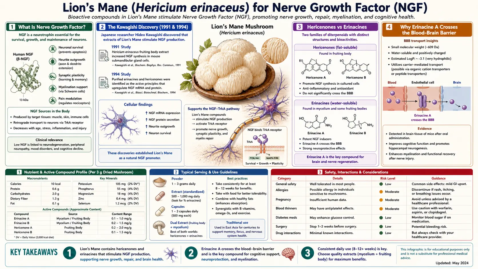
Lion's Mane (Hericium erinaceus) is one of several culinary-medicinal mushrooms shown in laboratory studies to stimulate the synthesis of Nerve Growth Factor (NGF), the foundational neurotrophin that Rita Levi-Montalcini and Stanley Cohen won the 1986 Nobel Prize in Physiology or Medicine for discovering. The Japanese chemist Hirokazu Kawagishi isolated the NGF-stimulating hericenones C, D and E from the fruit body in 1991 and erinacines A, B and C from the mycelium in 1994. Erinacine A (molecular weight 432.5) was shown in a 2021 rat study to cross the blood-brain barrier (BBB) by passive diffusion. Hericenones, by contrast, are concentrated in the white above-ground fruit body; no brain measurement of hericenones appears in the studies on this page, and the idea that they act peripherally on the enteric nervous system and indirectly support central NGF tone is a proposal. Because NGF is specifically required for the survival of cholinergic basal forebrain neurons — the exact population that dies progressively in Alzheimer's disease — this mushroom has accumulated more neurodegenerative-disease research attention than any other dietary fungus.
Table of Contents
- How Nerve Growth Factor Works
- The Kawagishi Discovery (1991 & 1994)
- Hericenones vs Erinacines
- Why Erinacine A Crosses the Blood-Brain Barrier
- Fruit Body vs Mycelium — The Product Selection Decision
- NGF and Cholinergic Basal Forebrain Neurons
- The Alzheimer's Connection
- Peripheral Nerve Regeneration
- Brain-Derived Neurotrophic Factor and Hippocampal Neurogenesis
- Practical Dose and Form for NGF Effects
- Cautions and Drug Interactions
- Key Research Papers
- Connections
- Featured Videos
How Nerve Growth Factor Works
Nerve Growth Factor (NGF) is a 26 kilodalton protein and the founding member of the neurotrophin family — a small set of secreted proteins (NGF, BDNF, NT-3, NT-4) that specific neuron populations require for survival, axon growth, dendritic arborization, and synaptic plasticity. NGF was discovered by Rita Levi-Montalcini and Stanley Cohen in the 1950s in the now-famous experiment in which mouse sarcoma cells transplanted into chick embryos caused massive nerve fiber outgrowth toward the tumor. The pair was awarded the 1986 Nobel Prize in Physiology or Medicine for the discovery, which fundamentally changed the understanding of how the nervous system is built and maintained.
NGF binds two receptors on neuronal surfaces: TrkA, the high-affinity tyrosine kinase receptor that drives survival, axon growth, and trophic responses; and p75NTR, the low-affinity receptor that can in some contexts drive apoptosis (programmed cell death) when NGF is scarce. The TrkA-expressing neurons that depend most acutely on NGF are cholinergic basal forebrain neurons in the medial septum, the diagonal band of Broca, and the nucleus basalis of Meynert — the same neurons that progressively die in Alzheimer's disease, producing the cholinergic deficit that the cholinesterase inhibitor class of Alzheimer's drugs (donepezil, rivastigmine, galantamine) attempts to compensate for symptomatically.
NGF cannot be administered as a drug. It is a large, polar protein that does not cross the blood-brain barrier and that is degraded rapidly in serum. Intranasal and intracerebroventricular NGF delivery have been tested in small clinical trials with modest results and significant side effects. The Lion's Mane discovery — that small-molecule mushroom-derived compounds can stimulate endogenous NGF synthesis in the brain — represents a fundamentally different and potentially more practical therapeutic approach.
The Kawagishi Discovery (1991 & 1994)
The Lion's Mane NGF story begins with one Japanese chemist: Hirokazu Kawagishi, then at Shizuoka University. Kawagishi was systematically screening edible mushrooms for compounds that would stimulate NGF synthesis in cultured mouse astroglial cells (1321N1 human astrocytoma cells in later work) when he found that ethanolic extracts of Lion's Mane fruit body produced a striking NGF response.
The 1991 paper (Kawagishi et al., Tetrahedron Letters) reported isolation of three novel aromatic compounds — hericenones C, D, and E — from the fruit body of Hericium erinaceum (later reclassified as Hericium erinaceus). A 1992 paper in Phytochemistry added hericenones F, G, and H. The hericenones share an aromatic backbone with a long acyl (fatty-acid) chain attached to a phenol, and they are highly fat-soluble: PubChem lists hericenone C (molecular weight 570.8) with a calculated XLogP of 11.2. Their NGF-stimulating activity was shown in cultured cells.
The breakthrough came three years later. In 1994, the same Kawagishi group reported isolation of a structurally distinct compound family — the erinacines — from the underground mycelial network of Lion's Mane. The first three members (erinacines A, B, and C) were cyathane-type diterpenoids with smaller molecular weights than the hericenones (PubChem: erinacine A 432.5, hericenone C 570.8). Erinacines D, E, F, G, H, I, J, and K followed in later papers. The 1994 paper was a chemistry and cell-assay study. Brain work in rats came later: a 2005 study reported that erinacine A increases catecholamine and NGF content in the central nervous system of rats (Shimbo 2005), and a 2021 study was the first to show that erinacine A itself crosses the blood-brain barrier of rats, by passive diffusion (Tsai PC 2021).
Hericenones vs Erinacines
The two compound families have fundamentally different chemistry and consequently different biological action:
- Hericenones — aromatic compounds with long acyl chains. Hericenone C has a molecular weight of 570.8 and is highly fat-soluble (PubChem XLogP 11.2). Concentrated in the fruit body (the visible white above-ground pom-pom). Stimulate NGF synthesis in cultured cells; no study on this page measures whether they reach the brain after oral intake. A proposed action on enteric and other peripheral neurons, with indirect support of central NGF tone through vagal signaling and reduction of systemic inflammation, is a hypothesis.
- Erinacines — cyathane-type diterpenes with terpenoid skeletons. Erinacine A has a molecular weight of 432.5 and a calculated XLogP of 2 (PubChem). Concentrated in the mycelium (the underground or substrate-bound vegetative root-like network). In rats, erinacine A taken by mouth reaches the brain, crossing the BBB by passive diffusion (Tsai PC 2021), and erinacine A increased NGF content in the rat central nervous system (Shimbo 2005).
The practical consequence is that the two compound families are complementary, not interchangeable. The fruit body alone misses the central erinacine A effect. The mycelium alone misses the peripheral hericenone effect (and may have lower erinacine content than expected if the artificial grain substrate doesn't trigger erinacine production). A dual extract that combines fruit body and mycelium is the format described as delivering both compound families.
This is not a marketing distinction — it is the central scientific finding of the Kawagishi work. Any Lion's Mane product label that does not specify whether the source is fruit body, mycelium, or both is providing incomplete information.
Why Erinacine A Crosses the Blood-Brain Barrier
The blood-brain barrier (BBB) is a selective permeability layer formed by tight junctions between brain capillary endothelial cells, supported by pericytes and astrocyte end-feet. It excludes most polar small molecules, all proteins, and most therapeutic drugs. Three properties predict whether a small molecule will cross the BBB by passive diffusion:
- Molecular weight under ~500 Da — the practical upper bound for efficient passive transcellular diffusion
- Lipophilicity (log P between 1 and 3) — molecules need enough lipid solubility to partition into the membrane bilayer but not so much that they get trapped there
- Low hydrogen-bond donor count and small polar surface area — reduces the energetic cost of stripping water from the molecule as it enters the membrane
Erinacine A's molecular weight is 432.5; PubChem lists a calculated XLogP of 2 and a topological polar surface area of 96.2. The direct evidence that it crosses comes from a 2021 rat study (Tsai PC 2021): after oral dosing of a mycelium extract, erinacine A was detected in the brain at 1 hour and reached its peak at 8 hours; its absolute bioavailability was estimated at 24.39%, and the unbound erinacine A fractions in brain and blood were close to a 1:1 ratio, supporting passive diffusion. The authors describe this as the first study to show that erinacine A can penetrate the blood-brain barrier of rats. Hericenone C, by comparison, is larger (570.8) and far more fat-soluble (XLogP 11.2, polar surface area 89.9); no brain measurement of hericenones appears in the studies on this page.
This is rare. Most small-molecule natural products either lack the lipophilicity to cross (most polyphenols, most water-soluble vitamins) or, if they do cross, lack a defined central pharmacology. Erinacine A combines BBB penetration with a specific, validated central effect (NGF synthesis induction in basal forebrain cholinergic neurons), which is what makes it pharmacologically interesting. The closest analogues are some of the alkaloids found in caffeine, nicotine, and the cannabinoid family — small lipophilic plant-derived molecules with specific central receptor targets.
Fruit Body vs Mycelium — The Product Selection Decision
The Lion's Mane supplement market is dominated by three product formats, with very different cost and quality profiles:
- Mycelium-on-grain ("MOG") — mycelium is grown on rice, oat, or sorghum substrate; at harvest, the entire mass (mycelium plus residual grain) is dried, ground, and sold as Lion's Mane powder. This is the cheapest format and dominates the online and discount-store supplement segment. Quality concerns: the residual grain can constitute 60-80% of the final product weight; beta-glucan content (often used as a fungal-content marker) can be low; and erinacine content is variable because erinacines may not be fully induced in artificial grain substrate. Many products do not disclose the MOG composition on the label. Third-party analyses have repeatedly found low actual mushroom content in this category.
- Fruit body extract — cultivated fruit bodies are extracted with hot water (for polysaccharides) and/or ethanol (for hericenones and other lipophilic compounds) and concentrated. This format carries the hericenones at validated concentrations but contains little to no erinacine A. The Mori 2009 mild-cognitive-impairment trial used a 96% fruit-body powder, and the cognitive benefits observed in that trial are attributable to this format. Cost is moderate.
- Dual extract (fruit body + mycelium) — combines extracted fruit body (for hericenones) with extracted mycelium-without-grain (for erinacines) in defined ratios. Costs more and contains both compound families. Some products disclose milligram-per-serving content for both hericenones and erinacines (or at least beta-glucan and triterpene content) and provide third-party testing.
For cognitive aims, the erinacine A that crosses the blood-brain barrier in rats comes from the mycelium, so it is found in a dual extract or a pure mycelium-without-grain extract, not in fruit body alone. For gut aims, the polysaccharides are the fraction of interest, and a hot-water fruit-body extract carries them. Fresh culinary Lion's Mane fruit body (available at well-stocked grocery stores in the U.S. since the late 2010s) is the whole-food form and has none of the supplement-grade quality concerns.
NGF and Cholinergic Basal Forebrain Neurons
NGF is most critical for one specific neuron population: the cholinergic basal forebrain neurons. These large projection neurons sit in three anatomically connected nuclei — the medial septum, the diagonal band of Broca, and the nucleus basalis of Meynert — and send acetylcholine-releasing axons throughout the hippocampus and cerebral cortex. They are the principal source of acetylcholine to the forebrain, and acetylcholine is the principal neurotransmitter underlying attention, working memory, and short-term encoding of new information.
These neurons express high levels of the TrkA NGF receptor and depend on NGF, retrogradely transported back from their cortical and hippocampal targets, for survival across adult life. When NGF supply is reduced experimentally (lesion of the projection target, anti-NGF antibody administration, or genetic knockout of NGF or TrkA), cholinergic basal forebrain neurons atrophy and eventually die, producing characteristic cognitive deficits.
In Alzheimer's disease, this exact neuron population progressively dies, producing the cholinergic deficit that the cholinesterase inhibitor drugs (donepezil, rivastigmine, galantamine) attempt to compensate for by reducing acetylcholine breakdown. The cholinesterase inhibitors do not slow the underlying neuron loss; they only extract more transmitter from each surviving neuron. A treatment that could preserve the basal forebrain cholinergic neurons themselves — by maintaining their NGF supply — would in principle modify the disease course rather than only its symptoms. This is the conceptual basis for the substantial preclinical attention Lion's Mane has received in Alzheimer's research.
The Alzheimer's Connection
One animal model used to study Lion's Mane in Alzheimer's disease is the APPswe/PS1dE9 transgenic mouse, in which amyloid-beta plaques are well established and visible at 6 months old. The Tsai-Teng et al. 2016 paper (Journal of Biomedical Science) gave 5-month-old female APPswe/PS1dE9 mice erinacine A-enriched Lion's Mane mycelium, or its ethanol extract, at 300 mg/kg/day by mouth for 30 days and found:
- Reduction in cerebral amyloid-beta plaque burden, in the non-compact part of the plaques
- Elevation of insulin-degrading enzyme (an amyloid-beta clearance enzyme) in the cerebral cortex
- Fewer plaque-activated microglia and astrocytes in the cerebral cortex and hippocampus
- A higher ratio of NGF to its precursor (proNGF)
- More hippocampal neurogenesis (new nerve cells)
- Recovery of nest building, a test of daily living skills in mice, after 81 days of the mycelium
These are mouse results.
Multiple subsequent animal studies have replicated subsets of these findings with various Lion's Mane preparations. The human translation remains preliminary but encouraging: the Mori 2009 trial in Japanese adults aged 50 to 80 with mild cognitive impairment showed progressive cognitive improvement on the Hasegawa Dementia Scale-Revised (HDS-R) at weeks 8, 12, and 16 of supplementation, with regression at week 20 (four weeks after stopping). This is exactly the dose-response pattern expected of a neurotrophic effect — benefit accumulates over weeks of administration and dissipates over weeks after discontinuation, as would be expected if the mechanism is sustained NGF support of vulnerable neurons rather than acute pharmacological effect.
A 2020 pilot double-blind placebo-controlled study in mild Alzheimer's disease tested three erinacine A-enriched mycelium capsules a day (350 mg per capsule, containing 5 mg/g erinacine A) for 49 weeks. Mini-Mental State Examination (MMSE) scores improved significantly in the mycelium group, while Cognitive Abilities Screening Instrument scores fell significantly in the placebo group. Four participants dropped out because of abdominal discomfort, nausea and skin rash; no other adverse events were reported (Li 2020).
A 2025 systematic review of Lion's Mane as a supplement included five randomized controlled trials and three pilot clinical trials that assessed memory and thinking. MMSE scores from one randomized trial and one pilot trial showed a combined weighted mean increase of 1.17 in the Lion's Mane groups. The review names stomach discomfort, headache and allergic reactions as potential side effects, which it describes as commonly unreported (Menon 2025).
Larger, longer randomized trials in early Alzheimer's disease and in mild cognitive impairment (the prodromal stage that converts to Alzheimer's at approximately 10-15% per year) are needed. The human evidence so far comes from small trials in mild cognitive impairment and mild Alzheimer's disease; it does not establish disease modification in established Alzheimer's. The Alzheimer's Disease page covers the broader nutritional and lifestyle approach to dementia prevention.
Peripheral Nerve Regeneration
NGF also drives axon growth and regeneration in the peripheral nervous system. The Wong et al. peroneal-nerve-crush studies (2011 and 2016) gave rats a daily oral aqueous extract of Lion's Mane after a crush injury of the peroneal nerve. In the 2011 study, which used an extract of fresh fruiting bodies, return of hind-limb function on walking-track analysis came earlier than in untreated controls, as it did with mecobalamin (the positive control), and regrowth of axons and reinnervation of motor endplates developed better. The 2016 study found higher activity of the Akt, MAPK, c-Jun and c-Fos signaling pathways in the dorsal root ganglia and more protein synthesis in the crushed nerves than in untreated controls.
The clinical translation to humans with peripheral neuropathy (diabetic, chemotherapy-induced, idiopathic small-fiber) is plausible but not yet well-tested. Some people report symptomatic benefit, but the neuropathy studies found for this page are in animals, and no randomized placebo-controlled trial in human peripheral neuropathy has been published. Standard neuropathy care centres on glycemic control, alcohol cessation, and B-vitamin status; Lion's Mane has not been tested in place of it. See the Peripheral Neuropathy page for the broader approach.
The proposed mechanism is the same NGF/BDNF effect demonstrated in cell culture and central animal models. Hericenones in the fruit body are proposed to contribute on the peripheral side, where crossing the BBB is not needed.
Brain-Derived Neurotrophic Factor and Hippocampal Neurogenesis
Beyond NGF, Lion's Mane preparations have been shown to upregulate Brain-Derived Neurotrophic Factor (BDNF) in the hippocampus. Brain-derived neurotrophic factor is the neurotrophin most closely associated with adult hippocampal neurogenesis — the ongoing production of new neurons in the subgranular zone of the dentate gyrus throughout adult life — and with synaptic plasticity in the hippocampus generally.
Brain-derived neurotrophic factor is a separate research story from NGF but overlaps with it functionally: both are TrkA/TrkB-family neurotrophins that support neuronal survival, growth, and plasticity. The hippocampal brain-derived neurotrophic factor effect is mechanistically relevant to several of Lion's Mane's documented benefits:
- The cognitive improvement in mild cognitive impairment (Mori 2009) likely reflects brain-derived neurotrophic factor-driven hippocampal plasticity in addition to cholinergic NGF support
- The antidepressant effects observed in Nagano 2010 and the Chiu 2018 erinacine-A mouse depression study fit the brain-derived neurotrophic factor-as-antidepressant-target framework that has dominated antidepressant research since Eric Nestler's work in the 1990s
- The hippocampal neurogenesis effect provides a plausible explanation for the slow onset of clinical benefit — newly born neurons take weeks to integrate functionally into the hippocampal circuit, matching the 8-16 week onset observed in human trials
This is the molecular bridge that links Lion's Mane's cognitive effects to its mood effects: both depend on the same neurotrophin-driven hippocampal plasticity, just expressed in different cognitive and affective domains. The Mood and Depression page develops the depression-specific evidence in more detail.
Practical Dose and Form for NGF Effects
The doses used in the human trials, and in the main mouse study:
- Mori 2009 (mild cognitive impairment trial) — 1,000 mg of 96% Lion's Mane fruit-body powder, three times daily with meals (3,000 mg/day total), for 16 weeks
- Nagano 2010 (menopausal mood trial) — cookies containing 0.5 g of Lion's Mane powder, four cookies per day (2 g/day total) for 4 weeks
- Saitsu 2019 (elderly cognitive trial) — 3.2 g/day of Lion's Mane fruit-body powder for 12 weeks
- Li 2020 (mild Alzheimer's disease pilot trial) — three 350 mg capsules of erinacine A-enriched mycelium (5 mg/g erinacine A) per day for 49 weeks
- Tsai-Teng et al. 2016 (APPswe/PS1dE9 mouse) — 300 mg/kg/day of erinacine-A-enriched mycelium or its ethanol extract for 30 days for the pathology tests, and the mycelium for 70-90 days for the behavior tests; a mouse dose, not a human one
These are the amounts and durations the trials used. The slow onset matters — readers expecting a stimulant-like acute response will not find one. The cognitive benefit observed in Mori 2009 was not measurable at the 4-week assessment; it appeared at week 8 and grew through week 16. A 2-week trial of any Lion's Mane preparation is not informative.
For people who can find it fresh, culinary Lion's Mane fruit body (sauteed in butter or olive oil) is the whole-food form — the texture resembles crab meat or scallop and the flavor is mild and savory. A 100 g fresh portion provides on the order of 20-25 g dry-weight equivalent fruit body. Fresh fruit body alone will not deliver the BBB-crossing erinacine A from mycelium, but it does deliver the full hericenone content along with the polysaccharides, beta-glucans, and other nutrients of the whole food.
Cautions and Drug Interactions
- Mushroom allergy — case reports of allergic contact dermatitis from handling raw Lion's Mane during cultivation exist (Maes 1999), though oral allergic reactions are uncommon. A serious oral case is on record: a 63-year-old man taking a Lion's Mane extract was admitted for severe acute respiratory failure (acute respiratory distress syndrome); his lymphocyte stimulation test reacted strongly to the extract, and he recovered with steroid pulse therapy under mechanical ventilation. The authors call it the first report suggesting a causal relationship between the syndrome and the extract (Nakatsugawa 2003). In the 2020 mild Alzheimer's trial, four participants dropped out because of abdominal discomfort, nausea and skin rash (Li 2020).
- Anticoagulant interaction — Lion's Mane has mild antiplatelet activity in vitro. This raises a theoretical concern for people on warfarin, clopidogrel, apixaban, rivaroxaban, or daily aspirin; unusual bruising or bleeding is the sign of an added bleeding tendency. Routine combination is described as generally safe.
- Diabetes medication interaction — Lion's Mane modestly lowers blood glucose in some animal studies. In people on insulin or sulfonylureas this raises the possibility of an additive glucose-lowering effect.
- Pregnancy and lactation — no specific safety data exist for concentrated supplements in pregnancy or breastfeeding; culinary fruit-body consumption (the same as eating any other mushroom) is presumed safe.
- Quality concerns with mycelium-on-grain products — as discussed above, many inexpensive Lion's Mane products are heavily diluted with grain substrate and may contain little actual mushroom material. Products differ in whether they specify dual-extract or fruit-body-and-mycelium-without-grain, whether they disclose milligram content of bioactive markers (beta-glucans, triterpenes, or named compounds), and whether they provide third-party testing certificates.
- No replacement for prescribed neurological care — Lion's Mane has been tested in small trials in mild cognitive impairment and mild Alzheimer's disease; it has not been tested as a substitute for cholinesterase inhibitors, memantine, or other prescribed therapy in established Alzheimer's disease.
Key Research Papers
- Kawagishi H et al. (1991). Hericenones C, D and E, stimulators of nerve growth factor (NGF)-synthesis, from the mushroom Hericium erinaceum. Tetrahedron Letters. — doi:10.1016/0040-4039(91)80039-9
- Kawagishi H et al. (1992). Chromans, hericenones F, G and H from the mushroom Hericium erinaceum. Phytochemistry. — doi:10.1016/0031-9422(92)80127-z
- Kawagishi H et al. (1994). Erinacines A, B and C, strong stimulators of nerve growth factor (NGF)-synthesis, from the mycelia of Hericium erinaceum. Tetrahedron Letters. — doi:10.1016/s0040-4039(00)76760-8
- Shimbo M, Kawagishi H, Yokogoshi H (2005). Erinacine A increases catecholamine and nerve growth factor content in the central nervous system of rats. Nutrition Research. — doi:10.1016/j.nutres.2005.06.001
- Tsai PC et al. (2021). Preclinical Bioavailability, Tissue Distribution, and Protein Binding Studies of Erinacine A, a Bioactive Compound from Hericium erinaceus Mycelia Using Validated LC-MS/MS Method. Molecules. — PubMed PMID: 34361662
- Mori K et al. (2008). Nerve growth factor-inducing activity of Hericium erinaceus in 1321N1 human astrocytoma cells. Biological & Pharmaceutical Bulletin. — PubMed PMID: 18758067
- Mori K et al. (2009). Improving effects of the mushroom Yamabushitake (Hericium erinaceus) on mild cognitive impairment. Phytotherapy Research. — PubMed PMID: 18844328
- Tsai-Teng T et al. (2016). Erinacine A-enriched Hericium erinaceus mycelium ameliorates Alzheimer's disease-related pathologies in APPswe/PS1dE9 transgenic mice. Journal of Biomedical Science. — PubMed PMID: 27350344
- Li IC et al. (2020). Prevention of Early Alzheimer's Disease by Erinacine A-Enriched Hericium erinaceus Mycelia Pilot Double-Blind Placebo-Controlled Study. Frontiers in Aging Neuroscience. — PubMed PMID: 32581767
- Menon A et al. (2025). Benefits, side effects, and uses of Hericium erinaceus as a supplement: a systematic review. Frontiers in Nutrition. — PubMed PMID: 40959699
- Wong KH et al. (2011). Peripheral nerve regeneration following crush injury to rat peroneal nerve by aqueous extract of medicinal mushroom Hericium erinaceus. Evidence-Based Complementary and Alternative Medicine. — PubMed PMID: 21941586
- Wong KH et al. (2016). Hericium erinaceus (Bull.: Fr.) Pers., a medicinal mushroom, activates peripheral nerve regeneration. Chinese Journal of Integrative Medicine. — PubMed PMID: 25159861
- Lai PL et al. (2013). Neurotrophic properties of the Lion's mane medicinal mushroom, Hericium erinaceus (Higher Basidiomycetes) from Malaysia. International Journal of Medicinal Mushrooms. — PubMed PMID: 24266378
- Friedman M (2015). Chemistry, Nutrition, and Health-Promoting Properties of Hericium erinaceus (Lion's Mane) Mushroom Fruiting Bodies and Mycelia and Their Bioactive Compounds. Journal of Agricultural and Food Chemistry. — PubMed PMID: 26244378
- Phan CW et al. (2015). Therapeutic potential of culinary-medicinal mushrooms for the management of neurodegenerative diseases: diversity, metabolite, and mechanism. Critical Reviews in Biotechnology. — PubMed PMID: 24654802
- Mori K et al. (2011). Effects of Hericium erinaceus on amyloid-beta(25-35) peptide-induced learning and memory deficits in mice. Biomedical Research. — PubMed PMID: 21383512
- Levi-Montalcini R (1987). The nerve growth factor 35 years later. Science. — PubMed PMID: 3306916
- Cohen S, Levi-Montalcini R, Hamburger V (1954). A nerve growth-stimulating factor isolated from sarcomas 37 and 180. PNAS. — PubMed PMID: 16589582
- Nakatsugawa M et al. (2003). Hericium erinaceum (yamabushitake) extract-induced acute respiratory distress syndrome monitored by serum surfactant proteins. Internal Medicine. — PubMed PMID: 14714963
- Maes MF, van Baar HM, van Ginkel CJ (1999). Occupational allergic contact dermatitis from the mushroom White Pom Pom (Hericium erinaceum). Contact Dermatitis. — PubMed PMID: 10344494
- PubChem Compound Summary: Erinacine A (CID 10410568). National Center for Biotechnology Information. — PubChem CID 10410568
- PubChem Compound Summary: Hericenone C (CID 15658905). National Center for Biotechnology Information. — PubChem CID 15658905
PubMed Topic Searches
Connections
- Lion's Mane Overview
- Lion's Mane Benefits Hub
- Lion's Mane for Cognitive Function
- Lion's Mane for Mood & Depression
- Lion's Mane for Gut Health
- Alzheimer's Disease
- Parkinson's Disease
- Peripheral Neuropathy
- Dementia
- Depression
- Reishi Mushroom
- Turkey Tail Mushroom
- Chaga Mushroom
- Omega-3 Fatty Acids
- Ashwagandha
- Gut-Brain Axis
- History