Blurred Vision
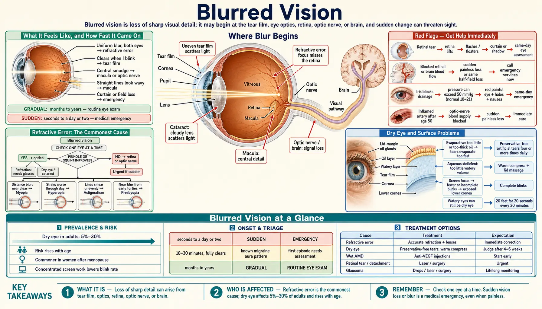
Table of Contents
- What It Feels Like, and How Fast It Came On
- Red Flags — Get Help Immediately
- Refractive Error: The Commonest Cause
- Dry Eye and Surface Problems
- Gradual Causes Inside the Eye
- Systemic Causes, Pregnancy, and Medications
- The Sudden and Dangerous Causes
- How Blurred Vision Is Evaluated
- What Helps, and What to Expect
- Connections
- Key Research Papers
- Featured Videos
What It Feels Like, and How Fast It Came On
Blurred vision means the detail has gone soft — edges lose their crispness, letters run together, faces flatten out. For most people the cause is ordinary and fixable, but it is also the phrase people reach for when something far more serious is happening. Each description points somewhere different:
- "Soft, like a camera out of focus." Uniform blur, both eyes, stable day to day — refractive error: you need glasses.
- "It clears when I blink." Blur that comes and goes with blinking, worse after screens and late in the day — a tear film problem.
- "A smudge in the middle of whatever I look at." Blur that sits in the center and follows your gaze — the macula or the optic nerve.
- "Straight lines look wavy." Door frames bowing, tile grout curving — the retina at the macula is being lifted or wrinkled.
- "A curtain came down over part of my vision." That is field loss, not blur. It is an emergency until proven otherwise.
Then check one eye at a time — many people with vision loss in one eye do not notice until they cover the good one.
Everything about urgency turns on the next question: how fast did it arrive? Gradual blur over months to years is usually structural and usually benign. The lens stiffens (presbyopia), then clouds (cataract); tear production declines and the macula thins. Most of it is correctable, and blur that is the same today as yesterday needs a routine eye exam, not an emergency room.
Sudden blur, over seconds to a day or two, is a different animal. Treatment windows are narrow, and retinal nerve tissue is central nervous system tissue: it does not regenerate. So, plainly: sudden loss or blurring of vision is a medical emergency. Do not wait to see whether it clears overnight, and do not assume that because it is painless it must be minor. Retinal artery occlusion, retinal detachment, giant-cell arteritis, optic neuritis, and stroke are all commonly painless. Painlessness is not reassurance.
One exception causes confusion. Blur lasting 10 to 30 minutes that then fully clears, often with a shimmering zigzag of light, is migraine aura, and it is benign if it follows a pattern you already know. A first-ever episode still needs assessment: transient vision loss in one eye from a carotid or cardiac source feels similar and warns of impending stroke.
Red Flags — Get Help Immediately
Get emergency care if blurred vision comes with any of the following. The list is short on purpose; every item on it can permanently take sight.
Call emergency services now
- Sudden vision loss in one or both eyes, painless or painful. Minutes matter.
- Any other stroke sign — face drooping, arm weakness, slurred speech, sudden confusion or imbalance. Losing the same half of the field in both eyes, so you keep bumping into things on one side, is stroke until proven otherwise.
- A new, severe, "worst-ever" headache, especially with vomiting or neck stiffness.
- Head or eye trauma, or any penetrating eye injury.
- Chemical splash. Irrigate continuously with clean water or saline on the way in.
Same-day emergency eye assessment
- A curtain, shadow, or veil moving across your vision, or a new blind area — retinal detachment.
- A sudden shower of new floaters or flashes of light, dozens of specks like a swarm of gnats — retinal tear or vitreous hemorrhage. A tear sealed with laser and a detachment needing surgery are often only days apart.
- Severe eye pain with a red eye, hazy cornea, fixed mid-dilated pupil, halos around lights, and nausea or vomiting — acute angle-closure glaucoma. Pressure can exceed 50 mmHg (normal roughly 10–21) and the optic nerve is damaged hour by hour.
- New temple headache, scalp tenderness, jaw ache on chewing, or unexplained fever and weight loss in anyone over 50 — giant-cell arteritis. Days of delay can cost sight in the second eye.
- Pain on moving the eye, with colors washed out in that eye — optic neuritis.
- A contact lens wearer with pain, light sensitivity, and discharge — possible corneal ulcer. Take the lenses out, keep them for culture, be seen today.
- Straight lines suddenly bending, or a new central gray spot — possible wet macular degeneration; injections work far better started early.
- Diabetes plus new floaters or a sudden dark veil — vitreous hemorrhage from proliferative retinopathy.
- Any infant or young child with blurred vision, who covers one eye or whose eye turns in or out — the visual system is still developing and delay costs permanent acuity.
For an eye-only symptom an eye emergency clinic is usually faster; with any neurological symptom, go to the emergency department.
Refractive Error: The Commonest Cause
Most blurred vision is simply light not landing in focus on the retina. Refractive error is not a disease, and it remains the leading cause of vision impairment worldwide — not because it is hard to fix, but because glasses do not reach everyone who needs them.
- Myopia (nearsightedness) — the eye is slightly too long, so distance blurs while near stays clear. High myopia, beyond about −6 diopters, raises lifetime risk of retinal detachment, myopic macular degeneration, and glaucoma.
- Hyperopia (farsightedness) — the eye is slightly too short. The focusing muscle compensates in youth, at the cost of strain, headache, and blur that worsens through the day; as that reserve declines, hidden hyperopia surfaces as blur that looks new.
- Astigmatism — the cornea is curved unevenly, so there is no single point of focus. Lines in one orientation blur more than others and point lights smear into streaks.
- Presbyopia — from the early forties the lens stiffens and cannot focus up close: the phone held farther away, small print that swims. Universal, normal, and solved by reading glasses.
Blur fully corrected by a pinhole, or by squinting, is almost certainly optical — refractive error, cataract, or dry eye — while blur a pinhole does not improve points to the retina or optic nerve.
Dry Eye and Surface Problems
The front surface of the tear film is the eye's most powerful refracting surface — it does more focusing work than the lens. When the film breaks up unevenly, blur builds while you stare, clears momentarily when you blink, and returns.
Dry eye disease is a disease of the tear film in which loss of tear-film homeostasis produces discomfort and visual disturbance; fluctuating blur is in the modern definition, not a footnote. Estimates put prevalence between 5% and 30% of adults, rising with age and commoner in women after menopause. The commoner evaporative type comes from lid-margin glands making too little or too thick an oil layer, so tears evaporate too fast. Aqueous-deficient dry eye means too little watery volume, classically in Sjögren's syndrome, which also dries the mouth.
Paradoxically, dry eye often causes watery eyes: the irritated surface triggers a flood of poor-quality reflex tears that does not fix the film. People routinely rule out dry eye because their eyes water, and that reasoning is backwards.
Blink rate falls sharply during concentrated screen work, and incomplete blinks leave the lower cornea exposed — blur that is worst at 4 p.m. and gone by morning. Blepharitis, conjunctivitis, pterygium, and corneal scarring also degrade the surface, as does keratoconus, in which the cornea thins into a cone, giving rapidly changing astigmatism in a young person whose glasses never quite work. Cross-linking can halt it if caught early.
Gradual Causes Inside the Eye
These are the slow, structural causes. None require an ambulance; all require an eye exam.
Cataract
The lens is a stack of transparent protein fibers that aggregate and yellow over decades, clouding it. The pattern is specific: haze, starbursts around oncoming headlights, muted colors, poor contrast so a gray step on a gray floor disappears, and reading that needs far more light. Cataract is the leading cause of reversible blindness worldwide. Nuclear cataract can cause a temporary myopic shift, so distance blurs while an older person reads without glasses again — a sign the lens is changing, not a good one. Posterior subcapsular cataract sits in the visual axis and causes disproportionate glare while chart acuity still looks acceptable. Surgery replaces the lens with a clear implant in 15 to 30 minutes under local anesthetic, and the decision rests on how much it interferes with your life.
Age-related macular degeneration
The macula is the small central patch of retina responsible for reading, faces, and fine detail. In dry AMD, drusen accumulate and the retinal pigment epithelium thins, giving gradual central blur and eventually a dense central blind spot; peripheral vision is preserved, so people can walk around but cannot read. In wet AMD, abnormal vessels grow beneath the retina and leak, and vision distorts and drops over days to weeks. Wet AMD is the urgent form: anti-VEGF injections now hold or improve acuity in most treated eyes, but only if treatment starts before the photoreceptors are lost. The Age-Related Eye Disease Study trials showed an antioxidant-and-zinc formulation slows progression in intermediate disease, with lutein and zeaxanthin replacing beta-carotene in the follow-up trial.
Glaucoma
Chronic open-angle glaucoma is the silent thief. It damages the optic nerve from the periphery inward, and because the two eyes' fields overlap and the brain fills in, people notice nothing until 30–40% of nerve fibers are gone. Reported blur is usually late-stage or a coexisting cataract, so the treatable window is entirely asymptomatic — the argument for screening exams.
Diabetic retinopathy and macular edema
Chronic high glucose damages retinal capillaries; they leak, and fluid collects in the macula. Diabetic macular edema is the commonest cause of vision loss in working-age adults with diabetes, and early retinopathy is asymptomatic — the argument for annual dilated exams. The Diabetes Control and Complications Trial showed intensive glycemic control substantially reduces the development and progression of retinopathy. Epiretinal membrane, macular hole, and central serous chorioretinopathy also blur central vision slowly.
Systemic Causes, Pregnancy, and Medications
Blood glucose swings. When glucose rises sharply, glucose and its metabolite sorbitol accumulate in the lens and draw water in osmotically; the lens swells and vision blurs, usually toward myopia. The same happens in reverse when very high sugars come down quickly, so newly diagnosed diabetics starting insulin often blur for days to weeks, typically clearing within four to six weeks. Do not buy new glasses until blood sugar has been stable for a month — a prescription measured on a swollen lens will be wrong once it settles. Low blood sugar also blurs vision, alongside sweating, tremor, and confusion.
Blood pressure and thyroid. Long-standing hypertension narrows retinal arterioles and causes hemorrhages and cotton-wool spots, usually silently, but a hypertensive emergency can blur vision abruptly with optic disc swelling — sudden blur with a systolic above roughly 180 and headache belongs in an emergency department. In Graves' disease, inflammation expands the fat and muscle inside the bony orbit, so the eyes protrude and thickened muscles restrict movement; in severe cases they compress the optic nerve, so worsening blur or dimming color in thyroid eye disease needs prompt review rather than reassurance.
Pregnancy. Mild refractive shifts and dry eye are common and usually resolve after delivery, which is why elective refractive surgery is deferred. But blurred vision, spots or flashing lights, and headache in the second half of pregnancy or postpartum are cardinal features of pre-eclampsia and need same-day obstetric assessment, with a blood pressure and urine check.
Nutritional and autoimmune. Severe vitamin A deficiency causes night blindness and, untreated, corneal drying and blindness; it is still seen after bariatric surgery and in severe malabsorption. Vitamin B12 deficiency can cause optic neuropathy with progressive central blur and color desaturation, sometimes before anemia appears. Multiple sclerosis commonly presents with optic neuritis, and Sjögren's syndrome, sarcoidosis, and lupus blur vision through dryness or uveitis.
Medications. Drug-induced blur is common and often fixed by changing the drug, so bring your complete list, eye drops and supplements included.
- Anticholinergics — oxybutynin, diphenhydramine, tricyclic antidepressants, scopolamine. They weaken the focusing muscle and dilate the pupil, and can precipitate acute angle closure in susceptible eyes.
- Corticosteroids in any form raise intraocular pressure and accelerate posterior subcapsular cataract, so long courses warrant pressure monitoring.
- Hydroxychloroquine — retinal toxicity is dose-dependent and largely preventable by screening; guidance keeps the daily dose at or below 5 mg/kg of actual body weight with regular OCT and visual fields. Damage, once established, does not reverse.
- Ethambutol and amiodarone cause optic neuropathy; topiramate can cause an acute myopic shift with angle closure in the first weeks.
- Alcohol blurs acutely and, with poor nutrition, contributes to optic neuropathy. Methanol poisoning from adulterated spirits causes rapid, often permanent blindness and is a poisoning emergency.
The Sudden and Dangerous Causes
These are the diagnoses the whole urgency framework exists to catch. Each is treatable, and each has a clock running.
Retinal detachment
The retina separates from the wall of the eye. The warning phase is a posterior vitreous detachment: the gel pulls away and produces a sudden burst of floaters and flashes. Usually harmless, but in a minority the gel tears the retina and it lifts — a curtain advancing from the periphery, then vision loss once the macula is reached. Risk factors are high myopia, prior cataract surgery, and trauma. A tear caught before detachment is often sealed with laser in minutes; once detached it needs surgery, and outcomes depend on whether the macula has come off yet.
Retinal artery occlusion — a stroke of the eye
Sudden, painless, profound loss of vision in one eye, typically within seconds. It is literally a stroke: the retina is brain tissue, and the same embolic sources — carotid plaque, atrial fibrillation, valve disease — are responsible. Outcomes are poor in most untreated eyes, so it is treated as a stroke equivalent needing immediate stroke evaluation. Amaurosis fugax — painless loss in one eye for minutes, like a shade coming down, then full recovery — is a transient ischemic attack of the eye and must be worked up urgently, not treated as a near miss.
Giant-cell arteritis
A vasculitis of medium and large arteries, essentially confined to people over 50. It occludes the arteries feeding the optic nerve, causing sudden, painless, often severe vision loss. Clues are new temporal headache, scalp tenderness, jaw claudication (aching in the jaw after chewing for a minute, the most specific single symptom), fever, weight loss, and the girdle stiffness of polymyalgia rheumatica. ESR and CRP are almost always raised. Untreated, many patients lose the second eye within days to weeks, so high-dose corticosteroids start on suspicion; biopsy stays positive for weeks afterwards, so treatment is never delayed for it.
Acute angle-closure glaucoma
In eyes with a naturally shallow anterior chamber — commoner in hyperopes, in people of East Asian descent, and in older women — the iris can abruptly block aqueous drainage, and pressure rockets. Expect severe one-sided eye and brow pain, a red eye, a hazy cornea, rainbow halos, a mid-dilated unreactive pupil, and prominent vomiting — which is why some patients get worked up for a stomach problem while the optic nerve dies. Treatment lowers pressure with drops and systemic agents, then a laser iridotomy creates a permanent bypass, usually in both eyes.
Optic neuritis
Inflammation of the optic nerve, most often in young adults and commoner in women. The triad is vision loss over hours to days, pain on eye movement, and reduced color saturation. The Optic Neuritis Treatment Trial showed intravenous methylprednisolone speeds recovery without changing final acuity, while oral prednisone alone at conventional dose gave no benefit and brought more recurrences. Most recover useful vision over weeks to months, and roughly half developed multiple sclerosis within 15 years of follow-up, with baseline brain MRI lesions the dominant predictor.
Stroke in the visual pathway
Occipital strokes produce a homonymous field defect — the same side of the field missing in both eyes. Patients rarely say "half my vision is gone"; they say "I keep bumping into door frames on the left." Because the macular representation is often spared, chart acuity can measure 20/20 while half the world is missing. Any new field defect respecting the vertical midline, or any visual symptom with weakness, numbness, or speech difficulty, needs emergency imaging.
Also urgent: retinal vein occlusion, uveitis, endophthalmitis after eye surgery, idiopathic intracranial hypertension, and orbital cellulitis.
How Blurred Vision Is Evaluated
The history covers onset and course; one eye or both; the pattern of loss; associated pain, redness, floaters, flashes, headache, or jaw and scalp pain; diabetes and recent HbA1c, blood pressure, autoimmune disease, migraine, prior eye surgery, and family glaucoma or AMD; every medication; contact lens habits; and any trauma or chemical exposure.
The examination then proceeds through:
- Visual acuity in each eye separately — the vital sign of the eye — plus pinhole acuity, which if it sharpens vision localizes the problem to the optics, and refraction, which separates "needs glasses" from everything else.
- Pupil testing, including the swinging-flashlight test for a relative afferent pupillary defect; a positive result points to the optic nerve or extensive retinal disease and excludes refractive error. Reduced color vision in one eye means the same.
- Visual field testing maps the shape of any loss — a defect respecting the vertical midline points to the brain, not the eye — and eye movements are checked for double vision.
- Slit-lamp examination of lids, tear-film break-up time, cornea with fluorescein staining, anterior chamber depth and inflammatory cells, and lens clarity, with intraocular pressure: normal roughly 10–21 mmHg, and 40–50 with a red painful eye means angle closure.
- Dilated fundus examination and optical coherence tomography — the only ways to properly assess the macula, optic disc, and peripheral retina. Expect blur and light sensitivity for four to six hours after dilation, so arrange a ride.
Tests beyond the eye: fasting glucose and HbA1c; ESR and CRP urgently in anyone over 50 with sudden vision loss, to support or argue against giant-cell arteritis; complete blood count and blood pressure; TSH; vitamin B12; MRI of brain and orbits with contrast for optic neuritis and MS plaques, plus aquaporin-4 and MOG antibodies if it is bilateral or recurrent; urgent head imaging for suspected stroke; and carotid ultrasound with echocardiogram to find the embolic source after retinal artery occlusion.
What Helps, and What to Expect
Self-care that genuinely works
- Get an accurate refraction and wear the correction. Unglamorous, and it fixes the majority of cases. Adults with no risk factors need an eye exam every one to two years; annually from age 60, annually with diabetes, and more often with glaucoma, high myopia, or AMD.
- Treat the tear film properly. Preservative-free artificial tears four or more times daily, since preserved drops irritate with frequent use, plus warm compresses with lid massage for gland dysfunction. Give any regimen four to six weeks before judging it.
- Fix the screen setup and the lighting. Screen top at or just below eye level, an arm's length away; look 20 feet away for 20 seconds every 20 minutes; blink completely; increase text size instead of leaning in. An older retina needs several times more light for the same task, so task lighting fixes much of "my vision is getting worse."
- Control blood glucose and blood pressure — the highest-value thing anyone with diabetes or hypertension can do for their sight. Expect a few weeks of fluctuating vision when control improves rapidly; that is the lens re-equilibrating, not damage.
- Stop smoking, which multiplies AMD risk, accelerates cataract, and worsens thyroid eye disease and dry eye. Wear UV-blocking sunglasses, since cumulative ultraviolet exposure contributes to cataract and pterygium.
- Eat for the retina. Leafy greens and colored vegetables supply lutein and zeaxanthin, which concentrate in the macula. An AREDS-type supplement is evidence-based for intermediate AMD and should be chosen with an eye doctor — the original beta-carotene formulation is not appropriate for smokers.
When treatment is warranted
Self-care does not fix a structural or vascular problem, and the treatments are cause-specific: cataract surgery once the blur interferes with driving, reading, or work; anti-VEGF injections for wet AMD, diabetic macular edema, and vein occlusion; drops, laser, or surgery to lower pressure in glaucoma; laser for a retinal tear and surgery for a detachment; intravenous methylprednisolone for significant optic neuritis; immediate corticosteroids for giant-cell arteritis; cross-linking for keratoconus; and prescription anti-inflammatory drops or punctal plugs for dry eye that tears alone do not control.
What to expect over time
Refractive blur is corrected immediately by the right lenses, and presbyopia plateaus after the fifties. Dry eye is usually managed rather than cured, but well-managed dry eye is genuinely comfortable and the fluctuating blur is among the first things to improve.
Diabetic retinopathy and glaucoma are lifelong: with monitoring and control most people keep functional vision for life, but sight already lost to glaucoma or advanced dry AMD does not return — which is why detection before symptoms is the whole game. Optic neuritis typically recovers substantially over two to six months. Where sight cannot be fully restored, low-vision rehabilitation — magnifiers, high-contrast materials, screen readers, eccentric-viewing training, task lighting — is effective and badly underused, so ask for a referral.
None of this replaces an examination. Vision is one of the few systems where a delay of hours changes the outcome, and where the most dangerous causes are often the least painful. If your vision changed suddenly, stop reading and get seen.
Connections
Key Research Papers
Key Research Papers
- Age-Related Eye Disease Study 2 Research Group. Lutein + zeaxanthin and omega-3 fatty acids for age-related macular degeneration: the AREDS2 randomized clinical trial. JAMA. 2013;309(19):2005-2015. — Search PubMed
- Beck RW, Cleary PA, Anderson MM, et al; Optic Neuritis Study Group. A randomized, controlled trial of corticosteroids in the treatment of acute optic neuritis. New England Journal of Medicine. 1992;326(9):581-588. — Search PubMed
- Optic Neuritis Study Group. Multiple sclerosis risk after optic neuritis: final optic neuritis treatment trial follow-up. Archives of Neurology. 2008;65(6):727-732. — PubMed PMID: 18541792
- Craig JP, Nichols KK, Akpek EK, et al. TFOS DEWS II Definition and Classification Report. The Ocular Surface. 2017;15(3):276-283. — PubMed PMID: 28736335
- Diabetes Control and Complications Trial Research Group. The effect of intensive treatment of diabetes on the development and progression of long-term complications in insulin-dependent diabetes mellitus. New England Journal of Medicine. 1993;329(14):977-986. — Search PubMed
- Hayreh SS, Zimmerman MB. Central retinal artery occlusion: visual outcome. American Journal of Ophthalmology. 2005;140(3):376-391. — Search PubMed
- Hayreh SS, Podhajsky PA, Zimmerman B. Ocular manifestations of giant cell arteritis. American Journal of Ophthalmology. 1998;125(4):509-520. — PubMed PMID: 9559737
- Hollands H, Johnson D, Brox AC, et al. Acute-onset floaters and flashes: is this patient at risk for retinal detachment? JAMA. 2009;302(20):2243-2249. — PubMed PMID: 20388891