Age-Related Macular Degeneration (AMD)
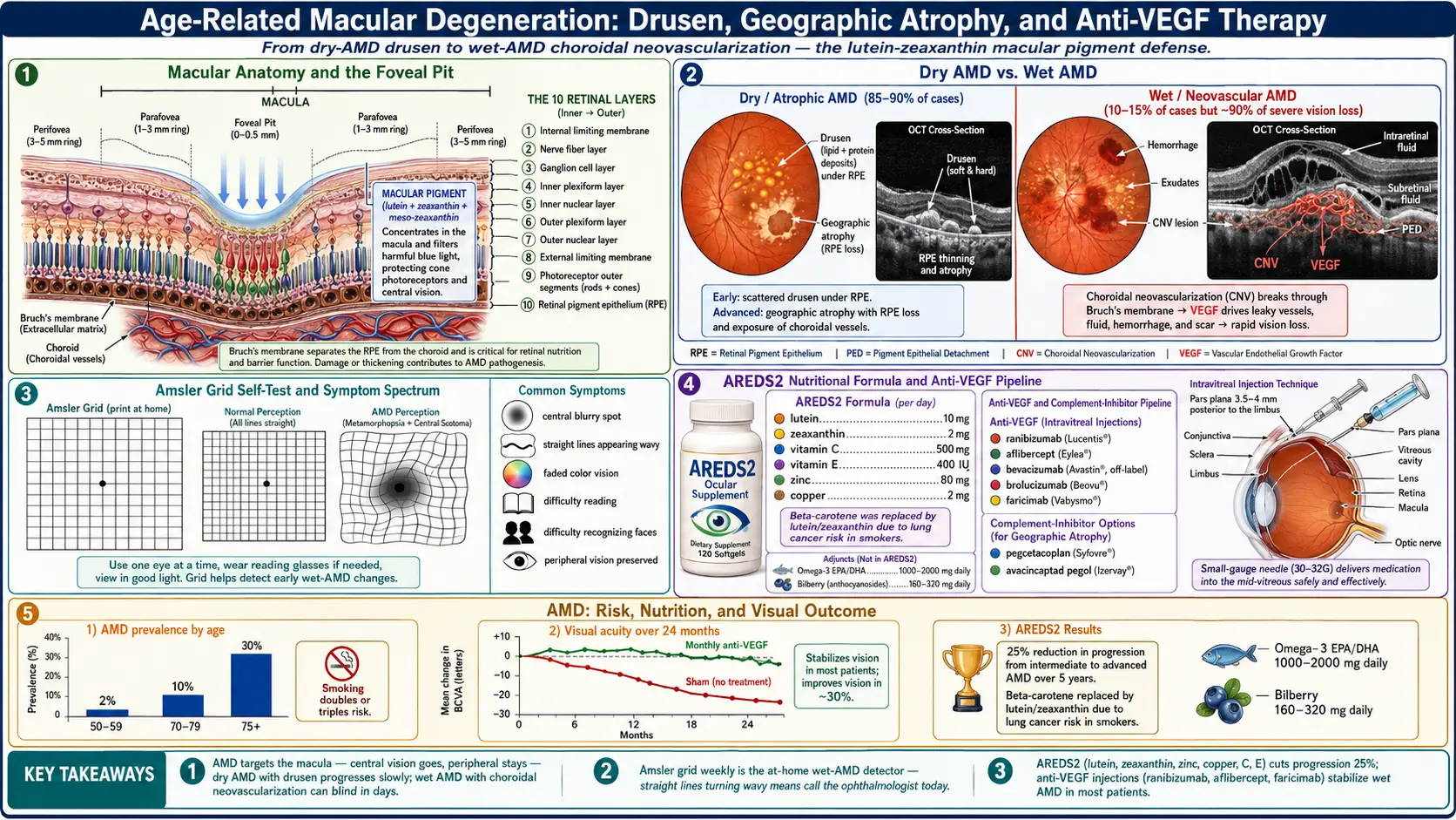
Table of Contents
1. Overview
Age-Related Macular Degeneration (AMD) is described in the research literature as the most frequent cause of irreversible blindness in older people in developed countries (Hageman 2005). It damages the macula, the central part of the retina used for reading, driving and recognizing faces.
AMD has two late forms. In the atrophic (dry) form, which a 2012 review puts at about 85–90% of cases, the earliest changes are large confluent drusen and changes in the retinal pigment epithelium (RPE); loss of the RPE then predicts progression to geographic atrophy (GA), the advanced form of dry AMD (Bhutto & Lutty 2012; Khanani 2023). GA is defined by sharply demarcated atrophic lesions of the outer retina, resulting from loss of photoreceptors, RPE and the underlying choriocapillaris (Fleckenstein 2018). Neovascular (wet) AMD, about 10–15% of cases in the same review, involves new blood vessels that grow from the choriocapillaris under the stimulus of vascular endothelial growth factor (VEGF) — choroidal neovascularization (CNV) — and photoreceptors die from leakage from these vessels and from scar formation (Bhutto & Lutty 2012).
The two forms progress differently. In GA, vision loss is gradual: the lesions produce dense, irreversible blind spots (scotomas) in the visual field, but visual acuity may at first decline only slightly if the fovea is spared. In wet AMD, vision loss is typically acute (Holz 2014).
2. Epidemiology
A 2014 meta-analysis of 39 population-based studies (129,664 people) projected that 196 million people worldwide would have AMD in 2020, rising to 288 million in 2040. Mapped to ages 45–85, the pooled prevalence was 8.01% for early AMD, 0.37% for late AMD and 8.69% for any AMD (Wong 2014). For the United States, a 2004 analysis estimated that 1.47% of people aged 40 and older — 1.75 million people — had neovascular AMD and/or geographic atrophy, that more than 7 million had drusen of 125 μm or larger and were therefore at substantial risk, and that the number with AMD would reach 2.95 million in 2020 (Friedman 2004).
Prevalence rises steeply with age: in the US analysis, more than 15% of white women older than 80 had neovascular AMD and/or geographic atrophy (Friedman 2004). In the Beaver Dam Eye Study, which followed 3,917 people aged 43–86 at baseline, the 15-year cumulative incidence was 14.3% for early AMD and 3.1% for late AMD; people aged 75 or older at baseline had a 15-year incidence of exudative (wet) AMD of 4.4%, compared with 0.4% in those aged 43–54, and an 8% 15-year incidence of late AMD (Klein 2007).
In the 2014 meta-analysis, early and any AMD were more prevalent in people of European ancestry than in Asian or African populations, late AMD was more prevalent in Europeans than Africans (0.5% vs 0.3%), and geographic atrophy was more prevalent in Europeans (1.11%) than in Africans (0.14%), Asians (0.21%) and Hispanics (0.16%); no significant difference by sex was found (Wong 2014). The US analysis likewise found AMD far more prevalent among white than among Black people (Friedman 2004). A systematic review of 17 epidemiological studies found that 13 reported a statistically significant association between smoking and AMD, with a two- to three-fold increased risk in current smokers compared with never-smokers, along with evidence of a dose–response, a temporal relationship and reversibility of effect; five studies found no association (Thornton 2005).
3. Pathophysiology
A 2012 review describes AMD as the loss of a mutually supporting relationship between the photoreceptors, the RPE, Bruch's membrane and the choriocapillaris. Which part of this complex is affected first appears to depend on the type of AMD, and in both forms the environment around the choriocapillaris is described as proinflammatory, with accumulation of complement components and molecules such as C-reactive protein (Bhutto & Lutty 2012).
Drusen Formation
Drusen are deposits beneath the RPE and are the hallmark lesion of AMD; complement activation has been implicated in their formation, and complement factor H accumulates within them (Hageman 2005). A 2013 expert classification grades drusen by size within two disc diameters of the fovea in people older than 55: small drusen (<63 μm) are considered normal aging with no clinically relevant increase in risk; medium drusen (63 to <125 μm) without pigmentary abnormalities are early AMD; large drusen, or pigmentary abnormalities with at least medium drusen, are intermediate AMD. Estimated 5-year risks of progressing to late AMD range from 0.5% for normal aging changes to 50% for the highest-risk intermediate group (Ferris 2013). In the Beaver Dam Eye Study, eyes with soft indistinct drusen at baseline developed late AMD more often than eyes without them (17.8% vs 1.2%) (Klein 2007).
RPE Atrophy — Dry AMD Pathway
In atrophic AMD, large confluent drusen and hyperpigmentation (presumed RPE dysfunction) appear to be the initial insult; resorption of drusen and loss of RPE can predict progression to geographic atrophy, and death of photoreceptors and choriocapillaris appears to be secondary to RPE loss (Bhutto & Lutty 2012). GA lesions typically appear first in the perifoveal macula, initially sparing the foveal center, and over time often expand and coalesce to include the fovea. Reported GA growth rates for whole study populations range from 0.53 to 2.6 mm² per year (median about 1.78 mm² per year), with wide variation between patients (Fleckenstein 2018).
Choroidal Neovascularization — Wet AMD Pathway
In wet AMD, loss of choriocapillaris may be the initial insult. Dying or dysfunctional choriocapillaris leaves the adjacent RPE hypoxic; the hypoxic cells produce angiogenic substances such as VEGF, which stimulate new vessels to grow from the choriocapillaris (CNV). The photoreceptors ultimately die from lack of nutrients, leakage of serum components from the new vessels, and scar formation (Bhutto & Lutty 2012). Untreated eyes in a sham-injection arm lost a mean of 10.4 letters of visual acuity over 12 months, and 62.2% lost fewer than 15 letters (Rosenfeld 2006).
Complement Pathway Dysregulation
Complement factor H, the major inhibitor of the alternative complement pathway, accumulates within drusen and is synthesized by the RPE (Hageman 2005). The CFH Y402H variant (a tyrosine-to-histidine change at amino acid 402) lies in a region of the protein that binds heparin and C-reactive protein (Klein 2005). Hageman and colleagues proposed that genetic variation in this regulator of the alternative complement pathway, combined with a triggering event such as infection, underlies a major proportion of AMD (Hageman 2005). The complement C3 inhibitor pegcetacoplan and the C5 inhibitor avacincaptad pegol both slowed GA lesion growth in trials (section 7), which is consistent with a role for complement in GA.
Oxidative Stress
The hypothesis that oxidative damage contributes to AMD motivated the AREDS antioxidant trial; its investigators cited observational and experimental data suggesting that antioxidant and/or zinc supplements might delay progression of AMD and vision loss (AREDS 2001). AREDS2 then tested the carotenoids lutein and zeaxanthin because observational data suggested that higher dietary intake might further reduce risk (AREDS2 2013). The trial results are in section 7.
4. Genetics
In the US twin study of 840 elderly male twins, additive genetic heritability was 0.46 for overall AMD grade, 0.67 for intermediate AMD and 0.71 for advanced AMD; the authors concluded that genetic factors explained 46% to 71% of the variation in overall disease severity, with environmental factors unique to each twin also contributing (Seddon 2005). A genome-wide association study of 16,144 patients and 17,832 controls identified 52 independently associated common and rare variants across 34 loci (Fritsche 2016). The first two major susceptibility regions identified were the CFH gene on chromosome 1 and a region on chromosome 10q26 (Klein 2005; Rivera 2005).
CFH Y402H — Chromosome 1
A 2005 genome-wide screen found a common variant in CFH strongly associated with AMD; in people homozygous for the risk allele, the likelihood of AMD was increased by a factor of 7.4 (95% CI 2.9 to 19). Resequencing found the Y402H change in linkage disequilibrium with the risk allele (Klein 2005). In a separate study of about 900 cases and 400 controls, one common at-risk CFH haplotype was present in 50% of cases and 29% of controls (odds ratio 2.46); homozygotes for it made up 24% of cases and 8% of controls (odds ratio 3.51), and several protective haplotypes were also found (Hageman 2005).
Chromosome 10q26 (LOC387715/ARMS2)
In two German case–control cohorts (1,166 cases, 945 controls), the strongest association on 10q26 (P=10−34) centered on a common coding polymorphism, Ala69Ser, in a then-hypothetical gene, LOC387715, which contributed to risk independently of CFH. At the time there was no functional information on the gene, which showed weak expression in the human retina (Rivera 2005). Later studies refer to this locus as ARMS2/HTRA1 (Merle 2015).
Additional Genetic Loci
The 2016 genome-wide study found that wet and dry AMD share most of their genetics, but identified a signal specific to wet AMD near MMP9; very rare coding variants in CFH, CFI and TIMP3, and a splice variant in SLC16A8, suggested causal roles for these genes (Fritsche 2016). The effects of the two major regions combine: homozygosity for the risk alleles at both CFH and LOC387715 carried an odds ratio of 57.6 (95% CI 37.2 to 89.0) compared with the baseline non-risk genotype in the German cohorts (Rivera 2005). Genes may also modify diet associations: in AREDS participants, higher Mediterranean-diet scores were associated with lower risk of advanced AMD among carriers of the CFH Y402H non-risk allele, but not among people homozygous for the risk allele (Merle 2015).
5. Clinical Presentation
Dry AMD
Under the 2013 classification, early and intermediate AMD are defined by drusen and pigmentary findings on examination rather than by symptoms (Ferris 2013). Geographic atrophy can cause significant deficits in reading, night vision and dark adaptation and produce dense, irreversible scotomas, while the initial decline in visual acuity may be relatively minor if the fovea is spared; best-corrected visual acuity does not correlate well with GA lesion size or progression (Holz 2014). Lesions typically start around the fovea and expand over time to include it (Fleckenstein 2018).
Wet AMD
In contrast to the gradual course of GA, vision loss in wet AMD is typically acute (Holz 2014). The Home Monitoring of the Eye (HOME) trial investigators note that visual acuity at the time of CNV treatment is the main predictor of the outcome of anti-VEGF treatment (AREDS2-HOME 2014).
Amsler Grid and Home Monitoring
People with dry AMD are commonly encouraged to use the Amsler grid, a simple chart for noticing distortion of straight lines (metamorphopsia). A 2023 meta-analysis of 10 studies (1,890 eyes) found a sensitivity of 71% and specificity of 63% for detecting wet AMD when the comparison group had dry AMD; the authors wrote that this sensitivity may be below levels typically recommended for monitoring and that at-risk patients typically should be encouraged to undergo regular eye examination regardless of Amsler grid results (Bjerager 2023). In the HOME trial (1,520 participants at high risk of CNV), participants given a home device using hyperacuity testing with telemonitoring lost fewer letters between baseline and CNV detection than those on standard care (median 4 vs 9 letters), and the trial was stopped early for efficacy (AREDS2-HOME 2014).
6. Diagnosis
Fundus Examination and Classification
The 2013 clinical classification is based on lesions seen on examination within two disc diameters of the fovea in people older than 55: no AMD, normal aging changes (small drusen), early AMD (medium drusen), intermediate AMD (large drusen or pigmentary abnormalities with at least medium drusen) and late AMD (neovascular AMD or geographic atrophy) (Ferris 2013).
Optical Coherence Tomography (OCT)
Spectral-domain OCT is among the imaging techniques used to detect GA (Holz 2014) and to measure retinal fluid in wet AMD: in the 5-year follow-up of the CATT trial, OCT found fluid in 83% of 555 imaged eyes (61% intraretinal, 38% subretinal, 36% beneath the RPE) (CATT 2016).
Fundus Fluorescein Angiography (FFA)
Fluorescein angiography is also used to detect GA (Holz 2014) and to classify and measure CNV lesions. The pivotal ranibizumab trials enrolled patients by angiographic lesion type — minimally classic or occult CNV in MARINA, predominantly classic CNV in ANCHOR (Rosenfeld 2006; Brown 2006) — and the CATT 5-year follow-up measured a mean total lesion area of 12.9 mm² by angiography in 467 eyes (CATT 2016).
Fundus Autofluorescence (FAF)
FAF imaging detects GA (Holz 2014), and abnormal FAF patterns in the retina around a lesion are among the characteristics that predict faster progression (Holz 2014; Fleckenstein 2018). The pivotal GA trials measured their primary endpoint, GA lesion area, with FAF (Heier 2023; Khanani 2023).
7. Treatment — Dry AMD
AREDS and AREDS2 Nutritional Supplementation
The Age-Related Eye Disease Study (AREDS), an 11-center double-masked trial, enrolled 3,640 participants aged 55–80 and randomly assigned them to daily tablets of antioxidants (vitamin C 500 mg, vitamin E 400 IU and beta-carotene 15 mg), zinc (80 mg as zinc oxide, with copper 2 mg as cupric oxide), antioxidants plus zinc, or placebo; average follow-up was 6.3 years. Compared with placebo, antioxidants plus zinc reduced the odds of developing advanced AMD (odds ratio 0.72; 99% CI 0.52–0.98); the odds ratios for zinc alone (0.75) and antioxidants alone (0.80) were not statistically significant. Participants with only extensive small drusen, non-extensive intermediate drusen or pigment abnormalities had a 1.3% 5-year probability of progression; when they were excluded, the odds ratio for antioxidants plus zinc was 0.66 (99% CI 0.47–0.91). Antioxidants plus zinc was the only formulation that significantly reduced at least moderate visual acuity loss (odds ratio 0.73), and no formulation was associated with a statistically significant serious adverse effect. The investigators concluded that people with extensive intermediate drusen, at least one large druse, non-central GA, or advanced AMD or vision loss from AMD in one eye, and without contraindications such as smoking, should consider a supplement of the kind tested (AREDS 2001).
AREDS2 (4,203 participants aged 50–85 with bilateral large drusen, or large drusen in one eye and advanced AMD in the other; median follow-up 5 years) tested adding lutein 10 mg plus zeaxanthin 2 mg, the omega-3 fatty acids DHA 350 mg plus EPA 650 mg, or both, to the AREDS formulation. The primary comparisons with placebo showed no statistically significant reduction in progression to advanced AMD (hazard ratio 0.90 for lutein plus zeaxanthin, P=.12; 0.97 for DHA plus EPA, P=.70; 0.89 for both, P=.10). A secondary randomization found no apparent effect on progression from removing beta-carotene or lowering the zinc dose. More lung cancers occurred with beta-carotene than without it (23 [2.0%] vs 11 [0.9%], nominal P=.04), mostly in former smokers. Because of this, the investigators concluded that lutein plus zeaxanthin could be an appropriate carotenoid substitute for beta-carotene in the AREDS formulation (AREDS2 2013).
Pegcetacoplan for Geographic Atrophy
Pegcetacoplan is a complement C3 inhibitor given by intravitreal injection (Liao 2020). The US Food and Drug Administration approved it for GA on 17 February 2023 (Drugs@FDA, NDA 217171), and the phase 3 report describes it as the first FDA-approved treatment for GA (Heier 2023). In the phase 2 FILLY trial (246 patients), monthly and every-other-month injections reduced the GA growth rate by 29% (P=0.008) and 20% (P=0.067) compared with sham over 12 months (Liao 2020). The phase 3 OAKS and DERBY trials enrolled 1,258 patients. At 12 months, monthly and every-other-month pegcetacoplan significantly slowed lesion growth in OAKS (by 21% and 16%), but in DERBY the 12% and 11% reductions did not reach statistical significance. At 24 months, growth was slowed by 22% and 18% in OAKS and by 19% and 16% in DERBY. There were no differences in the key secondary visual function endpoints at 24 months. New-onset exudative (wet) AMD by 24 months was reported in 11% and 8% of OAKS patients on monthly and every-other-month pegcetacoplan versus 2% on sham, and in 13% and 6% versus 4% in DERBY (Heier 2023).
Avacincaptad Pegol for Geographic Atrophy
Avacincaptad pegol is a complement C5 inhibitor given monthly by intravitreal injection; its investigators noted that C5 inhibition theoretically preserves C3 activity (Jaffe 2021). The FDA approved it for GA on 4 August 2023 (FDA, Novel Drug Approvals for 2023). In the phase 2/3 GATHER1 trial (286 participants), the mean rate of GA growth over 12 months was reduced by 27.4% with 2 mg and 27.8% with 4 mg compared with sham (Jaffe 2021). In the phase 3 GATHER2 trial (448 patients), monthly 2 mg slowed GA growth by 14% compared with sham over 12 months; macular neovascularization occurred in 7% of treated and 4% of sham patients (exudative in 5% vs 3%), and no endophthalmitis, intraocular inflammation or ischaemic optic neuropathy occurred over 12 months (Khanani 2023).
Low Vision Rehabilitation
In the Veterans Affairs Low Vision Intervention Trial, 126 patients with macular diseases and visual acuity between 20/100 and 20/500 in the better eye were randomized to an outpatient low-vision program (examination, devices and six weekly sessions with a low-vision therapist) or a waiting list. At 4 months the program group improved in reading ability and every other measured domain of visual function compared with controls, while the waiting-list group declined (Stelmack 2008).
8. Treatment — Wet AMD (Anti-VEGF)
Clinical trials established the efficacy of intravitreal anti-VEGF injections for neovascular AMD (CATT 2011), and the 5-year CATT follow-up describes anti-VEGF therapy as a major long-term therapeutic advance for the disease (CATT 2016).
Ranibizumab
Ranibizumab is a recombinant, humanized monoclonal antibody Fab fragment that neutralizes all active forms of VEGF-A (Rosenfeld 2006). The MARINA trial randomized 716 patients with minimally classic or occult CNV to monthly ranibizumab (0.3 or 0.5 mg) or sham injections for 2 years. At 12 months, 94.6% of the 0.5 mg group lost fewer than 15 letters, compared with 62.2% of the sham group, and 33.8% gained 15 or more letters, compared with 5.0%; the benefit was maintained at 24 months. Presumed endophthalmitis occurred in 1.0% and serious uveitis in 1.3% of ranibizumab patients over 24 months (Rosenfeld 2006). The ANCHOR trial (423 patients with predominantly classic CNV) compared ranibizumab with verteporfin photodynamic therapy: at 12 months, 96.4% of the 0.5 mg group lost fewer than 15 letters versus 64.3% with verteporfin, and 40.3% gained 15 or more letters versus 5.6% (Brown 2006).
Bevacizumab and the CATT Comparison
Bevacizumab has been used off-label for AMD. The CATT trial randomized 1,208 patients to ranibizumab or bevacizumab, monthly or as needed. At 1 year, the two drugs had equivalent effects on visual acuity when given on the same schedule (monthly: 8.0 letters gained with bevacizumab, 8.5 with ranibizumab). Rates of death, heart attack and stroke were similar, but serious systemic adverse events (mainly hospitalizations) were more frequent with bevacizumab (24.1% vs 19.0%), distributed across disease categories not previously identified as areas of concern; the investigators wrote that the differences require further study (CATT 2011).
Aflibercept
Aflibercept (VEGF Trap-Eye) was compared with monthly ranibizumab in the VIEW 1 and VIEW 2 trials (2,419 patients). Aflibercept 2 mg every 2 months after three monthly doses was non-inferior to monthly ranibizumab for maintaining vision at week 52 (95.1% and 95.6% in the two trials vs 94.4% with ranibizumab), with similar adverse events; the authors wrote that the every-2-month regimen offered the potential to reduce the risk from monthly injections and the burden of monthly monitoring (Heier 2012). A higher 8 mg dose was approved by the FDA on 18 August 2023 (Drugs@FDA, BLA 761355); in the PULSAR trial (1,011 patients), 8 mg every 12 or 16 weeks after three monthly doses was non-inferior to 2 mg every 8 weeks for visual acuity gain at week 48 (+6.7 and +6.2 vs +7.6 letters) (Lanzetta 2024).
Brolucizumab
Brolucizumab is a single-chain antibody fragment that inhibits VEGF-A. In the HAWK and HARRIER trials (1,817 patients), brolucizumab given every 12 weeks after three monthly loading doses (shortened to every 8 weeks if disease activity was present) was non-inferior to aflibercept every 8 weeks for visual acuity at week 48; 56% (HAWK) and 51% (HARRIER) of eyes on 6 mg stayed on 12-week dosing through week 48, and reductions in central subfield thickness were greater with brolucizumab (Dugel 2020). A post hoc review of the same trials by an independent safety committee found definite or probable intraocular inflammation (IOI) in 4.6% of brolucizumab-treated eyes, IOI with retinal vasculitis in 3.3%, and IOI with vasculitis and retinal vascular occlusion in 2.1%; 0.74% had at least moderate vision loss (15 or more letters) with IOI. The IOI rate in aflibercept-treated eyes was 1.1% (Monés 2021).
Faricimab
Faricimab is a bispecific antibody that inhibits both angiopoietin-2 (Ang-2) and VEGF-A (Heier 2022). A review describes the Ang/Tie2 pathway as complementing VEGF activity in retinal vascular disease by decreasing vascular integrity and increasing neovascularization and inflammatory signaling (Khanani 2021). In the TENAYA and LUCERNE trials (1,329 treatment-naive patients), faricimab 6 mg at intervals of up to every 16 weeks was non-inferior to aflibercept 2 mg every 8 weeks for visual acuity change averaged over weeks 40–48 (TENAYA +5.8 vs +5.1 letters; LUCERNE +6.6 vs +6.6), with comparable rates of ocular adverse events (Heier 2022). The FDA approved faricimab on 28 January 2022 for wet AMD and diabetic macular edema (FDA, Novel Drug Approvals for 2022).
Treat-and-Extend Protocol
In treat-and-extend regimens, the interval between injections is adjusted step by step according to disease activity. The ALTAIR trial (123 treatment-naive patients per group) compared two such aflibercept regimens with 2-week or 4-week interval adjustments after three monthly doses: over 52 weeks, patients received a mean of 7.2 and 6.9 injections and gained 9.0 and 8.4 letters, and outcomes were similar between the two regimens (Ohji 2020). The ARIES trial (271 patients) found an early-start treat-and-extend regimen non-inferior to a late-start regimen, with a mean of 12.0 versus 13.0 injections over 2 years (Mitchell 2021). Both trials compared treat-and-extend variants with each other, not with fixed monthly dosing.
Port Delivery System (PDS)
The Port Delivery System with ranibizumab is a surgically implanted, refillable ocular reservoir. The Archway phase 3 trial (418 patients previously treated with and responsive to anti-VEGF therapy) found the implant with ranibizumab 100 mg/mL, refilled every 24 weeks, non-inferior and equivalent to monthly ranibizumab injections for visual acuity (+0.2 vs +0.5 letters), and 98.4% of implant patients did not need supplemental treatment before the first refill. Prespecified ocular adverse events of special interest occurred in 19.0% of implant patients versus 6.0% on monthly injections, including endophthalmitis (1.6%), retinal detachment (0.8%), vitreous hemorrhage (5.2%), conjunctival erosion (2.4%) and conjunctival retraction (2.0%); most occurred within 1 month of implantation (Holekamp 2022).
9. Complications
- Severe vision loss: five years after starting anti-VEGF treatment in the CATT trials, 20% of eyes had visual acuity of 20/200 or worse, while 50% had 20/40 or better (CATT 2016).
- Reading and night vision: geographic atrophy can cause significant deficits in reading, night vision and dark adaptation, and dense, irreversible scotomas (Holz 2014).
- Depression: a systematic review found prevalence estimates of depressive symptoms in people with AMD ranging from 15.7% to 44%; case–control studies found depressive symptoms more common in people with AMD than without it, but not anxiety symptoms (Dawson 2014).
- Macular atrophy after wet AMD treatment: at 5 years in the CATT cohort, geographic atrophy was present in 41% of gradable eyes and was subfoveal in 17% (CATT 2016).
- Treatment-related events: endophthalmitis and serious uveitis with monthly ranibizumab (1.0% and 1.3% over 2 years; Rosenfeld 2006), intraocular inflammation with retinal vasculitis or occlusion with brolucizumab (Monés 2021), and new-onset wet AMD with complement inhibitors for GA (Heier 2023; Khanani 2023).
10. Prognosis
For wet AMD, the pivotal trials found that 94.6–96.4% of patients on monthly 0.5 mg ranibizumab lost fewer than 15 letters at 12 months, compared with 62.2% on sham injections and 64.3% on verteporfin, and 33.8–40.3% gained 15 or more letters (Rosenfeld 2006; Brown 2006). Longer term, in the CATT cohort, vision gains during the first 2 years were not maintained at 5 years: mean visual acuity was 3 letters below baseline and 11 letters below the 2-year level (CATT 2016). The authors of a 2024 gene-therapy study note that, because of undertreatment, many patients on anti-VEGF injections lose vision over time (Campochiaro 2024).
For geographic atrophy, the two approved complement inhibitors slowed lesion growth in trials — pegcetacoplan by 16–22% at 24 months and avacincaptad pegol by 14% at 12 months in their phase 3 trials — but pegcetacoplan showed no difference from sham in the key visual function endpoints at 24 months (Heier 2023; Khanani 2023). Characteristics replicated in several cohorts as predictors of GA progression include baseline lesion size, lesion location, multifocality, FAF patterns and the status of the fellow eye (Fleckenstein 2018).
11. Prevention and Risk Reduction
- Smoking: in a systematic review, 13 of 17 studies found a two- to three-fold increased risk of AMD in current smokers compared with never-smokers, with evidence that the effect is reversible (Thornton 2005).
- AREDS-type supplements: the AREDS benefit was found in people already at risk of advanced AMD; participants with only extensive small drusen, non-extensive intermediate drusen or pigment abnormalities had a 1.3% 5-year probability of progression (AREDS 2001). In AREDS2, adding lutein plus zeaxanthin, omega-3 fatty acids or both did not further reduce progression in the primary analyses (AREDS2 2013). The formulation the AREDS2 investigators proposed is the AREDS formulation (vitamin C 500 mg, vitamin E 400 IU, zinc 80 mg as zinc oxide, copper 2 mg as cupric oxide) with lutein 10 mg plus zeaxanthin 2 mg in place of beta-carotene 15 mg (AREDS 2001; AREDS2 2013).
- Beta-carotene and lung cancer: in AREDS2, more lung cancers occurred in participants assigned beta-carotene than in those not assigned it (2.0% vs 0.9%, nominal P=.04), mostly in former smokers; the AREDS investigators had listed smoking as a contraindication (AREDS 2001; AREDS2 2013).
- Mediterranean diet: among 2,525 AREDS participants followed for 13 years, a high alternate Mediterranean diet score (6–9, built from vegetables, fruit, legumes, whole grains, nuts, fish, red and processed meats, alcohol and the ratio of monounsaturated to saturated fat) was associated with a lower risk of progression to advanced AMD (hazard ratio 0.74; 95% CI 0.61–0.91); the association was absent in people homozygous for the CFH Y402H risk allele (Merle 2015).
- Dietary lutein and zeaxanthin: in a case–control analysis of 4,519 AREDS participants, the highest compared with the lowest fifth of dietary lutein/zeaxanthin intake was associated with lower odds of neovascular AMD (odds ratio 0.65), geographic atrophy (0.45) and large or extensive intermediate drusen (0.73) (AREDS Report No. 22, 2007).
- Early detection of wet AMD: in the HOME trial, home hyperacuity monitoring with telemonitoring led to better visual acuity at the time CNV was detected than standard care (AREDS2-HOME 2014); the Amsler grid alone detected 71% of wet AMD cases among people with dry AMD in a meta-analysis (Bjerager 2023).
12. Recent Research
Complement inhibition for geographic atrophy: the FDA approved pegcetacoplan for GA on 17 February 2023 (Drugs@FDA) and avacincaptad pegol on 4 August 2023 (FDA, Novel Drug Approvals for 2023). In OAKS, monthly pegcetacoplan slowed GA growth by 21% at 12 months; in DERBY the 12-month reduction (12%) was not statistically significant; at 24 months the reductions were 22% (OAKS) and 19% (DERBY), with no difference in key visual function endpoints (Heier 2023).
Faricimab dual VEGF-A/Ang-2 inhibition: in TENAYA and LUCERNE, faricimab at intervals of up to every 16 weeks matched aflibercept every 8 weeks for visual acuity at about one year; the investigators concluded that it could extend the time between treatments with sustained efficacy (Heier 2022).
High-dose aflibercept: aflibercept 8 mg, approved by the FDA on 18 August 2023 (Drugs@FDA), was non-inferior at every-12- and every-16-week maintenance dosing to 2 mg every 8 weeks in the PULSAR trial at week 48 (Lanzetta 2024).
Gene therapy for neovascular AMD: a phase 1/2a dose-escalation study gave 42 previously treated patients a single subretinal injection of an adeno-associated virus serotype 8 vector that expresses an anti-VEGF-A antibody fragment. At doses of 6×10¹⁰ genome copies or higher, most participants had sustained levels of the protein in the eye and stable or improved visual acuity with few or no supplemental injections over 2 years. One serious adverse event was considered possibly related (pigmentary changes in the macula with severe vision reduction at the highest dose), and asymptomatic pigmentary changes in the peripheral retina were seen, most often at higher doses (Campochiaro 2024).
RPE cell replacement and visual-cycle drugs: in a 2017 report, a sheet of RPE cells grown from a patient's own induced pluripotent stem cells was transplanted under the retina in one patient with neovascular AMD; at 1 year the sheet remained intact, visual acuity had neither improved nor worsened, and cystoid macular edema was present (Mandai 2017). The oral visual-cycle modulator emixustat did not reduce the growth rate of GA in a 24-month trial of 508 patients (1.69–1.84 mm²/year vs 1.69 mm²/year on placebo), and delayed dark adaptation occurred in 55% of treated patients (Rosenfeld 2018).
Key Research Papers
- Age-Related Eye Disease Study Research Group (2001). A randomized, placebo-controlled, clinical trial of high-dose supplementation with vitamins C and E, beta carotene, and zinc for age-related macular degeneration and vision loss: AREDS report no. 8. Arch Ophthalmol — PubMed PMID: 11594942
- Age-Related Eye Disease Study 2 Research Group (2013). Lutein + zeaxanthin and omega-3 fatty acids for age-related macular degeneration: the Age-Related Eye Disease Study 2 (AREDS2) randomized clinical trial. JAMA — PubMed PMID: 23644932
- Heier JS, Brown DM, Chong V, et al. (2012). Intravitreal aflibercept (VEGF trap-eye) in wet age-related macular degeneration. Ophthalmology — PubMed PMID: 23084240
- Rosenfeld PJ, Brown DM, Heier JS, et al. (2006). Ranibizumab for neovascular age-related macular degeneration. N Engl J Med — PubMed PMID: 17021318
- Heier JS, Khanani AM, Quezada Ruiz C, et al. (2022). Efficacy, durability, and safety of intravitreal faricimab up to every 16 weeks for neovascular age-related macular degeneration (TENAYA and LUCERNE): two randomised, double-masked, phase 3, non-inferiority trials. Lancet — PubMed PMID: 35085502
- Liao DS, Grossi FV, El Mehdi D, et al. (2020). Complement C3 Inhibitor Pegcetacoplan for Geographic Atrophy Secondary to Age-Related Macular Degeneration: A Randomized Phase 2 Trial. Ophthalmology — PubMed PMID: 31474439
- Klein R, Klein BE, Knudtson MD, et al. (2007). Fifteen-year cumulative incidence of age-related macular degeneration: the Beaver Dam Eye Study. Ophthalmology — PubMed PMID: 17270675
- Hageman GS, Anderson DH, Johnson LV, et al. (2005). A common haplotype in the complement regulatory gene factor H (HF1/CFH) predisposes individuals to age-related macular degeneration. Proc Natl Acad Sci U S A — PubMed PMID: 15870199
- Friedman DS, O'Colmain BJ, Muñoz B, et al. (2004). Prevalence of age-related macular degeneration in the United States. Arch Ophthalmol — PubMed PMID: 15078675
- Bhutto I, Lutty G (2012). Understanding age-related macular degeneration (AMD): relationships between the photoreceptor/retinal pigment epithelium/Bruch's membrane/choriocapillaris complex. Mol Aspects Med — PubMed PMID: 22542780
- Holz FG, Strauss EC, Schmitz-Valckenberg S, et al. (2014). Geographic atrophy: clinical features and potential therapeutic approaches. Ophthalmology — PubMed PMID: 24433969
- Brown DM, Kaiser PK, Michels M, et al. (2006). Ranibizumab versus verteporfin for neovascular age-related macular degeneration. N Engl J Med — PubMed PMID: 17021319
- Heier JS, Lad EM, Holz FG, et al. (2023). Pegcetacoplan for the treatment of geographic atrophy secondary to age-related macular degeneration (OAKS and DERBY): two multicentre, randomised, double-masked, sham-controlled, phase 3 trials. Lancet — PubMed PMID: 37865470
- Jaffe GJ, Westby K, Csaky KG, et al. (2021). C5 Inhibitor Avacincaptad Pegol for Geographic Atrophy Due to Age-Related Macular Degeneration: A Randomized Pivotal Phase 2/3 Trial. Ophthalmology — PubMed PMID: 32882310
- Khanani AM, Patel SS, Staurenghi G, et al. (2023). Efficacy and safety of avacincaptad pegol in patients with geographic atrophy (GATHER2): 12-month results from a randomised, double-masked, phase 3 trial. Lancet — PubMed PMID: 37696275
- CATT Research Group, Martin DF, Maguire MG, et al. (2011). Ranibizumab and bevacizumab for neovascular age-related macular degeneration. N Engl J Med — PubMed PMID: 21526923
- Comparison of Age-related Macular Degeneration Treatments Trials (CATT) Research Group, Maguire MG, Martin DF, et al. (2016). Five-Year Outcomes with Anti-Vascular Endothelial Growth Factor Treatment of Neovascular Age-Related Macular Degeneration: The Comparison of Age-Related Macular Degeneration Treatments Trials. Ophthalmology — PubMed PMID: 27156698
- Dugel PU, Koh A, Ogura Y, et al. (2020). HAWK and HARRIER: Phase 3, Multicenter, Randomized, Double-Masked Trials of Brolucizumab for Neovascular Age-Related Macular Degeneration. Ophthalmology — PubMed PMID: 30986442
- Monés J, Srivastava SK, Jaffe GJ, et al. (2021). Risk of Inflammation, Retinal Vasculitis, and Retinal Occlusion-Related Events with Brolucizumab: Post Hoc Review of HAWK and HARRIER. Ophthalmology — PubMed PMID: 33207259
- Holekamp NM, Campochiaro PA, Chang MA, et al. (2022). Archway Randomized Phase 3 Trial of the Port Delivery System with Ranibizumab for Neovascular Age-Related Macular Degeneration. Ophthalmology — PubMed PMID: 34597713
- Lanzetta P, Korobelnik JF, Heier JS, et al. (2024). Intravitreal aflibercept 8 mg in neovascular age-related macular degeneration (PULSAR): 48-week results from a randomised, double-masked, non-inferiority, phase 3 trial. Lancet — PubMed PMID: 38461841
- Ohji M, Takahashi K, Okada AA, et al. (2020). Efficacy and Safety of Intravitreal Aflibercept Treat-and-Extend Regimens in Exudative Age-Related Macular Degeneration: 52- and 96-Week Findings from ALTAIR : A Randomized Controlled Trial. Adv Ther — PubMed PMID: 32016788
- Mitchell P, Holz FG, Hykin P, et al. (2021). EFFICACY AND SAFETY OF INTRAVITREAL AFLIBERCEPT USING A TREAT-AND-EXTEND REGIMEN FOR NEOVASCULAR AGE-RELATED MACULAR DEGENERATION: The ARIES Study: A Randomized Clinical Trial. Retina — PubMed PMID: 33782365
- Wong WL, Su X, Li X, et al. (2014). Global prevalence of age-related macular degeneration and disease burden projection for 2020 and 2040: a systematic review and meta-analysis. Lancet Glob Health — PubMed PMID: 25104651
- Klein RJ, Zeiss C, Chew EY, et al. (2005). Complement factor H polymorphism in age-related macular degeneration. Science — PubMed PMID: 15761122
- Rivera A, Fisher SA, Fritsche LG, et al. (2005). Hypothetical LOC387715 is a second major susceptibility gene for age-related macular degeneration, contributing independently of complement factor H to disease risk. Hum Mol Genet — PubMed PMID: 16174643
- Fritsche LG, Igl W, Bailey JN, et al. (2016). A large genome-wide association study of age-related macular degeneration highlights contributions of rare and common variants. Nat Genet — PubMed PMID: 26691988
- Seddon JM, Cote J, Page WF, et al. (2005). The US twin study of age-related macular degeneration: relative roles of genetic and environmental influences. Arch Ophthalmol — PubMed PMID: 15767473
- Fleckenstein M, Mitchell P, Freund KB, et al. (2018). The Progression of Geographic Atrophy Secondary to Age-Related Macular Degeneration. Ophthalmology — PubMed PMID: 29110945
- Ferris FL, Wilkinson CP, Bird A, et al. (2013). Clinical classification of age-related macular degeneration. Ophthalmology — PubMed PMID: 23332590
- Thornton J, Edwards R, Mitchell P, et al. (2005). Smoking and age-related macular degeneration: a review of association. Eye (Lond) — PubMed PMID: 16151432
- Merle BM, Silver RE, Rosner B, et al. (2015). Adherence to a Mediterranean diet, genetic susceptibility, and progression to advanced macular degeneration: a prospective cohort study. Am J Clin Nutr — PubMed PMID: 26490493
- Age-Related Eye Disease Study Research Group, SanGiovanni JP, Chew EY, et al. (2007). The relationship of dietary carotenoid and vitamin A, E, and C intake with age-related macular degeneration in a case-control study: AREDS Report No. 22. Arch Ophthalmol — PubMed PMID: 17846363
- Dawson SR, Mallen CD, Gouldstone MB, et al. (2014). The prevalence of anxiety and depression in people with age-related macular degeneration: a systematic review of observational study data. BMC Ophthalmol — PubMed PMID: 24923726
- AREDS2-HOME Study Research Group, Chew EY, Clemons TE, et al. (2014). Randomized trial of a home monitoring system for early detection of choroidal neovascularization home monitoring of the Eye (HOME) study. Ophthalmology — PubMed PMID: 24211172
- Bjerager J, Schneider M, Potapenko I, et al. (2023). Diagnostic Accuracy of the Amsler Grid Test for Detecting Neovascular Age-Related Macular Degeneration: A Systematic Review and Meta-analysis. JAMA Ophthalmol — PubMed PMID: 36795396
- Stelmack JA, Tang XC, Reda DJ, et al. (2008). Outcomes of the Veterans Affairs Low Vision Intervention Trial (LOVIT). Arch Ophthalmol — PubMed PMID: 18474769
- Khanani AM, Russell MW, Aziz AA, et al. (2021). Angiopoietins as Potential Targets in Management of Retinal Disease. Clin Ophthalmol — PubMed PMID: 34511878
- Campochiaro PA, Avery R, Brown DM, et al. (2024). Gene therapy for neovascular age-related macular degeneration by subretinal delivery of RGX-314: a phase 1/2a dose-escalation study. Lancet — PubMed PMID: 38554726
- Mandai M, Watanabe A, Kurimoto Y, et al. (2017). Autologous Induced Stem-Cell-Derived Retinal Cells for Macular Degeneration. N Engl J Med — PubMed PMID: 28296613
- Rosenfeld PJ, Dugel PU, Holz FG, et al. (2018). Emixustat Hydrochloride for Geographic Atrophy Secondary to Age-Related Macular Degeneration: A Randomized Clinical Trial. Ophthalmology — PubMed PMID: 29716784
- U.S. Food and Drug Administration (2023). Novel Drug Approvals for 2023 (avacincaptad pegol, approved 8/4/2023). — fda.gov
- U.S. Food and Drug Administration (2022). Novel Drug Approvals for 2022 (faricimab, approved 1/28/2022). — fda.gov
- U.S. Food and Drug Administration. Drugs@FDA, NDA 217171 (pegcetacoplan intravitreal injection, original approval 02/17/2023). — Drugs@FDA
- U.S. Food and Drug Administration. Drugs@FDA, BLA 761355 (aflibercept 8 mg, original approval 08/18/2023). — Drugs@FDA
Connections
- Ophthalmology
- Retinal Imaging — drusen, OCT, and how AMD is staged
- Vision & the Retina — interactive animation
- Glaucoma
- Diabetic Retinopathy
- Cataracts
- Retinal Detachment
- Type 2 Diabetes
- Hypertension
- Vitamin C
- Vitamin E
- Zinc
- Depression
- Cigarette Smoke
- Retinal Imaging
- Herbs covered on this site that discuss this condition: Ginkgo Biloba (Ginkgo biloba) · Saffron · Chrysanthemum (Chrysanthemum morifolium)