Polymyalgia Rheumatica
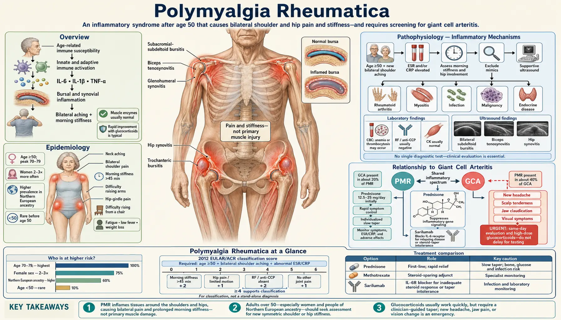
Table of Contents
- Overview
- Epidemiology
- Pathophysiology — Inflammatory Mechanisms
- Relationship to Giant Cell Arteritis
- Symptoms and Clinical Presentation
- Diagnosis and Diagnostic Criteria
- Treatment — Corticosteroids and Beyond
- Natural and Nutritional Approaches
- Disease Course and Prognosis
- Complications
- Prevention and Lifestyle
- Key Research Papers
- Connections
- Featured Videos
1. Overview
Polymyalgia rheumatica (PMR) is a common inflammatory condition that causes aching and stiffness in the proximal muscles — the shoulders, upper arms, neck, hips, and thighs. The name comes from the Greek: poly (many), myo (muscle), algos (pain). Despite the name, the pain actually originates from inflammation in the bursae and tendon sheaths around large joints rather than in the muscle tissue itself.
PMR is almost exclusively a disease of older adults. It virtually never occurs before age 50, and the average age at diagnosis is around 70. It is one of the most common inflammatory rheumatic diseases in people over 65, second only to rheumatoid arthritis. The hallmark of PMR is its remarkable, dramatic response to low-dose corticosteroids — most patients experience profound relief within 24 to 48 hours of starting prednisone, a response so characteristic that it is itself used as a diagnostic clue.
PMR is closely linked to giant cell arteritis (GCA), a vasculitis affecting large- and medium-sized arteries, particularly the temporal artery. About 15–20% of people with PMR will develop GCA at some point, and conversely, about 40–50% of GCA patients have concurrent PMR. Understanding this relationship is critical because GCA can cause sudden, irreversible vision loss if untreated, and symptoms that overlap with PMR can mask GCA's presence.
2. Epidemiology
PMR is predominantly a disease of Northern European ancestry. Incidence rates in Scandinavia and the United Kingdom are among the highest in the world — approximately 50–112 per 100,000 people aged 50 and older per year in some northern European populations. Rates are lower in Mediterranean countries and substantially lower in Asian and Hispanic populations, suggesting both genetic and environmental components.
Women are affected 2 to 3 times more often than men, a pattern common to many autoimmune and inflammatory conditions. A population study from Olmsted County, Minnesota, estimated the lifetime risk of PMR for US adults at 2.4% for women and 1.7% for men, making it the second most common inflammatory autoimmune rheumatic disease after rheumatoid arthritis. The peak incidence occurs in the 70–79 age group. PMR is rare under 50 and essentially unheard of in people under 40.
In the United States, an estimated 711,000 Americans have PMR at any given time, making it one of the most frequently diagnosed inflammatory rheumatic conditions in the elderly. Incidence increases with each decade after 50 and decreases again after age 80, possibly due to competing comorbidities limiting survival to diagnosis. Seasonal clustering in spring and early autumn, along with geographic clustering, suggests that environmental triggers — perhaps infectious agents — may initiate disease in genetically susceptible individuals.
PMR and GCA share similar epidemiology: both peak in the same age group, both are rare below 50, and both occur predominantly in people of Northern European ancestry. This epidemiologic overlap strongly supports a shared or closely related pathogenesis.
3. Pathophysiology — Inflammatory Mechanisms
The underlying cause of PMR remains incompletely understood, but current evidence points to a dysregulated innate immune response triggered by an environmental stimulus (likely infectious) in a genetically susceptible host. Rather than the classic B-cell and autoantibody-driven pathology of rheumatoid arthritis, PMR appears driven primarily by innate immune activation and cytokine-mediated systemic inflammation.
Interleukin-6 (IL-6) plays a central role. Serum interleukin-6 levels are markedly elevated in active PMR, correlate with disease activity, and fall rapidly with corticosteroid treatment. This finding explains why tocilizumab — an interleukin-6 receptor antagonist — has emerged as an effective steroid-sparing therapy in both PMR and the closely related GCA. IL-17A and IL-12/23 pathways also appear involved, particularly in patients who are refractory to steroids.
Pathological changes in PMR are found primarily in the periarticular structures rather than in muscles or synovium. Ultrasound and magnetic resonance imaging (MRI) studies consistently demonstrate:
- Subdeltoid and subacromial bursitis (shoulders) — nearly universal and highly characteristic
- Bicipital tenosynovitis at the shoulder
- Hip bursitis (trochanteric and iliopsoas bursae)
- Cervical interspinous bursitis explaining neck stiffness
- Mild synovitis of glenohumeral, hip, and knee joints in some patients
Genetic susceptibility is real. The strongest association is with HLA-DRB1*04 (HLA-DR4) alleles, shared with rheumatoid arthritis. However, unlike RA, PMR is seronegative (no rheumatoid factor, no anti-CCP antibodies), and human leukocyte antigen (HLA) associations in PMR are weaker and less consistently replicated across populations. Other candidate genes include those regulating innate immunity (NLRP1, PTPN22) and cytokine responses (IL-6, TNFA promoter polymorphisms).
The inflammatory cascade in PMR involves activation of CD4+ T-helper cells (particularly Th1 and Th17 subsets), macrophage activation, and dendritic cell maturation. These activated cells infiltrate the inflamed bursae and produce the elevated cytokines — interleukin-6, tumor necrosis factor-alpha, interleukin-1 beta (IL-1β) that drive the systemic symptoms of fatigue, weight loss, fever, and elevated acute-phase reactants (erythrocyte sedimentation rate (ESR), C-reactive protein (CRP), fibrinogen).
4. Relationship to Giant Cell Arteritis
The relationship between PMR and giant cell arteritis (GCA) is one of the most important concepts in understanding this condition. They are best thought of as two expressions of the same underlying disease spectrum rather than two entirely separate entities. Shared features include nearly identical epidemiology, overlapping (though not identical) human leukocyte antigen associations — most studies link GCA to HLA-DRB1*04 alleles, while the HLA-DRB1 link in PMR varies from one population to another — the same dramatic corticosteroid responsiveness, and overlapping histological and immunological findings.
GCA (temporal arteritis) is a granulomatous vasculitis affecting medium- and large-sized arteries, most characteristically the temporal artery but also the ophthalmic artery and the aorta and its branches. When GCA involves the ophthalmic artery, it can cause sudden, permanent vision loss — the most feared complication. This is a true ophthalmologic emergency requiring immediate high-dose prednisone (40–60 mg/day or intravenous (IV) methylprednisolone) rather than the low doses used for uncomplicated PMR.
The clinical overlap is substantial:
- Approximately 15–20% of PMR patients develop GCA during the course of their illness, either at diagnosis or at some point during follow-up
- Approximately 40–50% of GCA patients have concurrent PMR symptoms
- Some patients with "pure PMR" have subclinical large-vessel vasculitis detectable only on PET-CT or magnetic resonance imaging angiography, without symptoms of GCA
- GCA can develop after PMR has been in remission, so long-term vigilance is necessary
Red flag symptoms that suggest GCA in a PMR patient require urgent evaluation:
- New headache, particularly temporal or occipital
- Scalp tenderness (especially with combing hair)
- Jaw claudication (aching pain in the jaw with chewing)
- Visual disturbances: blurring, double vision, transient vision loss (amaurosis fugax)
- Tenderness or nodularity of the temporal artery on palpation
- Fever above 38°C without other explanation
- Limb claudication (arm or leg pain with use, suggesting large-vessel involvement)
Hospitals treat these symptoms in a person with PMR as urgent: evaluation includes temporal artery biopsy, color Doppler ultrasound of cranial arteries, and/or PET-CT, and higher-dose corticosteroids are usually started while results are awaited.
5. Symptoms and Clinical Presentation
PMR has a characteristic clinical pattern that, once recognized, is hard to mistake. The onset is typically subacute — developing over days to weeks — though some patients describe an almost overnight onset of severe stiffness. The core symptom cluster is:
Bilateral proximal muscle aching and stiffness affecting:
- Shoulder girdle (most common; virtually universal): pain and stiffness in the shoulders, upper arms, and sometimes the neck. Patients often struggle to lift their arms above shoulder height, put on a coat, or reach behind their back
- Pelvic girdle (hips and thighs): difficulty rising from a chair, climbing stairs, or getting out of a car. Hip pain may radiate to the buttocks or thighs, mimicking hip arthritis
- Neck: stiffness and aching, often making turning the head uncomfortable
Morning stiffness is the defining feature and is typically severe — lasting 45 minutes or more, often over an hour. This is far longer than the brief morning stiffness of osteoarthritis. Many patients describe being "frozen" when they wake up and needing an hour or more before they can move normally. Night pain that disturbs sleep is common, and rolling over in bed or getting up to use the bathroom can be agonizing.
Gel phenomenon: stiffness returns after any period of inactivity, not just in the morning. Sitting for a long car ride or movie produces renewed stiffness and difficulty getting up.
Systemic symptoms reflect the underlying inflammatory state:
- Profound fatigue, sometimes the most disabling symptom
- Low-grade fever and night sweats
- Unintentional weight loss (5–10 lbs is common)
- Depression and malaise
- Loss of appetite
What PMR does NOT typically cause:
- Distal joint involvement (fingers, wrists, ankles) is usually absent or minimal — this distinguishes it from rheumatoid arthritis
- True muscle weakness on neurological testing (grip strength, pushes) is not a feature; the "weakness" is functional, due to pain inhibition
- Skin rashes, mouth sores, or kidney involvement — features of lupus
- True muscle enzyme elevation (creatine kinase (CK), aldolase) — which would suggest polymyositis instead
The physical examination is often surprisingly unremarkable despite the patient's distress. There is restricted active range of motion at the shoulders and hips due to pain and stiffness, but passive range of motion (when the examiner moves the joint) is often preserved. This pattern is characteristic: the patient cannot raise the arm, but the physician can raise it for them without resistance. True joint swelling is uncommon in classical PMR, though mild knee effusions or wrist synovitis occurs in a minority.
6. Diagnosis and Diagnostic Criteria
There is no single diagnostic test for PMR. Diagnosis is clinical — based on the characteristic symptom pattern, the patient's age, elevated inflammatory markers, exclusion of mimicking conditions, and the dramatic response to corticosteroids.
Laboratory findings:
- erythrocyte sedimentation rate (ESR): typically elevated above 40 mm/h, often above 80 mm/h. Erythrocyte sedimentation rate above 50 mm/h in someone over 50 with proximal stiffness is a strong prompt for PMR. However, up to 20% of PMR patients have a normal erythrocyte sedimentation rate at presentation.
- C-reactive protein (CRP): elevated in virtually all active PMR patients and is actually more sensitive than erythrocyte sedimentation rate. A normal C-reactive protein is a reason to reconsider the diagnosis.
- Complete blood count (CBC): normocytic normochromic anemia of chronic disease is common. Platelet count is often elevated (reactive thrombocytosis).
- Liver function tests: mild elevation of alkaline phosphatase in about 20–30% of patients.
- Muscle enzymes (creatine kinase, aldolase): NORMAL. Elevation would point toward polymyositis or dermatomyositis instead.
- Rheumatoid factor (RF) and anti-CCP antibodies: NEGATIVE. Positive serology favors rheumatoid arthritis (RA) over PMR.
- Antinuclear antibody (ANA): typically negative; a strongly positive result points toward lupus or another connective tissue disease.
- Thyroid function (thyroid-stimulating hormone, TSH): checked to exclude hypothyroidism, which can mimic PMR's fatigue and stiffness.
ACR/EULAR 2012 Classification Criteria for PMR:
Developed for research classification but widely used clinically, these criteria require: age ≥ 50, bilateral shoulder aching, and abnormal C-reactive protein or erythrocyte sedimentation rate. If those three are present, a point-scoring system (0–6 without ultrasound, 0–8 with ultrasound) determines classification:
- Morning stiffness > 45 min: 2 points
- Hip pain or limited hip range of motion: 1 point
- Absence of RF or anti-CCP: 2 points
- Absence of peripheral joint involvement: 1 point
- Ultrasound: at least 1 shoulder with subdeltoid bursitis and/or biceps tenosynovitis and/or glenohumeral synovitis PLUS at least 1 hip with hip synovitis and/or trochanteric bursitis: 1 point
- Ultrasound: both shoulders with subdeltoid bursitis, biceps tenosynovitis, or glenohumeral synovitis: 1 point
A score of ≥ 4 (without ultrasound) or ≥ 5 (with ultrasound) classifies as PMR. Sensitivity is 68% without ultrasound, 66% with ultrasound; specificity is 78% without and 81% with. Specificity was higher (88%) for telling PMR apart from shoulder conditions and lower (65%) for telling it apart from rheumatoid arthritis, and the authors state that these criteria are not meant for diagnostic purposes.
The role of ultrasound has grown substantially. High-resolution musculoskeletal ultrasound can demonstrate subdeltoid bursitis, bicipital tenosynovitis, and hip bursitis — the characteristic periarticular inflammation of PMR — far earlier and more specifically than plain X-rays. Ultrasound is now recommended as a first-line imaging test when the diagnosis is uncertain.
Conditions to exclude (differential diagnosis):
- Rheumatoid arthritis: check RF, anti-CCP; rheumatoid arthritis involves distal joints (fingers, wrists); rheumatoid arthritis is usually seronegative in the elderly ("seronegative rheumatoid arthritis of the elderly" can closely mimic PMR)
- Hypothyroidism: check thyroid-stimulating hormone; can cause aching, fatigue, stiffness
- Malignancy: particularly lymphoma and myeloma can cause erythrocyte sedimentation rate elevation and aching; check complete blood count, protein electrophoresis; imaging if clinically suspicious
- Polymyositis/dermatomyositis: true muscle weakness and elevated creatine kinase; check muscle enzymes, EMG/MRI if uncertain
- Fibromyalgia: widespread pain but erythrocyte sedimentation rate/C-reactive protein normal; no response to corticosteroids
- Rotator cuff disease: usually unilateral; erythrocyte sedimentation rate normal; responds to local injection not systemic steroids
- Remitting seronegative symmetrical synovitis with pitting edema (RS3PE): a PMR variant with dramatic hand and foot swelling; responds well to steroids
7. Treatment — Corticosteroids and Beyond
The treatment of PMR is one of the most gratifying in medicine. The dramatic, near-complete response to corticosteroids within 24 to 48 hours is both therapeutic and diagnostic. When a patient takes their first dose of prednisone and wakes up the next morning able to move without agony for the first time in months, the relief can be overwhelming — and it confirms the diagnosis more convincingly than any laboratory test.
Standard initial corticosteroid dosing:
- Prednisone 15–20 mg/day as a single morning dose is the standard starting regimen for uncomplicated PMR (no GCA features)
- This is deliberately lower than the 40–60 mg/day used for GCA, reflecting PMR's lower risk of irreversible organ damage
- If there is NO meaningful improvement within one week of starting prednisone at 15–20 mg/day, the diagnosis of PMR is usually reconsidered
- Modified-release prednisone (a delayed-release, or modified-release, prednisone tablet): taken at bedtime (10 pm), it releases its dose about 4 hours later (~2 am) to target the early-morning inflammatory surge. In the CAPRA-1 trial — carried out in 288 people with rheumatoid arthritis, not PMR — the duration of morning stiffness fell by 22.7% with the bedtime tablet versus 0.4% with immediate-release prednisone taken in the morning; the bedtime group's stiffness fell by a mean of 44 minutes, and the 29-minute difference between the groups was not statistically significant
Tapering strategy: Steroid tapering in PMR is slow and guided by symptoms and inflammatory markers (C-reactive protein/erythrocyte sedimentation rate). Too-rapid tapering is the most common cause of relapse. The BSR/BHPR guidelines (2010) describe the following schedule; the 2015 EULAR/ACR recommendations also cover starting doses and tapering:
- Reduce prednisone by 2.5 mg every 2–4 weeks to 10 mg/day
- Then reduce by 1 mg every 4–8 weeks (or slower)
- Total treatment duration: typically 1–2 years, though some patients require 3–6 years
- Monitor erythrocyte sedimentation rate and C-reactive protein at each taper step; rising markers before symptoms often herald relapse
- If relapse occurs, return to the last effective dose (not necessarily to the starting dose)
Relapse rates are high. Approximately 30–50% of patients relapse at least once during tapering. Factors associated with increased relapse risk include: female sex, higher initial erythrocyte sedimentation rate, peripheral arthritis at presentation, and presence of HLA-DRB1*04.
Tocilizumab (interleukin-6 receptor blockade) as steroid-sparing therapy:
The GiACTA trial (PMID 28745999) demonstrated that tocilizumab subcutaneous (162 mg weekly or every 2 weeks) significantly increased sustained GCA remission rates compared with prednisone alone and allowed substantially lower cumulative steroid doses. While GiACTA enrolled GCA patients, many had concurrent PMR, and the interleukin-6 pathway is central to both conditions. Tocilizumab is now approved by the U.S. Food and Drug Administration (FDA) for GCA and is increasingly used off-label in refractory PMR or as a steroid-sparing agent in patients with high steroid-related toxicity risk (osteoporosis, diabetes, hypertension, glaucoma).
A trial in PMR itself has since been published: in 101 French patients whose PMR stayed active on 10 mg or more of prednisone a day, intravenous tocilizumab every 4 weeks for 24 weeks met the trial's main goal (low disease activity together with a reduced prednisone dose) in 67.3% versus 31.4% on placebo, and 49.0% versus 19.6% were no longer taking prednisone at 24 weeks. Infections were the most frequent side effect (46.9% vs 39.2%).
Sarilumab (a second interleukin-6 receptor blocker):
In the SAPHYR trial (2023), 118 people whose PMR flared while their steroid dose was being lowered received either sarilumab 200 mg injections twice a month plus a 14-week prednisone taper, or placebo plus a 52-week taper. Sustained remission at one year was 28% versus 10%, and the median total prednisone taken over the year was 777 mg versus 2,044 mg. A low white-blood-cell count (neutropenia, 15% vs 0%), joint pain (15% vs 5%) and diarrhea (12% vs 2%) were more common with sarilumab. The current US label lists PMR in adults who have had an inadequate response to corticosteroids or who cannot tolerate a corticosteroid taper.
Methotrexate has been studied as a steroid-sparing agent in PMR with modest evidence. Several small randomized trials suggest it can reduce cumulative steroid dose and relapse risk, but evidence is less robust than for GCA/tocilizumab. It may be considered in patients with frequent relapses or steroid intolerance.
Monitoring during treatment:
- Erythrocyte sedimentation rate and C-reactive protein at each clinic visit (every 4–8 weeks during tapering)
- Blood pressure and blood glucose (corticosteroid side effects)
- Bone density (DEXA scan at baseline and after 12 months)
- Calcium and vitamin D supplementation throughout steroid therapy
- Bisphosphonate therapy if DEXA shows significant bone loss
- Eye pressure monitoring if patient has glaucoma or family history
- Vigilance for GCA symptoms at every visit
8. Natural and Nutritional Approaches
Natural approaches in PMR are not replacements for corticosteroid therapy — no natural product comes close to matching the rapid, profound anti-inflammatory effect of prednisone in PMR. However, nutritional strategies can meaningfully address the secondary effects of both the disease and its treatment, particularly bone loss, cardiovascular risk, and the metabolic effects of long-term corticosteroids.
Vitamin D and Calcium — essential during steroid therapy:
Corticosteroids impair calcium absorption in the gut and increase urinary calcium excretion, directly causing bone loss. This is the most predictable and serious long-term side effect of PMR treatment. The 2017 American College of Rheumatology (ACR) guideline on steroid-related osteoporosis recommends calcium and vitamin D for adults on long-term corticosteroids (3 months or more) at low fracture risk, and calcium and vitamin D plus an osteoporosis medicine (an oral bisphosphonate preferred) at moderate-to-high fracture risk. Many rheumatologists co-prescribe a bisphosphonate (alendronate, risedronate) from the start of steroid therapy if the patient already has bone density below the normal range.
Omega-3 fatty acids:
Fish oil (eicosapentaenoic acid (EPA) and docosahexaenoic acid (DHA), 2–4 g/day) has documented anti-inflammatory properties and may reduce the baseline inflammatory burden. While no large randomized trial has tested omega-3s specifically in PMR, they have demonstrated benefit in rheumatoid arthritis at similar doses. They also reduce cardiovascular risk — relevant because PMR patients are older adults already at elevated cardiovascular baseline risk, which is further increased by chronic steroid use (blood pressure, glucose, lipids).
Mediterranean-style diet:
A Mediterranean diet (olive oil, fish, legumes, vegetables, fruits, whole grains; limited red meat and ultra-processed foods) reduces systemic inflammatory markers (C-reactive protein, interleukin-6) in older adults and reduces cardiovascular risk. For PMR patients on chronic corticosteroids, reducing cardiovascular risk is particularly important. The diet's anti-inflammatory polyphenol and omega-3 content may support lower background inflammation, potentially facilitating steroid tapering.
Curcumin (turmeric):
Curcumin inhibits NF-κB and reduces interleukin-6 and tumor necrosis factor-alpha in laboratory models. Clinical evidence in inflammatory arthritis is preliminary but promising. Doses of 500–1,000 mg of phospholipid-complexed or nanoparticle curcumin have shown modest anti-inflammatory effects. It is not a substitute for prednisone; some practitioners describe it as an add-on for patients who want to reduce their steroid dose and have a low inflammatory burden.
Bone-protective lifestyle measures:
- Weight-bearing exercise (walking, resistance training) to stimulate bone formation
- Fall prevention strategies (balance exercises, home hazard assessment) — critical given that PMR patients are older adults at elevated fall and fracture risk
- Limiting alcohol (alcohol suppresses bone formation and adds cardiovascular risk)
- Not smoking (smoking accelerates bone loss and worsens cardiovascular risk)
Ginger:
Gingerols and shogaols inhibit prostaglandin and leukotriene synthesis, with mild anti-inflammatory and analgesic properties. Culinary amounts of ginger in food are safe for everyone; concentrated ginger supplements (1–2 g/day) may provide modest adjunct benefit. They can thin the blood slightly, and concentrated ginger may add to the bleeding effect of anticoagulant (blood-thinning) medicines.
9. Disease Course and Prognosis
PMR is not a life-threatening condition in itself, and its prognosis for functional outcome is generally good when properly treated. However, it follows a variable and often relapsing course that requires patience from both patient and physician.
Duration of treatment: Most guidelines recommend a minimum treatment duration of 12–24 months, with many patients requiring 2–3 years or longer. Approximately 25–30% of patients can successfully discontinue steroids within 2 years without relapse. Another 40–50% achieve remission but only after 3–6 years of treatment. A minority (<20%) remain steroid-dependent long-term and require ongoing low-dose prednisone.
Relapse prediction: Several factors are associated with more prolonged or relapsing disease:
- Female sex
- Higher erythrocyte sedimentation rate at diagnosis
- Peripheral arthritis at onset
- Rapid steroid taper (less than 12 months)
- HLA-DRB1*04 genotype
- Persistent elevated C-reactive protein despite clinical improvement
Quality of life: When PMR is well controlled, most patients maintain normal or near-normal functional capacity. The disease does not cause joint destruction (unlike rheumatoid arthritis), and the muscles are not permanently damaged. Patients who achieve stable remission typically have no residual disability attributable to the PMR itself. The main long-term quality-of-life concerns relate to steroid side effects rather than the disease itself.
Mortality: Overall survival in PMR patients is similar to the general age-matched population when GCA does not develop. The presence of GCA, particularly with vascular complications, worsens prognosis. Patients with PMR who develop aortic aneurysm (a known GCA complication) face significantly elevated mortality from aortic dissection. This is another reason why screening for large-vessel involvement with periodic imaging in patients with known GCA-PMR overlap has been advocated by some experts.
10. Complications
The complications of PMR fall into two categories: those arising from the disease itself and those arising from its treatment.
Disease-related complications:
- Giant cell arteritis development: the most feared disease complication. As noted above, approximately 15–20% of PMR patients develop GCA, which can cause vision loss, stroke, or aortic aneurysm. Regular surveillance for GCA symptoms is essential throughout the entire duration of PMR.
- Functional disability from undertreated disease: prolonged untreated PMR causes severe functional limitation. Patients who cannot lift their arms or rise from a chair unassisted may become dependent, fall, and develop deconditioning and its own cascade of complications.
Treatment-related complications (corticosteroids):
- Osteoporosis and fractures: the most common serious steroid complication, affecting approximately 30–50% of PMR patients on long-term steroids. Corticosteroids suppress osteoblast function and impair gut calcium absorption, causing rapid bone loss (greatest in the first 6–12 months). Vertebral compression fractures and hip fractures can occur. Prevention requires calcium, vitamin D, and often bisphosphonate co-prescription from the start.
- Adrenal insufficiency: chronic exogenous corticosteroid use suppresses the hypothalamic-pituitary-adrenal axis. If steroids are stopped too abruptly, the adrenal glands may not produce sufficient cortisol, causing adrenal crisis (severe fatigue, hypotension, nausea, vomiting) — a medical emergency. This is why steroid tapering is always gradual, and patients are commonly given a steroid alert card to carry.
- Diabetes mellitus: corticosteroids raise blood glucose, particularly postprandial. Patients with pre-existing prediabetes or family history are at highest risk. Fasting glucose and hemoglobin A1c (HbA1c) are checked at regular intervals.
- Hypertension: corticosteroids promote sodium and water retention, raising blood pressure. Blood pressure is checked at each visit.
- Cataract and glaucoma: particularly with doses above 10 mg/day for prolonged periods. Annual eye checks are common practice.
- Infection susceptibility: corticosteroids broadly suppress immune function. Pneumococcal and influenza vaccines are part of usual care for people on long-term corticosteroids, and close contact with people who have active infections carries extra risk.
- Skin changes, weight gain, and cushingoid features: thinning skin, easy bruising, central weight gain, and moon face are dose-dependent and often improve as the dose tapers.
- Mood changes: corticosteroids can cause euphoria at high doses and depression or irritability as doses are reduced. Rarely, psychosis at high doses.
11. Prevention and Lifestyle
PMR cannot currently be prevented — there are no known strategies to prevent its development in at-risk individuals. However, several lifestyle measures can meaningfully reduce the burden of disease and the complications of treatment.
Protecting bone health during treatment:
- The 2017 ACR guideline recommends calcium and vitamin D for adults on long-term corticosteroids at low fracture risk, with an added osteoporosis medicine (an oral bisphosphonate preferred) at moderate-to-high risk
- The 2022 ACR guideline strongly recommends assessing fracture risk — including a bone density (DEXA) scan with a check for spine fractures — as soon as possible after corticosteroids are started for more than 3 months; repeat scans are commonly done yearly while on steroids
- For adults at medium, high or very high fracture risk, the 2022 ACR guideline strongly recommends an osteoporosis medicine — bisphosphonates (alendronate, risedronate, zoledronate), denosumab or parathyroid-hormone-type drugs
- Regular weight-bearing exercise — even a daily 30-minute walk — stimulates bone formation and reduces fracture risk
Cardiovascular protection:
- Blood pressure, blood glucose, and lipids are all worsened by corticosteroids and are checked during treatment
- Mediterranean diet reduces cardiovascular inflammatory burden
- Aerobic exercise usually becomes possible again as PMR symptoms allow (often markedly improved on prednisone)
- Not smoking
Staying vigilant for GCA:
- The warning signs of GCA (new headache, jaw pain with chewing, scalp tenderness, visual changes) are signs that hospitals treat as needing same-day evaluation
- Vision changes are an emergency sign — GCA-related vision loss can become permanent within hours if untreated
Patient education and shared decision-making:
- Understanding that PMR is a long-term condition requiring patience during tapering significantly improves adherence and outcomes
- Knowing that mild symptoms during tapering do not always mean a full relapse — watchful waiting with repeat labs is sometimes appropriate rather than immediately increasing the dose
- Joining a patient support group (Vasculitis UK, Polymyalgia Rheumatica and GCA UK) provides practical experience-sharing from others who have navigated the tapering journey
Key Research Papers
- Dasgupta B, Borg FA, Hassan N, et al., 2010 — BSR and BHPR guidelines for the management of polymyalgia rheumatica. Rheumatology (Oxford). 2010;49(1):186-190; foundational clinical practice guidance establishing diagnostic criteria, tapering protocols, and monitoring standards widely used by UK rheumatologists. — PubMed PMID: 19910443
- Dejaco C, Singh YP, Perel P, et al., 2015 — 2015 recommendations for the management of polymyalgia rheumatica: a European League Against Rheumatism/American College of Rheumatology collaborative initiative; international consensus recommendations covering initial dosing, tapering strategy, steroid-sparing agents, and monitoring in PMR. Arthritis Rheumatol. 2015;67(10):2569-2580. — PubMed PMID: 26352874
- Stone JH et al., 2017 — GiACTA trial: Tocilizumab in giant cell arteritis; landmark randomized controlled trial demonstrating superiority of tocilizumab plus prednisone over prednisone alone in achieving sustained GCA remission and reducing cumulative corticosteroid exposure. N Engl J Med. — PubMed PMID: 28745999
- Salvarani C, Cantini F, Boiardi L, Hunder GG, 2002 — Polymyalgia rheumatica and giant-cell arteritis; comprehensive review covering epidemiology, pathogenesis, clinical features, and treatment; a foundational reference for the condition. N Engl J Med. 2002;347(4):261-271. — PubMed PMID: 12140303
- Macchioni P, Boiardi L, Catanoso M, Pazzola G, Salvarani C, 2014 — Performance of the new 2012 EULAR/ACR classification criteria for polymyalgia rheumatica: comparison with the previous criteria in a single-centre study; demonstrated the added diagnostic value of musculoskeletal ultrasound in meeting the classification criteria. Ann Rheum Dis. 2014;73(6):1190-1193. — PubMed PMID: 24297384
- — Epidemiology of polymyalgia rheumatica and giant cell arteritis; comprehensive review of incidence rates, geographic and ethnic variations, temporal trends, and the epidemiological links between PMR and GCA. — Search PubMed
- — HLA-DRB1 and polymyalgia rheumatica-giant cell arteritis; delineated the HLA class II genetic susceptibility loci shared between PMR and GCA, including the DRB1*04 shared epitope association. — Search PubMed
- Dasgupta B, Cimmino MA, Maradit-Kremers H, et al., 2012 — 2012 provisional classification criteria for polymyalgia rheumatica: a European League Against Rheumatism/American College of Rheumatology collaborative initiative; development and validation of the scoring algorithm that now defines PMR for research classification, including the added value of ultrasound findings. Ann Rheum Dis. 2012;71(4):484-492. — PubMed PMID: 22388996
- Hellmich B, Agueda A, Monti S, et al., 2020 — 2018 update of the EULAR recommendations for the management of large vessel vasculitis; covering both GCA and Takayasu's arteritis with specific guidance on imaging modalities, treatment escalation, and monitoring in patients with PMR-GCA overlap. Ann Rheum Dis. 2020;79(1):19-30. — PubMed PMID: 31270110
- Buttgereit F, Doering G, Schaeffler A, et al., 2008 — Efficacy of modified-release versus standard prednisone to reduce duration of morning stiffness of the joints in rheumatoid arthritis (CAPRA-1): a double-blind, randomised controlled trial; a rheumatoid-arthritis trial showing that timing corticosteroid release to the nocturnal inflammatory peak reduces morning stiffness duration. Lancet. 2008;371(9608):205-214. — PubMed PMID: 18207016
- Devauchelle-Pensec V, Carvajal-Alegria G, Dernis E, et al., 2022 — Effect of tocilizumab on disease activity in patients with active polymyalgia rheumatica receiving glucocorticoid therapy: a randomized clinical trial. JAMA. 2022;328(11):1053-1062. — PubMed PMID: 36125471
- Spiera RF, Unizony S, Warrington KJ, et al., 2023 — Sarilumab for relapse of polymyalgia rheumatica during glucocorticoid taper (SAPHYR trial). N Engl J Med. 2023;389(14):1263-1272. — PubMed PMID: 37792612
- Buckley L, Guyatt G, Fink HA, et al., 2017 — 2017 American College of Rheumatology guideline for the prevention and treatment of glucocorticoid-induced osteoporosis. Arthritis Rheumatol. 2017;69(8):1521-1537. — PubMed PMID: 28585373
- Humphrey MB, Russell L, Danila MI, et al., 2023 — 2022 American College of Rheumatology guideline for the prevention and treatment of glucocorticoid-induced osteoporosis. Arthritis Rheumatol. 2023;75(12):2088-2102. — PubMed PMID: 37845798
- Crowson CS, Matteson EL, Myasoedova E, et al., 2011 — The lifetime risk of adult-onset rheumatoid arthritis and other inflammatory autoimmune rheumatic diseases. Arthritis Rheum. 2011;63(3):633-639. — PubMed PMID: 21360492
PubMed Topic Searches
13. Connections
- Giant Cell Arteritis
- Vasculitis
- Polyarteritis Nodosa
- Rheumatoid Arthritis
- Arthritis
- Osteoporosis
- Lupus
- Gout
- Ankylosing Spondylitis
- Sjögren's Syndrome
- Raynaud's Disease
- Osteoarthritis
- Ehlers-Danlos Syndrome
- Vitamin D3 — bone health during steroid therapy
- Calcium — osteoporosis prevention