Polyarteritis Nodosa
- Overview
- Epidemiology
- Pathophysiology
- Clinical Manifestations
- Hepatitis B-Associated PAN
- Diagnosis
- ACR Classification Criteria
- Treatment
- Prognosis and Monitoring
- An Inherited Form: Deficiency of Adenosine Deaminase 2
- Key Research Papers
- Connections
- Featured Videos
Overview
Polyarteritis nodosa (PAN) is a medium-vessel necrotizing vasculitis affecting medium and small muscular arteries. It is defined by transmural inflammatory destruction of arterial walls — progressing to fibrinoid necrosis, aneurysm formation, thrombosis, and downstream ischemia. The most critical diagnostic distinction from other systemic vasculitides is that PAN is antineutrophil cytoplasmic antibody-negative: antineutrophil cytoplasmic antibodies (p-ANCA and c-ANCA) are absent in true PAN, which separates it entirely from microscopic polyangiitis (MPA), granulomatosis with polyangiitis (GPA), and eosinophilic granulomatosis with polyangiitis (EGPA). A positive antineutrophil cytoplasmic antibody (ANCA) result effectively rules out PAN and redirects the diagnostic workup toward the antineutrophil cytoplasmic antibody-associated vasculitides (AAV).
PAN spares capillaries, venules, and arterioles — this anatomical restriction has profound clinical consequences. Because the glomerulus is supplied by capillaries, PAN does not cause glomerulonephritis. Renal involvement in PAN is entirely ischemic in mechanism: stenosis or infarction of the renal arteries produces renovascular hypertension and renal cortical infarcts, but there is no proliferative glomerulonephritis, no red cell casts in the urine, and typically no heavy proteinuria — features that would otherwise suggest an antineutrophil cytoplasmic antibody vasculitis. Similarly, PAN spares pulmonary capillaries, so pulmonary capillaritis and diffuse alveolar hemorrhage do not occur. Pulmonary involvement is absent in classic PAN — another key distinguishing feature.
PAN holds the distinction of being the first vasculitis ever described in medicine: Adolf Kussmaul and Rudolf Maier published the seminal case report in 1866, naming the condition "periarteritis nodosa" for the palpable nodules felt along inflamed arteries. Historically, hepatitis B virus (HBV) infection was identified as a causative trigger in 30–50% of PAN cases; widespread hepatitis B vaccination has since reduced hepatitis B virus-associated PAN to fewer than 10% of cases in developed nations. The 2012 revised Chapel Hill Consensus Conference nomenclature and later classification work have substantially narrowed PAN's definition — many cases previously labeled "PAN" have been reclassified as MPA or other antineutrophil cytoplasmic antibody vasculitides as antineutrophil cytoplasmic antibody testing became routine. True idiopathic PAN is now recognized as a comparatively uncommon disease.
Epidemiology
PAN is a rare condition with an estimated annual incidence of 2–9 cases per million population in Western countries. It can occur at any age but peaks between 40 and 60 years. There is a slight male predominance, with a male-to-female ratio of approximately 1.5:1. In regions where hepatitis B vaccination coverage remains incomplete — including parts of sub-Saharan Africa, Southeast Asia, and Eastern Europe — hepatitis B virus-associated PAN remains proportionally more common and continues to affect younger adults in keeping with the epidemiology of hepatitis B virus infection in those populations.
Beyond hepatitis B virus, a small number of well-recognized associations exist. Hairy cell leukemia-associated PAN is a rare but established entity in which the vasculitis may predate or follow the lymphoproliferative diagnosis; for this reason a search for a blood disorder (hematologic workup) is part of evaluating PAN that has no clear infectious trigger. Single-organ or limited forms of PAN — most notably cutaneous PAN (isolated skin involvement with livedo reticularis, subcutaneous nodules, and skin ulcers without systemic features) and appendiceal PAN (an incidental finding at appendectomy) — carry a considerably better prognosis than systemic PAN and rarely require systemic immunosuppression. Isolated cutaneous PAN may follow streptococcal infection in children and adolescents.
Pathophysiology
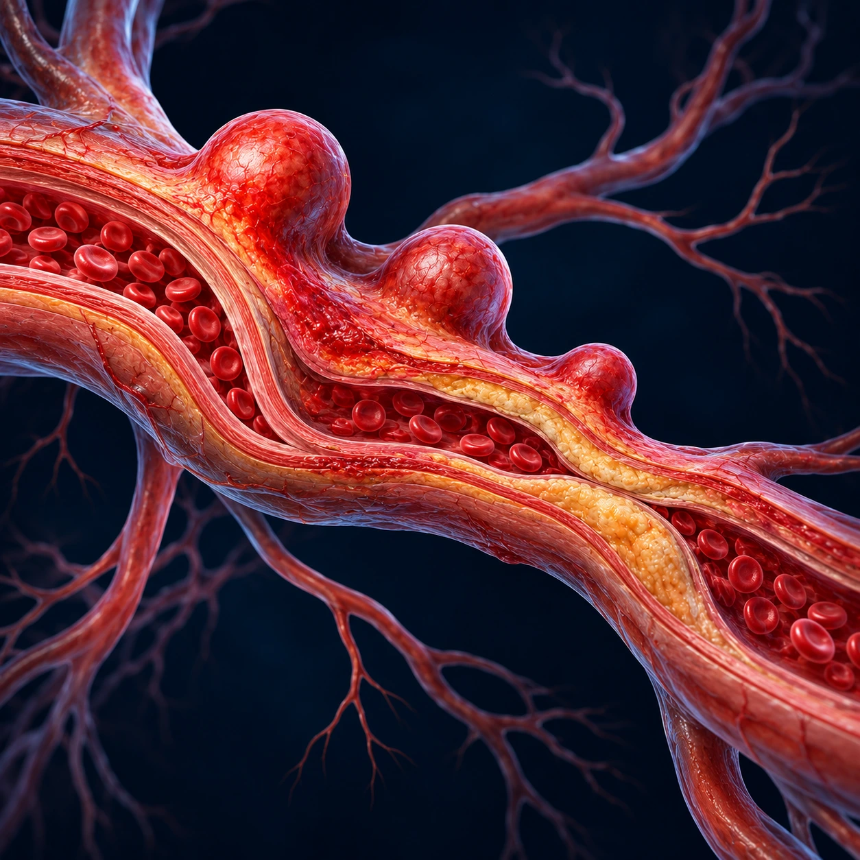
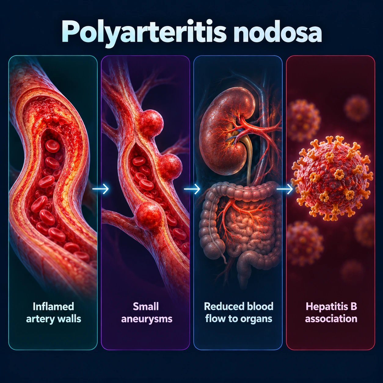
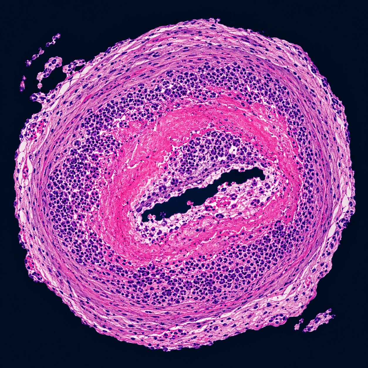
The core pathologic lesion in PAN is segmental fibrinoid necrosis of medium arterial walls. The inflammatory process begins with transmural neutrophilic infiltration of the vessel wall — neutrophils flood all layers of the artery, releasing proteases and reactive oxygen species that destroy the internal elastic lamina and media. This progresses to coagulative necrosis of the vessel wall (fibrinoid necrosis), structurally weakening the artery at points of maximal hemodynamic stress — particularly at bifurcations and branch points. The weakened wall dilates to form microaneurysms, typically 1–5 mm in diameter; these are the radiographic hallmark of PAN visible on conventional or computed tomography (CT) angiography as saccular outpouchings along the mesenteric and renal arterial trees. Superimposed thrombosis at the aneurysm neck or at sites of stenosis produces distal arterial occlusion and end-organ ischemia or infarction. The segmental, skip-lesion distribution — a defining histological feature — means that consecutive segments of the same artery alternate between inflamed/necrotic and normal, explaining why portions of a single organ (e.g., kidney, bowel) can be infarcted while adjacent tissue remains viable.
In hepatitis B virus-associated PAN, the pathogenic mechanism is immune complex deposition rather than direct viral cytopathic effect. Circulating hepatitis B surface antigen (HBsAg) forms immune complexes with anti-HBs antibodies; these complexes deposit in medium artery walls, activate the complement cascade, and trigger the same transmural necrotizing inflammation seen in idiopathic PAN. The timing is characteristic: vasculitis typically develops within the first year of hepatitis B virus infection, when circulating HBsAg is abundant and the immune response is generating antibodies against it. This immune complex mechanism also explains why elimination of hepatitis B virus replication through antiviral therapy — rather than indefinite immunosuppression — can produce sustained vasculitis remission: removing the antigen source eliminates the driver of immune complex formation.
Mononeuritis multiplex, the most clinically distinctive neurological feature of PAN, arises from vasculitis of the epineurial (nutrient) arteries supplying peripheral nerves. The epineurial arteries are medium-sized muscular arteries — squarely within PAN's target range — and their occlusion by necrotizing vasculitis produces focal nerve ischemia. Unlike length-dependent polyneuropathy, where dying-back from the feet occurs symmetrically, mononeuritis multiplex presents as asymmetric deficits that develop in a stepwise fashion: the peroneal nerve is infarcted at one point in time (producing unilateral foot drop), then the ulnar or radial nerve at another, then a different nerve again, each event marking a new arterial occlusion. Over time the cumulative picture may superficially resemble a symmetric polyneuropathy, but careful history reveals the stepwise, non-length-dependent evolution that points to a vasculitic etiology.
Clinical Manifestations
Constitutional symptoms are the most common presenting feature and are present in the majority of patients. Weight loss (present in approximately 70% of cases) is the most frequent constitutional symptom and is often striking — patients commonly lose 5–15 kg over weeks to months before diagnosis. Fever, profound fatigue, myalgia, and diffuse arthralgia accompany weight loss in most cases. These nonspecific symptoms frequently lead to a broad initial differential including malignancy, infection, and inflammatory bowel disease, causing diagnostic delays of months to years. Elevated acute-phase reactants (erythrocyte sedimentation rate (ESR) often 60–120 mm/hr, C-reactive protein (CRP) markedly elevated) are invariably present and may be the laboratory finding that prompts rheumatologic evaluation.
Peripheral neuropathy (mononeuritis multiplex) is the most common specific clinical manifestation, present in 50–75% of patients. Its asymmetric, stepwise character is diagnostically important. Common presentations include unilateral foot drop from peroneal nerve ischemia — the patient reports sudden inability to dorsiflex the foot, with a classic slapping gait — and wrist drop from radial nerve involvement. Sensory symptoms (burning, numbness, or allodynia in the nerve's territory) typically accompany or precede motor deficits. Because axonal injury from ischemia may be permanent, early diagnosis and treatment are essential to limit neurological deficit. Electrodiagnostic studies confirm axonal mononeuritis multiplex and help distinguish PAN neuropathy from demyelinating neuropathies or compression neuropathies. Renal involvement is driven entirely by medium artery ischemia: renovascular hypertension (from renal artery stenosis), renal cortical infarction causing flank pain and hematuria, and ultimately ischemic nephropathy contributing to renal insufficiency. The defining absence is glomerulonephritis — there are no red cell casts on urinalysis, no nephrotic-range proteinuria, and no crescentic changes on biopsy. This profile directly contradicts antineutrophil cytoplasmic antibody vasculitis renal disease and is a critical differentiating point at the bedside.
Gastrointestinal involvement is present in 30–50% of patients and carries the highest mortality risk of any PAN manifestation. Mesenteric artery vasculitis produces postprandial abdominal pain (intestinal angina) as the ischemic bowel fails to hyperemia adequately with meals; patients learn to eat less to avoid pain and lose further weight. More severe ischemia causes gastrointestinal (GI) bleeding, segmental bowel infarction, and perforation — surgical emergencies with high mortality. Hepatic artery involvement can produce liver infarction or rupture of a hepatic artery aneurysm (rare but catastrophic). Appendiceal vasculitis, when present, is usually discovered incidentally at appendectomy performed for presumed appendicitis. PAN can cause acute abdominal presentations: an acute abdomen in a patient with unexplained systemic illness and weight loss raises the possibility of mesenteric vasculitis.
Cutaneous manifestations include livedo reticularis (a lace-like, violaceous mottling of the skin that reflects dermal arteriolar compromise), tender subcutaneous nodules overlying inflamed arteries (most prominent over the lower legs and feet), skin ulcers from cutaneous infarction, and digital gangrene in severe disease. Testicular involvement — orchitis from testicular artery vasculitis producing scrotal pain and swelling — is a pathognomonic association. Unexplained orchitis that does not respond to antibiotics in a middle-aged man is a recognized pointer toward PAN; testicular biopsy may confirm the vasculitic diagnosis. Hypertension is present in 25–50% of patients, attributable to renovascular disease. Cardiac involvement (coronary arteritis → myocardial infarction, pericarditis) and central nervous system (CNS) involvement (cerebral vasculitis → stroke, encephalopathy) occur in a minority of cases but contribute significantly to morbidity and mortality when present.
Hepatitis B-Associated PAN
Hepatitis B-associated PAN is a clinically and mechanistically distinct entity that requires a fundamentally different treatment strategy from idiopathic PAN. HBV-PAN once constituted 30–50% of all PAN diagnoses; universal hepatitis B vaccination programs — which have dramatically reduced new hepatitis B virus infections in developed nations since the 1990s — have reduced this proportion to fewer than 10% in high-income countries, though it remains more prevalent in regions where vaccination coverage is incomplete. The pathogenic mechanism is immune complex-mediated: circulating hepatitis B virus surface antigen (HBsAg) combines with anti-HBs antibodies to form immune complexes, which deposit in the walls of medium arteries, activate complement, and trigger the necrotizing vasculitis cascade. Vasculitis typically emerges within the first year of hepatitis B virus infection, when antigen-antibody dynamics are most active. Testing for hepatitis B virus (HBsAg, anti-HBc total, HBV DNA) is standard in every newly diagnosed case of PAN because the diagnosis of HBV-PAN completely changes the treatment algorithm.
Treatment of HBV-PAN prioritizes viral eradication over long-term immunosuppression. Prolonged immunosuppression in hepatitis B virus infection accelerates viral replication, precipitates hepatitis flares, and risks progression to cirrhosis and hepatocellular carcinoma — making the standard idiopathic PAN regimen of long-term cyclophosphamide and corticosteroids directly harmful if applied to HBV-PAN without modification. The accepted regimen consists of three components: (1) a short initial course of prednisone to suppress the most dangerous organ-threatening inflammation, followed by rapid taper (typically 2–4 weeks); (2) antiviral therapy — tenofovir or entecavir are preferred over the older lamivudine due to superior resistance profiles and potency; and (3) plasma exchange (plasmapheresis) to physically remove circulating immune complexes and HBsAg from the bloodstream, accelerating the immunological clearance process. As antiviral therapy suppresses HBV DNA and drives HBeAg-to-anti-HBe seroconversion (loss of viral replication markers), the immune complex burden falls, complement activation ceases, and the vasculitis enters sustained remission — uniquely achievable without ongoing immunosuppression. Hepatitis B vaccination serves as primary prevention for HBV-PAN and is recommended for all unvaccinated household contacts and sexual partners of hepatitis B virus-positive individuals.
Diagnosis
The diagnostic workup for PAN begins with establishing the antineutrophil cytoplasmic antibody-negative status and ruling out alternative diagnoses. Antineutrophil cytoplasmic antibody testing (both p-ANCA/MPO-ANCA and c-ANCA/PR3-ANCA by ELISA) is negative in PAN — a positive result effectively reassigns the diagnosis to the antineutrophil cytoplasmic antibody-associated vasculitides (MPA for p-ANCA/MPO, GPA for c-ANCA/PR3, or EGPA). Hepatitis B virus serology — HBsAg, total anti-HBc, and HBV DNA quantification — is part of every PAN workup, as the diagnosis of HBV-PAN has direct treatment implications. Hepatitis C serology is also checked because hepatitis C virus-associated cryoglobulinemic vasculitis can closely mimic PAN clinically, including mononeuritis multiplex and renal involvement, but is a different entity managed with antivirals (and rituximab for severe cases).
Laboratory findings in PAN are nonspecific but characteristically show leukocytosis with neutrophilia, normochromic normocytic anemia of chronic disease, thrombocytosis, markedly elevated erythrocyte sedimentation rate and C-reactive protein, and elevated acute-phase proteins. Renal function (creatinine, blood urea nitrogen, BUN) may be elevated if renovascular disease has compromised renal perfusion. Urinalysis is critically informative: PAN-related renal disease produces hematuria from renal cortical infarction but notably lacks the active urinary sediment of glomerulonephritis — no red cell casts, no white cell casts, and proteinuria below 1 g/day. The presence of red cell casts or >1 g/day proteinuria points the workup toward antineutrophil cytoplasmic antibody-associated GN. Tissue biopsy of a clinically affected organ provides definitive histological diagnosis. Combined sural nerve and muscle biopsy (gastrocnemius) substantially increases diagnostic yield over either alone — the combination approach is standard when mononeuritis multiplex is present. Biopsy shows necrotizing vasculitis of medium and small arteries with transmural neutrophilic infiltration, fibrinoid necrosis, and disruption of the internal elastic lamina; healing lesions may show intimal proliferation and fibrosis.
Angiography (conventional digital subtraction angiography, or computed tomography angiography) of the mesenteric and renal arteries demonstrates the radiographic hallmark of PAN: microaneurysms (typically 1–5 mm) at arterial branch points, accompanied by segmental stenoses and the "beading" appearance of alternating aneurysmal dilatation and stenosis. This angiographic pattern is considered pathognomonic for medium-vessel vasculitis and is reserved for cases where biopsy is not feasible, results are inconclusive, or when visceral involvement is suspected clinically. Renal biopsy is generally not performed in PAN because the microaneurysms create a risk of catastrophic hemorrhage; the combination of negative antineutrophil cytoplasmic antibody, absent urinary sediment, and characteristic angiographic findings is typically sufficient for diagnosis without renal tissue sampling. Fibromuscular dysplasia and atherosclerosis are the main alternative explanations for angiographic abnormalities that are ruled out — FMD produces a characteristic "string of beads" in the mid-renal artery without systemic inflammation.
ACR Classification Criteria
The American College of Rheumatology 1990 classification criteria for polyarteritis nodosa were developed to distinguish PAN from other vasculitides in research cohorts. Classification (not diagnosis) requires at least 3 of the following 10 criteria, yielding a sensitivity of 82% and specificity of 87%:
- Weight loss of 4 kg or more since illness began, not due to dieting or other factors
- Livedo reticularis — mottled reticular pattern over portions of the extremities or torso
- Testicular pain or tenderness — not due to infection, trauma, or other cause
- Myalgias, weakness, or leg tenderness
- Mononeuropathy or polyneuropathy
- Diastolic blood pressure greater than 90 mmHg (new hypertension)
- Elevated blood urea nitrogen (>40 mg/dL) or creatinine (>1.5 mg/dL), not due to dehydration or obstruction
- Presence of hepatitis B surface antigen (HBsAg) or antibody in serum
- Arteriographic abnormality — aneurysm or occlusion of visceral arteries not due to arteriosclerosis, fibromuscular dysplasia, or other noninflammatory causes
- Biopsy of small or medium artery containing polymorphonuclear cells (PMNs) in the arterial wall
These 1990 criteria predate routine antineutrophil cytoplasmic antibody testing and the 2012 revised Chapel Hill Consensus Conference nomenclature, which formally separated MPA from PAN. The 2022 ACR/EULAR classification criteria cover the antineutrophil cytoplasmic antibody-associated vasculitides only (granulomatosis with polyangiitis, microscopic polyangiitis and eosinophilic granulomatosis with polyangiitis); they contain no new criteria for PAN, so the 1990 criteria remain the published PAN classification set. In clinical practice (as opposed to research classification), a diagnosis of PAN requires excluding antineutrophil cytoplasmic antibody vasculitides, demonstrating pathological or angiographic evidence of medium-vessel vasculitis, and confirming the absence of capillaritis features (no GN, no pulmonary hemorrhage).
Treatment
Treatment of non-HBV (idiopathic) PAN is guided by disease severity, assessed using the Five-Factor Score (FFS). For mild-to-moderate disease (FFS 0), high-dose corticosteroids alone are the initial treatment: prednisone 1 mg/kg/day (typically 60–80 mg/day in adults), tapered gradually over 12–18 months as disease activity is controlled. This regimen achieves remission in the majority of patients with FFS-0 disease, and additional immunosuppression may not be required. The original 1996 Five-Factor Score assigned one point each for: proteinuria greater than 1 g/day, renal insufficiency (creatinine >1.58 mg/dL), cardiomyopathy, gastrointestinal involvement, and central nervous system involvement. The 2009 revision (Guillevin 2011, built from 1,108 patients in the French Vasculitis Study Group database) scores one point each for age over 65 years, heart symptoms, gastrointestinal involvement and kidney failure (stabilized peak creatinine ≥150 µmol/L), plus one point for the absence of ear, nose and throat symptoms (a factor that applies to granulomatosis with polyangiitis and eosinophilic granulomatosis with polyangiitis); only kidney failure, not proteinuria or blood in the urine, was kept. Patients with FFS ≥ 1 — particularly those with gastrointestinal ischemia, renal infarction, mononeuritis multiplex affecting multiple nerve territories, or central nervous system vasculitis — are usually given cyclophosphamide (CYC) in addition to induce remission more rapidly and prevent irreversible organ damage.
When cyclophosphamide is used, intravenous (IV) pulse CYC is generally given rather than daily oral CYC. The CYCLOPS trial (de Groot 2009; 149 patients with antineutrophil cytoplasmic antibody-associated vasculitis, not PAN — its results are applied to PAN because both are necrotizing vasculitides) found the same remission rate at 9 months with pulse and daily oral CYC (88.1% vs 87.7%) at about half the cumulative dose (8.2 g vs 15.9 g) and with less leukopenia (low white blood cell count); the trial was not powered to compare relapse, and relapse after remission was more frequent in the pulse group (13 vs 6 patients). A lower cumulative dose matters because bladder toxicity (hemorrhagic cystitis, bladder cancer) and gonadotoxicity rise with total cyclophosphamide exposure. The standard pulse regimen is 15 mg/kg intravenous every 2–3 weeks for 6–9 pulses. After remission induction with cyclophosphamide, transition to a maintenance agent — azathioprine (2 mg/kg/day) or methotrexate (20–25 mg/week) — is standard to prevent relapse while minimizing CYC cumulative toxicity. Maintenance is typically continued for 18–24 months after achieving remission. Biologic agents — including rituximab and tumor necrosis factor (TNF) inhibitors — are not currently standard of care for PAN (the inherited PAN-like disease DADA2, described below, is the exception for TNF inhibitors) (in contrast to their established role in GPA and MPA); evidence for their use in PAN is limited to small case series and expert opinion. Hepatitis B virus-associated PAN requires the distinct antiviral-centered regimen described in the dedicated section above; long-term cyclophosphamide is contraindicated without concomitant antiviral therapy.
The 2021 American College of Rheumatology/Vasculitis Foundation guideline (Chung 2021), written for systemic PAN not related to hepatitis B, makes 16 recommendations, most graded conditional because good evidence is scarce. They support early treatment of severe PAN with cyclophosphamide plus glucocorticoids, limiting toxicity by keeping long-term exposure to both as short as possible, and the use of imaging and tissue biopsy to make the diagnosis.
Adjunctive management is essential. Renovascular hypertension is treated with tight blood pressure control, usually with angiotensin-converting enzyme inhibitors (ACE inhibitors) or angiotensin receptor blockers (ARBs) (used with care in severe bilateral renal artery stenosis). Anticoagulation may be considered for thrombotic complications at aneurysm sites, balancing thrombotic risk against hemorrhage risk at aneurysm walls. Prophylaxis against opportunistic infections (Pneumocystis jirovecii pneumonia with trimethoprim-sulfamethoxazole; gastric protection with proton pump inhibitors; calcium and vitamin D supplementation for steroid osteoporosis) is standard alongside immunosuppressive regimens. Physical and occupational therapy are important for rehabilitation of mononeuritis multiplex deficits, given that axonal regeneration and functional recovery occur over months to years following successful vasculitis treatment.
Prognosis and Monitoring
The natural history of untreated PAN is severe: historical series from the pre-treatment era reported 5-year survival rates as low as 13%. Modern immunosuppressive treatment has transformed outcomes, with 5-year survival now exceeding 80% in most contemporary cohorts. The Five-Factor Score remains the most validated prognostic tool: with the 2009 revised score (Guillevin 2011), 5-year mortality was 9% for a score of 0, 21% for a score of 1 and 40% for a score of 2 or more, and 86 of the 349 PAN patients in that database (24.6%) died. The leading causes of death in PAN are: uncontrolled vasculitis with visceral ischemia (gastrointestinal perforation, renal failure, cardiac ischemia); treatment-related complications (serious infections from immunosuppression, bladder toxicity from cyclophosphamide); and cardiovascular events from chronic hypertension and accelerated atherosclerosis driven by years of systemic inflammation. Relapse is less common in PAN than in antineutrophil cytoplasmic antibody-associated vasculitides — a single course of treatment achieves long-term remission in many patients, particularly in HBV-PAN where viral clearance removes the antigenic stimulus entirely. Mononeuritis multiplex deficits may recover partially or completely over 1–3 years following successful treatment, reflecting slow axonal regeneration, which is why physical therapy is a long-term part of recovery.
The largest cohort of well-defined PAN comes from the French Vasculitis Study Group (Pagnoux 2010): 348 patients diagnosed between 1963 and 2005, mean age 51 at diagnosis. General symptoms such as fever and weight loss were present in 93%, nerve involvement in 79%, skin involvement in 50%, abdominal pain in 36% and high blood pressure in 35%. Over a mean follow-up of about 5.7 years (68 months), 24.7% died — 34.1% of those with hepatitis B-related PAN versus 19.6% of the others — and 21.8% relapsed, more often in PAN not related to hepatitis B (28% versus 10.6%). Age over 65, high blood pressure and gut involvement needing surgery or a surgeon's assessment predicted death.
Disease monitoring is systematic and lifelong given the risk of relapse and treatment complications. At each follow-up visit: erythrocyte sedimentation rate and C-reactive protein (sensitive markers of active vasculitis); renal function (blood urea nitrogen, creatinine) and urinalysis (to detect emerging GN if the diagnosis were to shift or a superimposed process develop); blood pressure measurement (target below 130/80 mmHg); assessment for new neurological deficits (new foot drop, wrist drop, or sensory changes suggesting new nerve ischemia); and symptom review for gastrointestinal or testicular involvement. Angiographic follow-up (repeat computed tomography or MR angiography) is indicated at 12–18 months to document resolution or stabilization of microaneurysms; persistent or enlarging aneurysms suggest ongoing active disease and prompt treatment reassessment. In HBV-PAN, serial HBV DNA quantification and liver function tests guide antiviral therapy duration and confirm viral suppression. Cardiovascular risk factor management — lipid control, blood pressure, smoking cessation, exercise — is integral to long-term follow-up given the additive cardiovascular burden of chronic vasculitis and steroid-related metabolic effects.
An Inherited Form: Deficiency of Adenosine Deaminase 2
In 2014 two research groups reported in the same journal issue that recessive (inherited from both parents) loss-of-function mutations in the gene CECR1, which makes the enzyme adenosine deaminase 2 (ADA2), cause a PAN-like vasculopathy. Navon Elkan and colleagues found the mutations in six families with several cases of systemic and skin PAN, with onset in childhood in most cases; Zhou and colleagues described intermittent fevers, early-onset lacunar strokes, a livedo rash, an enlarged liver and spleen and systemic vasculopathy, and found low ADA2 levels and low ADA2 enzyme activity in the blood of all nine patients. The condition is now called deficiency of adenosine deaminase 2 (DADA2).
A 2023 review (Grim 2023) describes DADA2 as a single-gene vasculitis syndrome confirmed by low ADA2 enzyme activity and genetic testing. Its symptoms range from skin vasculitis and PAN-like lesions to stroke, and it can affect the gut and kidneys. Blood problems appear early — low antibody levels (hypogammaglobulinemia), low lymphocyte counts, pure red cell aplasia or low counts of all blood cells. Unlike ordinary PAN, tumor necrosis factor inhibitors help control the inflammatory symptoms, and a stem-cell (bone marrow) transplant may be needed for blood-count problems, vasculopathy or immune deficiency that do not respond.
Key Research Papers
- Lightfoot RW Jr, et al. The American College of Rheumatology 1990 criteria for the classification of polyarteritis nodosa. Arthritis Rheum. 1990;33(8):1088–1093. — PubMed PMID: 1975174
- Guillevin L, Mahr A, Callard P, Godmer P, et al. Hepatitis B virus-associated polyarteritis nodosa: clinical characteristics, outcome, and impact of treatment in 115 patients. Medicine (Baltimore). 2005;84(5):313–322. — PubMed PMID: 16148731
- Jennette JC, et al. 2012 revised International Chapel Hill Consensus Conference Nomenclature of Vasculitides. Arthritis Rheum. 2013;65(1):1–11. — PubMed PMID: 23045170
- Pagnoux C, et al. Azathioprine or methotrexate maintenance for ANCA-associated vasculitis. N Engl J Med. 2008;359(26):2790–2803. — PubMed PMID: 19109574
- Guillevin L, et al. A prospective, multicenter, randomized trial comparing steroids and pulse cyclophosphamide versus steroids and oral cyclophosphamide in the treatment of generalized Wegener's granulomatosis. Arthritis Rheum. 1997;40(12):2187–2198. — PubMed PMID: 9416856
- Guillevin L, et al. The five-factor score revisited: assessment of prognoses of systemic necrotizing vasculitides based on the French Vasculitis Study Group (FVSG) cohort. Medicine (Baltimore). 2011;90(1):19–27. — PubMed PMID: 21200183
- Samson M, et al. Long-term outcomes of 118 patients with eosinophilic granulomatosis with polyangiitis (Churg-Strauss syndrome) enrolled in two prospective trials. J Autoimmun. 2013;43:60–69. — PubMed PMID: 23590801
- Hernández-Rodríguez J, Alba MA, Prieto-González S, Cid MC. Diagnosis and classification of polyarteritis nodosa. J Autoimmun. 2014;48-49:84–89. — PubMed PMID: 24485157
- Chung SA, Gorelik M, Langford CA, Maz M, et al. 2021 American College of Rheumatology/Vasculitis Foundation Guideline for the Management of Polyarteritis Nodosa. Arthritis Care Res (Hoboken). 2021;73(8):1061–1070. — PubMed PMID: 34235889
- de Groot K, Harper L, Jayne DR, Flores Suarez LF, et al. Pulse versus daily oral cyclophosphamide for induction of remission in antineutrophil cytoplasmic antibody-associated vasculitis: a randomized trial. Ann Intern Med. 2009;150(10):670–680. — PubMed PMID: 19451574
- Pagnoux C, Seror R, Henegar C, Mahr A, et al. Clinical features and outcomes in 348 patients with polyarteritis nodosa: a systematic retrospective study of patients diagnosed between 1963 and 2005 and entered into the French Vasculitis Study Group Database. Arthritis Rheum. 2010;62(2):616–626. — PubMed PMID: 20112401
- Navon Elkan P, Pierce SB, Segel R, Walsh T, et al. Mutant adenosine deaminase 2 in a polyarteritis nodosa vasculopathy. N Engl J Med. 2014;370(10):921–931. — PubMed PMID: 24552285
- Zhou Q, Yang D, Ombrello AK, Zavialov AV, et al. Early-onset stroke and vasculopathy associated with mutations in ADA2. N Engl J Med. 2014;370(10):911–920. — PubMed PMID: 24552284
- Grim A, Veiga KR, Saad N. Deficiency of adenosine deaminase 2: clinical manifestations, diagnosis, and treatment. Rheum Dis Clin North Am. 2023;49(4):773–787. — PubMed PMID: 37821195