⚠ Evidence warning: Clinical trials or other evidence do not support the claims that warm water soothes mast cells, that the Schroth method is transformative for kyphoscoliotic Ehlers-Danlos syndrome, and that a 25% restart rule prevents flares. This page keeps it as a report of what people, websites or other media say.
Physical Therapy and Joint Protection in Ehlers-Danlos Syndrome (EDS)
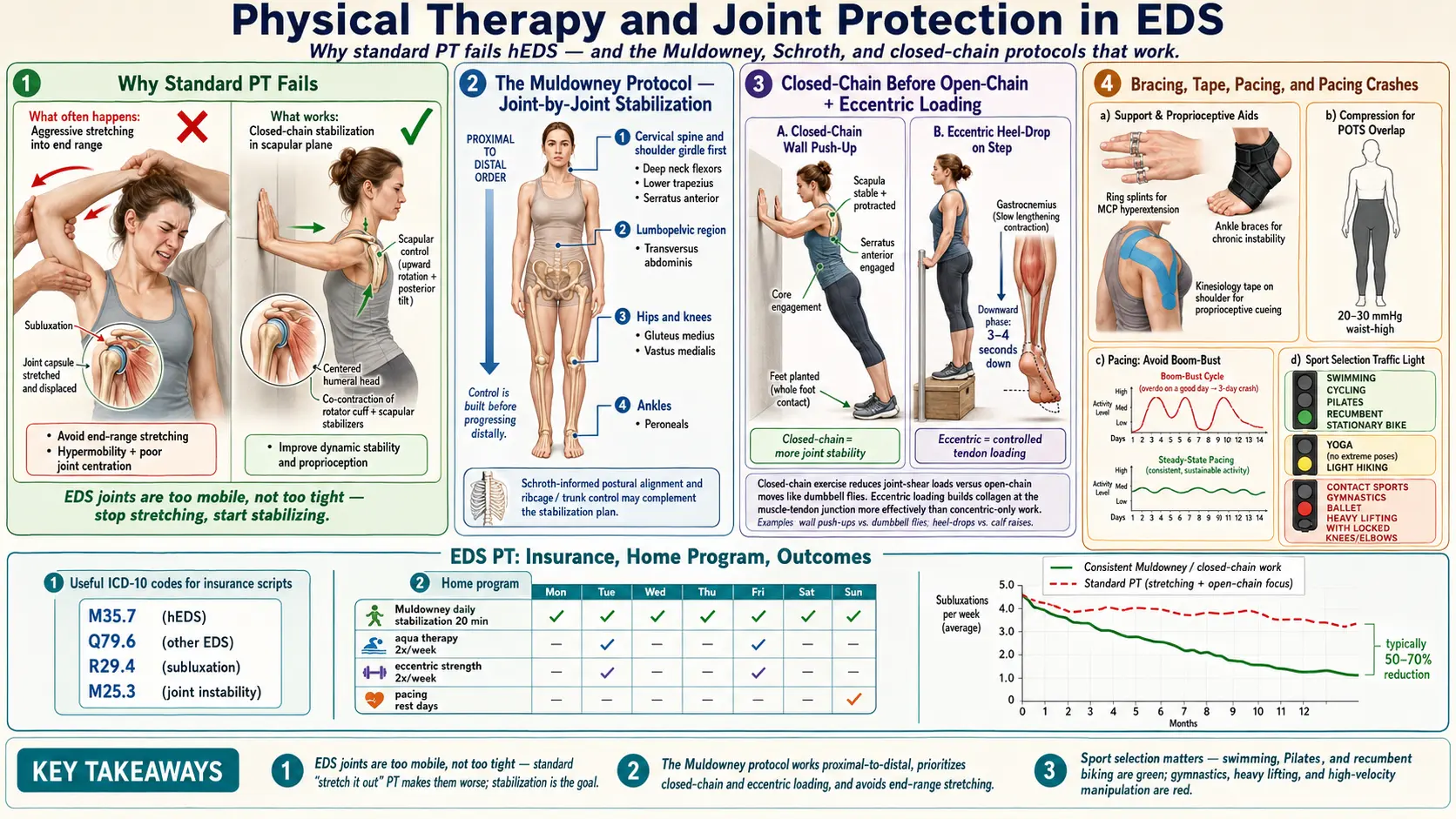
Table of Contents
- Why Standard Physical Therapy Fails Many People With Hypermobile Ehlers-Danlos Syndrome
- The Muldowney Protocol
- The Schroth Method for Kyphoscoliotic Ehlers-Danlos Syndrome
- Joint-by-Joint Stabilization Principles
- Closed-Chain Before Open-Chain
- Eccentric-Focused Loading
- Aqua Therapy
- Pilates and Reformer Work
- Bracing, Splints, and Orthotics
- Kinesiology Tape and Compression Garments
- Pacing and the Post-Activity Crash
- Treatments and Activities Practitioners Raise Concerns About
- Finding a Physical Therapist Familiar With Ehlers-Danlos Syndrome
- Insurance, ICD-10 Codes, and Scripts
- The Home Program
- Ergonomics — Desk, Sleep, and Daily Life
- Sports Grouped by Reported Joint Risk
- Return to Exercise After a Flare
- What the Research Shows About Exercise in Ehlers-Danlos Syndrome
- Key Research Papers
- Connections
- Featured Videos
Why Standard Physical Therapy Fails Many People With Hypermobile Ehlers-Danlos Syndrome
Many people with hypermobile Ehlers-Danlos syndrome (hEDS) report that a previous round of physical therapy left them worse. That report does not mean they are imagining it, weak, or failing. Generic orthopedic physical therapy is built for a population whose joints need more mobility and more stretch. In this condition they do not: ligaments and joint capsules are already lax — the problem is not range of motion, it is control of the range a person has. Pushing someone with this condition into end-range hamstring stretches, pec-minor stretches, or aggressive hip flexor lengthening is described as prying apart tissue that is already too loose. The short-term relief of a "good stretch" is described as being followed by days of subluxations, flares, and cascading pain.
The international physical therapy guideline for joint hypermobility syndrome and hypermobile Ehlers-Danlos syndrome states that many physical therapists are not familiar with the condition's diagnostic criteria, how common it is, how it usually presents, or how it is managed. It also describes the evidence on management as "limited in size and quality", so that treatment decisions rest on theory and that limited research (Engelbert 2017).
The second failure mode practitioners describe is strengthening without proprioception. A trainer prescribes squats, rows, and deadlifts; the patient's form looks off but tolerable; three weeks later the knees buckle inward, the low back spasms, or a shoulder pops out reaching for a coffee mug. The muscles grew stronger but the brain never learned where the joint actually was in space. Proprioception — joint position sense — has been measured in this group. A study of 32 women with the hypermobility type and 32 matched healthy women found significantly poorer position sense at the knee (P = 0.018) but no significant difference at the shoulder (Rombaut 2010). A 2013 review of 5 studies and 254 people with benign joint hypermobility syndrome, a closely related diagnosis, found poorer position sense in the lower limb (p < 0.001) and the finger (p < 0.001), no significant difference at the shoulder (p = 0.10), and called the upper-limb evidence unclear (Smith 2013). Strength without position sense is a loaded gun with a broken sight.
Practitioners describe a good physical therapy program for this condition as built on three premises. First, no stretching, because the tissue is already stretchy. Second, training the small stabilizing muscles that cross individual joints before adding any whole-body load. Third, retraining the brain-to-joint signal with balance and proprioceptive drills in every single session. The programs described as working — Muldowney, Levine-style (the graded training published by Fu and Levine for postural orthostatic tachycardia syndrome), and the protocols emerging from The Ehlers-Danlos Society — all share this backbone.
The Muldowney Protocol
Kevin Muldowney, a Rhode Island physical therapist, published Living Life to the Fullest with Ehlers-Danlos Syndrome with Kathleen Muldowney in 2015 after a decade of trial-and-error with patients with this condition. This page summarizes the protocol as a four-phase graded program that takes months, not weeks, and that asks for consistency over intensity. Kevin Muldowney's 2019 conference slides do not use these four phase names: they describe "a layered approach beginning with muscle activation and ending with high level exercises", split by book chapter into the low back and hips, the neck down to the hand, and the feet up to the knee, with numbered levels.
Phase 1 — Closed-chain stabilization. The patient lies on a mat or sits supported; the therapist works one joint at a time, teaching the surrounding stabilizers to fire in isolation. Hip stabilization comes first because the pelvis is the platform for everything above and below. Supine bridges with a block between the knees, clamshells with a resistance band, and single-leg stands against a wall are the early building blocks. This phase uses no barbell, no free weights, no cardio machines. Weeks, not days.
Phase 2 — Isometric hold work. Once each joint can be moved through a small controlled arc, the work shifts to holding position against gentle resistance. Isometrics (muscle contracts without joint movement) build tendon stiffness and neural drive without stressing lax capsules. Examples are wall-sits at a shallow angle, glute-bridge holds, and scapular retraction holds with a band. Holds run 10 to 30 seconds, repeated through the day.
Phase 3 — Controlled mid-range movement. Now the patient works through the middle of each joint's range, stopping well before end-range. Mid-range squats to a chair (not a deep squat), mid-range rows, mid-range step-ups. Slow eccentrics, with a pause at the bottom. This is where most patients with this condition fail when they try to skip ahead from a gym program — they go straight to full-range lifts without the stabilization base.
Phase 4 — Return to activity. Only after the earlier phases hold up does the patient reintroduce sport, hiking, dance, or recreational lifting. The point is not to avoid activity forever; it is to earn the right to do it without subluxating.
The Muldowney book is not a substitute for a therapist, but it is a realistic roadmap patients can bring to their physical therapist to set expectations. A therapist who has never heard of it is treated by some patients as a yellow flag.
The Schroth Method for Kyphoscoliotic Ehlers-Danlos Syndrome
The Schroth Method was developed in Germany in the 1920s for idiopathic scoliosis and remains the most evidence-based conservative approach for spinal curvature. For patients with kyphoscoliotic Ehlers-Danlos syndrome or hypermobile Ehlers-Danlos syndrome with secondary scoliosis, a Schroth-certified physical therapist can be transformative. The method uses three-dimensional corrective breathing, mirror-guided postural re-training, and isometric holds in a corrected posture to de-rotate and stabilize the spine. Certification is real and checkable — the Barcelona Scoliosis Physical Therapy School (BSPTS) and Schroth Best Practice keep directories. Schroth is not general physical therapy for Ehlers-Danlos syndrome; it is specific to scoliotic patterns.
Joint-by-Joint Stabilization Principles
Practitioners describe the most useful clinical habit in this condition as mapping each person's own instability. Every body is different. Instead of a generic "full-body strengthening" program, a good physical therapist walks through each quadrant and identifies which joints sublux, which pop, which ache at night, and which feel stable. Common hot spots:
- Shoulders. Often the worst offender. The glenohumeral joint is a shallow socket held in by the rotator cuff and capsule. In this condition both are lax. Programs train serratus anterior, lower trapezius, and the rotator cuff in tiny ranges with low load. Wall slides, scapular retraction against a band, and prone Y-T-W holds are staples.
- Pelvis and sacroiliac joint. Unilateral sacroiliac pain, "clunking" when rolling over in bed, a sense the hip is "out." Gluteus medius and deep core (transversus abdominis, pelvic floor) are the targets. A sacroiliac belt during flares can be a game-changer.
- Knees. Patellar subluxation, especially medial-to-lateral tracking problems. Vastus medialis oblique and gluteus medius work together to hold alignment. Terminal knee extensions with a band, step-downs, and single-leg bridges.
- Ankles. Chronic sprains, rolled ankles, weak peroneals. Single-leg balance on a firm surface, then foam, then wobble board. Towel scrunches and resisted eversion.
- Cervical spine. Craniocervical instability is a serious subgroup — practitioners describe imaging and a specialist opinion as coming before any loading of the neck. For uncomplicated cervical hypermobility, deep neck flexor training with a pressure biofeedback cuff is the foundation.
- Temporomandibular joint (the jaw joint). Jaw subluxation, clicking, and locking. A physical therapist or dentist trained in this joint can teach tongue-posture and controlled mouth-opening drills. Wide yawns, big bites, and gum are reported to aggravate it.
The rule described across every joint is the same: train the stabilizers, not the mobilizers. Proprioception is retrained with wobble boards, foam pads, half-dome balance trainers, and eyes-closed balance drills. Passive end-range stretches are left out, because they lengthen tissue that needs to stay short.
Closed-Chain Before Open-Chain
A closed-chain exercise is one in which the working hand or foot is fixed against a surface — push-ups, squats, step-ups, planks. Open-chain exercises move the limb through the air — bench press, leg extensions, dumbbell curls. Closed-chain movements co-contract the muscles on both sides of a joint, which produces joint compression and stability. Open-chain movements shear the joint. For hypermobile Ehlers-Danlos syndrome, closed-chain is the default and open-chain is earned later, if at all.
Practical translation: a wall push-up is safer than a dumbbell bench press. A wall-supported squat is safer than a leg-extension machine. A plank is safer than a sit-up. The usual order is closed-chain first, then partial open-chain with band resistance, with free weights added only when the joint has learned control.
Eccentric-Focused Loading
Eccentric contractions — the lengthening phase of a movement — are described as building tendon stiffness and connective-tissue quality better than concentric work alone. For hypermobile Ehlers-Danlos syndrome, this is held to matter because collagen is the problem; the aim is to give the tendons every possible signal to remodel toward stiffness rather than slack. Programs cue a 3- to 4-second lowering phase on every rep: lowering slowly out of a squat, from a wall push-up, on a step-down. Concentric (lifting) phases stay in the 1- to 2-second range. In people with Achilles and patellar tendon injuries (not this condition), a 2013 systematic review found limited (Achilles) and conflicting (patellar) evidence that clinical outcomes are better with eccentric loading than with other loading programs, and greater evidence for heavy slow resistance in the patellar tendon (Malliaras 2013).
Aqua Therapy
Warm-water therapy pools (typically 90 to 94 F) are among the single best environments for exercise in this condition. Buoyancy offloads gravity — a neck-deep patient bears roughly 10% of their body weight — which allows movement without compressing painful joints. The warm water soothes mast cells in patients with the postural orthostatic tachycardia syndrome (POTS)/mast cell activation syndrome (MCAS) triad, and water resistance is gentle and three-dimensional, matching the drag to the speed of movement. Walking forward, backward, and sideways in chest-deep water; gentle flutter kicks holding the pool edge; and light arm sweeps against the water are common starting points. Very cold pools are reported to trigger muscle guarding and postural orthostatic tachycardia syndrome symptoms. Therapy pools are often found at local hospitals and community recreation centers; Medicare often covers aqua therapy when prescribed as physical therapy.
Pilates and Reformer Work
Pilates — particularly reformer-based Pilates with an instructor trained in hypermobility — is one of the few movement disciplines that naturally matches the needs of this condition. The reformer provides spring resistance that is graduated and controllable, supports the spine in a range of positions, and encourages small-muscle control before whole-body movement. Much depends on the instructor. A general Pilates teacher may cue "lengthen, lengthen, lengthen" and push a client into end-range positions that wreck a lax spine. Patients describe looking for instructors with formal Pilates certification who advertise hypermobility experience.
For patients with craniocervical instability or suspected craniocervical instability, practitioners raise concerns about aggressive spinal flexion movements (roll-ups, neck curls, chest-lifts) and describe involving a physical therapist guided by a neurosurgeon before any loaded spinal flexion.
Bracing, Splints, and Orthotics
Bracing is controversial in this condition because prolonged passive support weakens the muscles around the joint. The usual frame is situational and time-limited. Bracing is described as appropriate:
- During an acute flare or subluxation, to restore proprioceptive input and offload pain.
- During specific high-demand tasks (long walks, travel, carrying groceries) in patients whose joints still fail with those loads.
- As a training wheel while physical therapy progresses, with an explicit plan to wean.
Bracing is not described as a permanent substitute for stabilization training. Specific options patients find useful:
- Custom ankle braces (lace-up or articulated) for chronic lateral instability.
- Sacroiliac belts for sacroiliac flares — worn during activity, removed at rest.
- Cervical collars (soft foam or Philadelphia) for neck flares only — long-term daily wear weakens deep neck flexors and can worsen instability.
- Finger ring splints — inexpensive plastic figure-eight splints (often used for trialing) and custom-sized, jewelry-grade, durable silver ring splints. They prevent hyperextension at the middle and end finger joints and are described as the single highest-value intervention for hand-dominant work such as typing, writing, and cooking.
- Custom foot orthotics for patients with pes planus, over-pronation, or posterior tibial tendon strain.
Kinesiology Tape and Compression Garments
Kinesiology tape does not structurally hold a joint in place — it provides sensory feedback. For people with this condition and impaired proprioception, that tactile input can be enough to help the nervous system recruit the right stabilizer at the right moment. Patellar tracking tape, shoulder-posture tape, and sacroiliac-joint tape are common applications. Typical wear is 2 to 4 days per application.
Compression garments serve double duty. For patients with co-morbid postural orthostatic tachycardia syndrome, medical-grade compression (20 to 30 mmHg) reduces venous pooling; see the postural orthostatic tachycardia syndrome compression and exercise program for specifics. For hypermobile Ehlers-Danlos syndrome, the same garment provides continuous proprioceptive input to the skin and soft tissues, subtly improving joint position sense. Patients use medical-grade compression garments and, for lighter-duty daytime wear, lighter athletic compression clothing. Waist-high or thigh-high is more effective than knee-high for postural orthostatic tachycardia syndrome support. Abdominal binders add splanchnic compression for patients with significant gastrointestinal symptoms.
Pacing and the Post-Activity Crash
Many people with this condition have overlapping postural orthostatic tachycardia syndrome, myalgic encephalomyelitis/chronic fatigue syndrome-like fatigue, or fibromyalgia, and will flare for days after a single overzealous workout. The approach described is graded, heart-rate-aware pacing — the same framework detailed in the fibromyalgia exercise pacing article and the postural orthostatic tachycardia syndrome exercise program. Short, frequent sessions beat one long workout. People describe stopping while they still have gas in the tank, tracking heart rate, and capping intensity below the level that triggers post-exertional malaise.
Treatments and Activities Practitioners Raise Concerns About
- Chiropractic high-velocity low-amplitude (HVLA) manipulation. The thrust techniques chiropractors use on a stable neck or back can cause frank dislocations and vertebral artery injury in this condition. Soft tissue work, instrument-assisted techniques, and gentle mobilization are described as possibly acceptable with a practitioner who knows Ehlers-Danlos syndrome — but practitioners describe thrusts, especially cervical, as excluded altogether.
- Aggressive or contortion-style yoga. Going deeper into a pose is the wrong direction. Yoga for this condition is described as working with an instructor who actively cues people to shorten instead of lengthen, staying well shy of end range, and skipping inversions when craniocervical instability is a concern.
- End-range ballet, gymnastics, or cheerleading past adolescence without medical supervision. Many people with this condition were praised for their flexibility as children; continuing those disciplines into adulthood accelerates joint damage.
- Acupuncture directly into lax joint capsules is an uncommon but real source of bleeding or joint aggravation. Acupuncture in muscle bellies is generally considered fine; needling near a joint line is where the concern lies.
- Competitive powerlifting and contact sports carry a high risk-to-reward ratio for these joints. Strength work in this condition is described as following the Muldowney approach, not the one-rep-max approach.
Finding a Physical Therapist Familiar With Ehlers-Danlos Syndrome
The therapist is described as the single biggest determinant of success in physical therapy for this condition. Ways people find one:
- The Ehlers-Danlos Society Healthcare Professionals Directory (ehlers-danlos.com) — searchable by specialty and country.
- Pain-medicine and physical therapy practices that specialize in Ehlers-Danlos syndrome, and the referral networks around them.
- Patient groups on social media for local recommendations.
- A phone interview with a candidate therapist before the first appointment. Questions people ask: "Have you treated hypermobile Ehlers-Danlos syndrome before? Are you familiar with the Muldowney protocol? Do you use end-range stretching in your programs?" The answer patients look for to the last is "rarely, and not for this population."
Insurance, ICD-10 Codes, and Scripts
Insurance often caps physical therapy visits per year (20 to 30 is common), so getting the prescription approved on the first pass matters. Relevant ICD-10 codes:
- Q79.6 — Ehlers-Danlos syndromes (a category heading since the update below, not itself a billable code).
- Q79.60 — Ehlers-Danlos syndrome, unspecified (added with the ICD-10-CM 2020 update, effective October 1, 2019).
- Q79.62 — hypermobile Ehlers-Danlos syndrome (added with the same update).
- M35.7 — Hypermobility syndrome.
- M25.3 — Other instability of joint (useful as a secondary code).
- M62.81 — Muscle weakness, generalized.
- R26.81 — Unsteadiness on feet (for balance/proprioception work).
Prescriptions often include the specific joint codes that are flaring (e.g., M25.312 for left shoulder instability) plus a goal statement like "proprioceptive retraining and stabilization, joint protection education." Pure "chronic pain" codes trigger denials more often than specific instability codes. When visits run out, some people turn to cash-pay packages with a physical therapist who knows Ehlers-Danlos syndrome — often cheaper per hour than insurance co-pays once deductibles are counted.
The Home Program
The home program is where recovery actually happens. Clinic visits teach technique; the work between visits creates adaptation. What programs typically lay out:
- 20 to 30 minutes per day, scheduled like medication. A 90-minute Saturday session does not replace six 15-minute weekday sessions.
- Priorities, in order: core (transversus abdominis, pelvic floor, deep spinal stabilizers); gluteals (medius, maximus); scapular stabilizers (lower trap, serratus, rhomboids). These three complexes anchor everything else.
- Bed-based isometrics on flare days. Programs scale down on bad days rather than skip them. Glute squeezes, quad sets, supine diaphragmatic breathing, and gentle scapular retractions can all be done flat on the back.
- Session logs. A simple checkbox calendar is enough. Patterns — "my shoulders always feel worst after a gap of more than two days" — emerge within weeks.
- Slow progression. When an exercise goes well for 10 to 14 days, programs add one variable (reps, hold time, or a slightly harder variation) — not three variables at once.
Ergonomics — Desk, Sleep, and Daily Life
Exercise is two percent of the day. The other 98 percent decides whether the joints survive. Ergonomic changes described for this condition:
- Chair with full arm support so the shoulders can let go of the keyboard. Armrests at elbow height, not an inch below.
- Standing desk with an anti-fatigue mat. Alternating sit and stand every 30 to 45 minutes. Knees kept unlocked when standing — micro-bends protect hypermobile knees.
- External keyboard and monitor raised to eye level. Laptop-on-lap hunching is described as crushing the cervical spine.
- Sleep setup. A medium-firm mattress; a pillow that fills the gap between shoulder and ear without tipping the head; a pillow between the knees for side-sleepers; a small lumbar roll or pillow under the low back for back-sleepers. Stomach-sleeping is described as hyper-rotating the neck.
- Kitchen and bathroom. Lever door handles, bigger-grip utensils, electric can opener, jar openers. Anything that reduces thumb and wrist strain on a lax thumb-base joint is described as worth the money.
- Driving. Seat upright, lumbar support, ten-and-two grip (not white-knuckle), breaks every 60 to 90 minutes.
Sports Grouped by Reported Joint Risk
Generally described as safe. Swimming (especially backstroke and sidestroke, without aggressive butterfly). Stationary cycling and recumbent elliptical. Walking, including Nordic walking with poles. Tai Chi and Qigong. Hatha yoga with a hypermobility-aware teacher. Aqua therapy.
Described as needing caution. Moderate-load weight training with strict form and a trainer who understands hypermobility. Hiking on smooth terrain with trekking poles. Recreational dance with no deep back-bends or aggressive turns. Road cycling on good roads (wrist and neck strain on drop bars is the concern).
Described as carrying the highest risk, and often given up or strongly limited. Contact sports (football, rugby, hockey, martial arts with throws). Gymnastics, cheerleading, and ballet past adolescence without medical clearance. Competitive powerlifting. High-rep Olympic lifting of the kind used in group fitness competitions. Trampolines. Running long distances on hard surfaces (short, soft-surface runs may be tolerable).
Return to Exercise After a Flare
When a flare resolves, the temptation is to pick up where one left off. Practitioners describe instead restarting at roughly 25% of pre-flare volume and intensity and rebuilding over two to three weeks. A four-day flare can cost a week of deconditioning; pushing back to 100% on day one triggers the next flare within 72 hours. This is described as the single most common self-sabotage pattern in rehabilitation for this condition, and people who use the 25% rule commit to it before the flare starts.
What the Research Shows About Exercise in Ehlers-Danlos Syndrome
Most of this page rests on practitioners' experience, because the research is still small. A 2022 systematic review of exercise and rehabilitation in adults with Ehlers-Danlos syndrome and hypermobility spectrum disorders found 10 studies with 330 participants, 5 of them randomized controlled trials. All 10 showed some improvement in a physical or psychological outcome after the program. One side effect, not serious, was possibly linked to the exercise. Only 2 of the 5 randomized trials were rated high quality with a low risk of bias, and most of the other studies were rated at critical risk of bias. The authors conclude that exercise and rehabilitation may be beneficial and that properly sized, rigorous trials are needed (Buryk-Iggers 2022).
Key Research Papers
- Rombaut L, De Paepe A, Malfait F, Cools A, Calders P. Joint position sense and vibratory perception sense in patients with Ehlers-Danlos syndrome type III (hypermobility type). Clin Rheumatol. 2010;29(3):289-95. — PubMed PMID: 19937459
- Palmer S, Bailey S, Barker L, Barney L, Elliott A. The effectiveness of therapeutic exercise for joint hypermobility syndrome: a systematic review. Physiotherapy. 2014;100(3):220-227. — doi:10.1016/j.physio.2013.09.002
- Castori M. Ehlers-Danlos syndrome, hypermobility type: an underdiagnosed hereditary connective tissue disorder with mucocutaneous, articular, and systemic manifestations. ISRN Dermatol. 2012;2012:751768. — doi:10.5402/2012/751768
- Keer R, Simmonds J. Joint protection and physical rehabilitation of the adult with hypermobility syndrome. Curr Opin Rheumatol. 2011. — doi:10.1097/bor.0b013e328342d3af
- Smith TO, Jerman E, Easton V, Bacon H, Armon K, Poland F, Macgregor AJ. Do people with benign joint hypermobility syndrome (BJHS) have reduced joint proprioception? A systematic review and meta-analysis. Rheumatol Int. 2013;33(11):2709-16. — PubMed PMID: 23728275
- Malliaras P, Barton CJ, Reeves ND, Langberg H. Achilles and patellar tendinopathy loading programmes: a systematic review comparing clinical outcomes and identifying potential mechanisms for effectiveness. Sports Med. 2013;43(4):267-86. — PubMed PMID: 23494258
- Engelbert RH, Juul-Kristensen B, Pacey V, de Wandele I, Smeenk S, Woinarosky N, Sabo S, Scheper MC, Russek L, Simmonds JV. The evidence-based rationale for physical therapy treatment of children, adolescents, and adults diagnosed with joint hypermobility syndrome/hypermobile Ehlers Danlos syndrome. Am J Med Genet C Semin Med Genet. 2017;175(1):158-167. — PubMed PMID: 28306230
- Buryk-Iggers S, Mittal N, Santa Mina D, Adams SC, Englesakis M, Rachinsky M, Lopez-Hernandez L, Hussey L, McGillis L, McLean L, Laflamme C, Rozenberg D, Clarke H. Exercise and rehabilitation in people with Ehlers-Danlos syndrome: a systematic review. Arch Rehabil Res Clin Transl. 2022;4(2):100189. — PubMed PMID: 35756986
PubMed Topic Searches
For further reading, the following PubMed topic searches return current peer-reviewed work on rehabilitation and joint protection in Ehlers-Danlos Syndrome:
- PubMed: Ehlers-Danlos and physical therapy
- PubMed: Hypermobility, proprioception, and rehabilitation
- PubMed: Hypermobility and closed-chain exercise
- PubMed: Ehlers-Danlos and aquatic therapy
- PubMed: Schroth method and kyphoscoliotic curves
- PubMed: Hypermobility, bracing, and splinting
- PubMed: Ehlers-Danlos and craniocervical instability
- PubMed: Eccentric exercise and tendon collagen remodeling
Connections
- Rheumatology
- Ehlers-Danlos Syndrome
- Hypermobile Ehlers-Danlos Syndrome and 2017 Diagnostic Criteria
- Classical and Vascular Ehlers-Danlos Syndrome
- Craniocervical Instability and AAI
- Gastrointestinal Involvement in Ehlers-Danlos Syndrome
- Postural Orthostatic Tachycardia Syndrome, Mast Cell Activation Syndrome and the Ehlers-Danlos Syndrome Triad
- Pain Management in hEDS
- Pediatric Ehlers-Danlos Syndrome and Transition of Care
- Pregnancy and Ehlers-Danlos Syndrome
- Postural Orthostatic Tachycardia Syndrome
- Postural Orthostatic Tachycardia Syndrome Compression and Exercise Program
- Fibromyalgia
- Exercise Pacing and Graded Movement
- Osteoporosis
- Arthritis
- Collagen
- Stress Management