Adult-onset Still's Disease
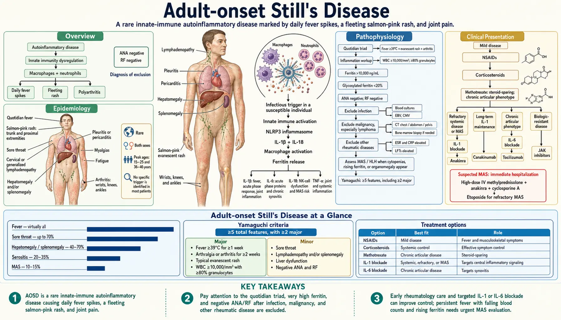
Adult-onset Still's Disease (AOSD) is a rare autoinflammatory condition defined by its dramatic daily fever spikes, a salmon-pink fleeting rash, and joint pain. Because it mimics infection and cancer so closely, reaching a diagnosis is a process of careful exclusion — ruling out other culprits before the label fits. The disease belongs to the innate immune system gone awry, not the antibody-driven autoimmune diseases most people have heard of, which is why the usual markers like antinuclear antibody (ANA) and rheumatoid factor come back negative. With modern biologic therapies targeting the exact inflammatory messengers that drive it, outcomes have improved enormously.
- Overview
- Epidemiology
- Pathophysiology
- Clinical Presentation
- Diagnosis
- Treatment
- Disease Course Patterns
- Macrophage Activation Syndrome (MAS)
- Prognosis
- Key Research Papers
- Connections
Overview
Adult-onset Still's Disease is the adult counterpart of systemic juvenile idiopathic arthritis (sJIA), sharing the same fundamental biology but appearing after age 16. The condition was first described in children by Sir George Frederic Still in 1897 and later recognized in adults by Eric Bywaters in the 1970s. It belongs to the family of autoinflammatory diseases — conditions driven by dysregulation of the innate immune system rather than by autoantibodies, which is why both antinuclear antibody (ANA) and rheumatoid factor (RF) tests are typically negative.
The clinical hallmark is the quotidian triad: daily fever spikes reaching at least 39°C (102.2°F), a transient salmon-pink evanescent rash that appears during fever peaks and fades within hours, and polyarthritis. Patients are often remarkably well between fever spikes, which can mislead clinicians into dismissing the severity of the illness.
Peak incidence occurs in a bimodal age distribution of 15–25 years and 36–46 years, though AOSD can appear at any adult age. Because no single test confirms it, AOSD remains a diagnosis of exclusion — infection, malignancy (especially lymphoma), and other connective tissue diseases must all be ruled out before the diagnosis is made. Serum ferritin is a useful clue: levels above 10,000 ng/mL are found in roughly 70% of active AOSD cases and, while not specific, are a powerful prompt to consider the diagnosis.
Epidemiology
AOSD is genuinely rare. Population-based studies estimate an annual incidence of 0.16 to 0.4 per 100,000 persons, with a prevalence of roughly 1 per 100,000. Both sexes are affected, with a modest female predominance (approximately 60% female across series, though some cohorts show near-equal sex ratios).
The bimodal age distribution — peaking at 15–25 years and again at 36–46 years — has been confirmed across multiple ethnic populations in Europe, Asia, and North America. The reason for two peaks is not fully understood but may reflect distinct immunological windows of susceptibility.
Human leukocyte antigen (HLA) associations have been inconsistent across studies. Human leukocyte antigen-B17, B18, B35, and DR2 have been reported in some series, but no single human leukocyte antigen allele is diagnostic or required. This contrasts with strongly human leukocyte antigen-linked rheumatic diseases such as ankylosing spondylitis (human leukocyte antigen-B27) or rheumatoid arthritis (HLA-DR4).
Infectious triggers are suspected in a subset of patients. Case reports and small series link AOSD onset to:
- Epstein–Barr virus (Epstein-Barr virus, EBV)
- Cytomegalovirus (CMV)
- Rubella virus and live rubella vaccine
- Influenza and parainfluenza viruses
- Yersinia enterocolitica
- Mycoplasma pneumoniae
The prevailing hypothesis is that an infectious trigger activates innate immune pathways in genetically susceptible individuals, initiating a self-perpetuating inflammatory cycle. However, in most patients no specific trigger is ever identified.
Pathophysiology
AOSD is fundamentally an autoinflammatory disease — a disorder of innate immunity — rather than a classic autoimmune disease driven by autoreactive T cells or autoantibodies. This distinction matters practically: seronegative results (negative antinuclear antibody, negative RF) are expected and do not argue against the diagnosis.
Central Cytokines
Macrophages and neutrophils are the primary effector cells. Key inflammatory mediators include:
- IL-18: The defining cytokine of AOSD. Levels are extraordinarily elevated — 10 to 100 times above normal — and correlate with disease activity. IL-18 drives natural killer (NK) cell activation; when natural killer cell function is impaired or overwhelmed, unchecked macrophage activation ensues, setting the stage for macrophage activation syndrome (MAS).
- Interleukin-1 beta (IL-1β): Produced through NLRP3 inflammasome activation; drives fever, acute phase response, and joint inflammation. The dramatic clinical responses seen with interleukin-1 (IL-1) blockade confirm its central role.
- Interleukin-6 (IL-6): Amplifies the inflammatory cascade, drives hepatic acute phase protein production (ferritin, C-reactive protein, CRP), and contributes to the chronic arthritis phenotype.
- Tumor necrosis factor alpha (TNF-α): Contributes to joint inflammation and systemic inflammation, though tumor necrosis factor (TNF) blockade is less effective in AOSD than interleukin-1 or interleukin-6 blockade.
Inflammasome and Ferritin
The NLRP3 inflammasome is constitutively activated in AOSD macrophages, generating mature interleukin-1 beta and IL-18. Activated macrophages also release massive quantities of ferritin — the iron-storage protein — as a direct acute phase reactant. Ferritin levels above 10,000 ng/mL are found in approximately 70% of active AOSD patients and are considered a pathognomonic clue in the right clinical context (though similar elevations occur in sepsis, HLH, and certain lymphomas).
A particularly useful refinement is the glycosylated ferritin fraction: normally 50–80% of circulating ferritin is glycosylated by the liver, but in AOSD the liver cannot glycosylate the massively overproduced ferritin rapidly enough, so the glycosylated fraction falls below 20%. Combined with a ferritin level five times normal, a glycosylated ferritin of 20% or less had a sensitivity of 43% and specificity of 93% for AOSD, and combined with any ferritin above normal, 70.5% and 83.2% (Fautrel 2001) — most useful when the result is low, supporting the diagnosis.
Macrophage Polarization and MAS Risk
M1 macrophage polarization dominates the active disease state. The failure of IL-18 to properly activate natural killer cells — combined with impaired perforin-mediated cytotoxicity — prevents normal shutdown of macrophage activation, creating a feedback loop. When this loop escapes regulatory control, macrophage activation syndrome (MAS) occurs: a cytokine storm with hemophagocytosis, pancytopenia, and life-threatening organ dysfunction.
Clinical Presentation
AOSD typically announces itself with a cluster of dramatic but seemingly unconnected symptoms. Patients and their families often spend weeks to months seeking a diagnosis before a rheumatologist recognizes the pattern.
The Quotidian Fever
Fever is present in virtually all patients and is the most reliable feature. It is quotidian (once-daily, sometimes twice-daily), spiking to at least 39°C (102.2°F), usually in the late afternoon or evening. Critically, patients often feel relatively well — even normal — between spikes. A patient who is febrile to 40°C one hour and afebrile the next should raise immediate suspicion for AOSD.
The Evanescent Rash
The rash is salmon-pink, macular or maculopapular, and evanescent — appearing during fever peaks and resolving within hours as the fever breaks. It typically appears on the trunk, proximal extremities, and occasionally the face. Because it fades so quickly, patients (and physicians) may never witness it during a clinic visit; asking the patient to photograph the rash during a fever spike at home is diagnostically valuable. Koebner phenomenon (rash provoked by pressure or scratching) is present in some patients.
Arthritis
Arthritis or arthralgia is present in the vast majority of patients. The most commonly affected joints are the wrists, knees, and ankles, followed by elbows, shoulders, hips, and small joints of the hands and feet. Early in the course the arthritis may be relatively mild and overlooked in the face of dramatic systemic features, but in the chronic articular disease phenotype it can become erosive and debilitating.
Additional Features (Yamaguchi Minor Criteria)
- Sore throat: Present in up to 70% of patients at onset; may be due to pharyngeal lymphoid activation rather than infection.
- Lymphadenopathy: Generalized or cervical, sometimes striking enough to prompt lymphoma workup.
- Hepatomegaly and/or splenomegaly: Occur in 40–70% of cases; transaminase elevation accompanies hepatomegaly.
- Abnormal liver function tests (LFTs): Transaminase elevations are common; rarely, severe liver involvement can cause acute liver failure, especially with MAS.
- Seronegative serology: Negative antinuclear antibody and RF are minor criteria precisely because their negativity supports AOSD over typical autoimmune disease.
Other Manifestations
Serositis — pleuritis or pericarditis — occurs in 20–35% of patients and can cause chest pain, dyspnea, or a friction rub. Myalgias are common during fever spikes. Weight loss and profound fatigue affect quality of life significantly during active disease phases.
Diagnosis
No single test confirms AOSD. Diagnosis relies on fulfilling clinical criteria after an active exclusion process.
Yamaguchi Criteria (1992)
The most widely used classification criteria require 5 or more features, with at least 2 being major criteria. Sensitivity is 96.2%; specificity is 92.1% (Yamaguchi 1992).
Major criteria:
- Fever ≥39°C, quotidian, lasting ≥1 week
- Arthralgia or arthritis lasting ≥2 weeks
- Typical rash (salmon-colored, evanescent, on trunk/extremities)
- WBC ≥10,000/mm³ with ≥80% granulocytes
Minor criteria:
- Sore throat
- Lymphadenopathy and/or splenomegaly
- Liver dysfunction (elevated transaminases or lactate dehydrogenase, LDH)
- Negative antinuclear antibody and RF
Exclusion criteria (must be absent): infections (especially sepsis, Epstein-Barr virus), malignancies (especially lymphoma), and other rheumatic diseases.
Fautrel Criteria (2002)
An alternative set requiring either 2 major criteria or 1 major + 2 minor criteria, with glycosylated ferritin <20% as a major criterion. These criteria perform comparably and are preferred in some centers.
Laboratory Investigation
- Serum ferritin: The most important single test. Levels >10,000 ng/mL occur in ~70% of active AOSD; glycosylated ferritin <20% has ~93% specificity.
- Complete blood count (CBC): Leukocytosis (WBC ≥10,000) with marked neutrophilia (often >80%) is characteristic. Anemia of chronic inflammation is common.
- Erythrocyte sedimentation rate (ESR), C-reactive protein: Markedly elevated in nearly all active cases.
- LFTs: Transaminase elevation in 50–75% of patients.
- ANA, RF: Should be negative (or only weakly positive) by definition.
Exclusion Workup
Before accepting the AOSD label, the following must be evaluated:
- Infection: Blood cultures (×3), throat culture, serologies for Epstein-Barr virus, cytomegalovirus, parvovirus B19, rubella, hepatitis B and C, human immunodeficiency virus (HIV), Yersinia, Mycoplasma.
- Malignancy: Lymphoma can mimic AOSD with high ferritin and lymphadenopathy — computed tomography (CT) scan of chest/abdomen/pelvis, positron emission tomography/computed tomography (PET-CT) if concern persists; bone marrow biopsy if needed.
- Other rheumatic diseases: systemic lupus erythematosus (SLE) (anti-dsDNA, complement), systemic vasculitis, adult-onset sJIA overlap.
- MAS/HLH: In patients with cytopenias, rising ferritin, and organomegaly, HLH criteria must be assessed; bone marrow biopsy demonstrating hemophagocytosis confirms concurrent MAS.
Treatment
Treatment follows a step-up approach based on disease severity, response, and the presence of life-threatening complications.
Step 1 — Nonsteroidal Anti-Inflammatory Drugs
Nonsteroidal anti-inflammatory drugs (ibuprofen 600–800 mg three times daily, naproxen, or indomethacin) are the initial treatment for mild disease. They can control fever and musculoskeletal symptoms in approximately 20% of patients — those with milder disease or monocyclic courses. They have no impact on the underlying inflammatory cascade and do not prevent MAS.
Step 2 — Corticosteroids
Prednisone (0.5–1 mg/kg/day, typically 40–60 mg/day in adults) controls symptoms effectively but carries long-term toxicity with chronic use. Most patients require corticosteroids at some point, and corticosteroid tapering frequently triggers flares. This reliance on steroids is one of the strongest arguments for early biologic initiation in moderate-to-severe disease.
Step 3 — Conventional DMARDs
Methotrexate (10–25 mg/week subcutaneously or orally) is the most commonly used steroid-sparing agent; it is particularly effective for the chronic articular phenotype. Hydroxychloroquine is sometimes added for milder cases. Cyclosporine plays a specific role in MAS management.
Step 4 — Biologics (Refractory Disease)
Interleukin-1 blockade is the cornerstone of biologic therapy for AOSD:
- Anakinra (Kineret): 100 mg daily subcutaneously; the fastest-acting interleukin-1 receptor antagonist. Produces dramatic responses within days in many patients. Drug of choice for refractory disease, MAS, and severe systemic presentations. Used off-label for AOSD.
- Canakinumab (Ilaris): interleukin-1 beta monoclonal antibody, 150–300 mg every 4–8 weeks subcutaneously; approved for sJIA, used off-label in AOSD. More convenient dosing than anakinra for long-term maintenance.
- Rilonacept: interleukin-1 trap; used in some refractory cases.
Interleukin-6 blockade with tocilizumab (Actemra, 8 mg/kg intravenous (IV) every 4 weeks) is particularly effective for patients with the chronic articular phenotype where interleukin-6 drives synovitis. Less effective for the acute systemic phenotype compared to interleukin-1 blockade.
JAK inhibitors (baricitinib, tofacitinib) are emerging as options for refractory or biologic-resistant AOSD, especially in the chronic articular phenotype, based on case series and small open-label studies.
MAS-Specific Treatment
Macrophage activation syndrome requires immediate, aggressive intervention: high-dose intravenous methylprednisolone (500–1000 mg/day for 3 days), intravenous anakinra escalated to 2–4 mg/kg/day or higher doses, and cyclosporine A (3–5 mg/kg/day). Etoposide is reserved for refractory MAS not responding to the above.
Investigational Therapies
Tadekinig alfa (recombinant IL-18 binding protein) directly neutralizes the disease-defining cytokine and showed early signs of efficacy in an open-label phase II trial in active AOSD whose primary endpoint was safety (Gabay 2018), potentially becoming the most pathophysiologically targeted treatment available.
Disease Course Patterns
AOSD does not follow a single predictable trajectory. Three main patterns have been described, and the course may not be apparent until 6–12 months after onset:
Monocyclic (20–30%)
A single episode lasting weeks to months — sometimes up to 1–2 years — followed by complete remission without recurrence. This is the best-case outcome. Patients in this group often do well with nonsteroidal anti-inflammatory drugs (NSAIDs) or a short course of corticosteroids alone.
Polycyclic / Intermittent (20–30%)
Recurring flares separated by periods of complete clinical and serological remission. Each flare can be triggered by minor infections, stress, or may occur without identifiable cause. Patients may require nonsteroidal anti-inflammatory drugs or short corticosteroid courses during each episode; some are managed with interleukin-1 blockade during flares only.
Chronic Articular (40–50%)
Persistent, ongoing inflammatory arthritis that continues after the systemic features (fever, rash) resolve or diminish. This is the most disabling course and accounts for most long-term joint damage and disability. These patients typically require continuous DMARD or biologic therapy. Wrist and hip involvement are particularly associated with radiographic damage over time. Predictors of chronic course include polyarthritis at onset, prolonged fever before treatment, and delay in diagnosis.
MAS Risk Across All Phenotypes
Macrophage activation syndrome can complicate any disease phenotype and any treatment phase. It is not restricted to the acute systemic presentation. Vigilance for early MAS signals — falling platelet count or hemoglobin in a patient with rising ferritin and persisting fever — is essential throughout the disease course.
Macrophage Activation Syndrome (MAS)
MAS is the most feared complication of AOSD, occurring in 10–15% of patients and carrying a mortality of 8–20% even with treatment. It represents a form of secondary hemophagocytic lymphohistiocytosis (HLH) — a cytokine storm in which macrophages engulf red blood cells, white blood cells, and platelets within bone marrow and lymphoid organs.
Clinical Features
MAS should be suspected when an AOSD patient develops:
- Persisting or worsening fever despite treatment
- Rapidly rising serum ferritin (often exceeding 50,000–100,000 ng/mL)
- Falling platelet count, hemoglobin, and/or WBC (cytopenias in a disease usually associated with leukocytosis — a paradoxically falling WBC is a red flag)
- Enlarging splenomegaly
- Elevated D-dimer and fibrin degradation products; falling fibrinogen (consumptive coagulopathy)
- Rising lactate dehydrogenase and transaminases
Diagnostic Criteria
MAS in AOSD can be diagnosed using either the HLH-2004 criteria (≥5 of 8 features including hemophagocytosis on bone marrow biopsy) or the 2016 MAS-in-sJIA criteria (validated in systemic JIA and applicable to AOSD). A ferritin above 684 ng/mL plus any 2 of: thrombocytopenia, elevated AST, elevated triglycerides, or low fibrinogen, is highly sensitive for MAS.
Pathogenesis
IL-18 drives natural killer cell dysfunction. Normally, natural killer cells kill virus-infected cells and activated macrophages via perforin/granzyme pathways, providing a brake on macrophage activation. In MAS, perforin-mediated cytotoxicity is impaired (either genetically predisposed or functionally overwhelmed by IL-18), allowing macrophages to proliferate unchecked. The resulting cytokine storm — with interleukin-6, IL-1β, IFN-γ, and tumor necrosis factor alpha all amplified — causes the multi-organ dysfunction seen clinically.
Treatment
MAS requires immediate hospitalization and aggressive intervention:
- High-dose intravenous methylprednisolone: 500–1000 mg/day for 3 days, followed by oral prednisone.
- Anakinra: Escalated to 2–10 mg/kg/day intravenous or SC; IL-1 blockade directly addresses the upstream driver. Some centers use anakinra at very high doses (up to 10 mg/kg/day) for life-threatening MAS.
- Cyclosporine A: 3–5 mg/kg/day; inhibits T cell activation and dampens IFN-γ production; has a long track record in pediatric HLH/MAS.
- Etoposide: Reserved for truly refractory MAS; cytotoxic agent that eliminates activated macrophages and T cells. Reserved for critical situations due to toxicity.
- Tadekinig alfa (IL-18 blood pressure, BP): Investigational; directly neutralizes IL-18; growing evidence base for MAS complicating AOSD and sJIA.
Monitoring during MAS treatment: daily ferritin, complete blood count, fibrinogen, LFTs, and D-dimer. Normalization of ferritin and rising platelet count are the earliest indicators of treatment response.
Prognosis
The overall prognosis for AOSD has improved substantially in the biologic era. Key prognostic points:
- Monocyclic course: Approximately one-third of patients experience a single self-limited episode and achieve durable remission off all therapy.
- Chronic articular disease: The dominant long-term burden. Persistent synovitis can lead to radiographic joint destruction, particularly at the wrists (carpal ankylosis is a classic AOSD complication) and hips. Interleukin-6 blockade and MTX have substantially reduced this risk.
- MAS: The primary acute mortality risk. Mortality has declined from historical rates of 20–30% to current rates of 8–20% with modern management, though it remains the most dangerous complication. Survivors may have recurrent MAS episodes.
- Biologic therapies: The availability of anakinra and canakinumab has transformed the outlook for refractory AOSD — many patients who would previously have required permanent high-dose corticosteroids achieve steroid-free remission.
- Cancer risk: Unlike rheumatoid arthritis (which carries an elevated lymphoma risk), AOSD has not been shown to increase malignancy risk in long-term follow-up studies.
- Quality of life: Active disease substantially impairs quality of life — fatigue, missed work, and the psychological burden of an uncertain diagnosis phase are significant. Early recognition and targeted therapy minimize this burden.
Predictors of worse long-term outcome include onset with polyarthritis, delay to diagnosis, hip involvement, and development of MAS. Conversely, monocyclic onset and rapid serological response to interleukin-1 blockade predict favorable outcomes.
Key Research Papers
- Yamaguchi M, Ohta A, Tsunematsu T, et al. (1992). Preliminary criteria for classification of adult Still's disease. The Journal of Rheumatology — PubMed PMID: 1578458
- Fautrel B, Zing E, Golmard JL, et al. (2002). Proposal for a new set of classification criteria for adult-onset still disease. Medicine — PubMed PMID: 11997716
- Fautrel B, Le Moël G, Saint-Marcoux B, et al. (2001). Diagnostic value of ferritin and glycosylated ferritin in adult onset Still's disease. The Journal of Rheumatology — PubMed PMID: 11246670
- Fautrel B, Mitrovic S, De Matteis A, et al. (2024). EULAR/PReS recommendations for the diagnosis and management of Still's disease, comprising systemic juvenile idiopathic arthritis and adult-onset Still's disease. Annals of the Rheumatic Diseases — PubMed PMID: 39317417
- Nordström D, Knight A, Luukkainen R, et al. (2012). Beneficial effect of interleukin 1 inhibition with anakinra in adult-onset Still's disease. An open, randomized, multicenter study. The Journal of Rheumatology — PubMed PMID: 22859346
- Kedor C, Listing J, Zernicke J, et al. (2020). Canakinumab for Treatment of Adult-Onset Still's Disease to Achieve Reduction of Arthritic Manifestation (CONSIDER): phase II, randomised, double-blind, placebo-controlled, multicentre, investigator-initiated trial. Annals of the Rheumatic Diseases — PubMed PMID: 32404342
- Kaneko Y, Kameda H, Ikeda K, et al. (2018). Tocilizumab in patients with adult-onset still's disease refractory to glucocorticoid treatment: a randomised, double-blind, placebo-controlled phase III trial. Annals of the Rheumatic Diseases — PubMed PMID: 30279267
- Gabay C, Fautrel B, Rech J, et al. (2018). Open-label, multicentre, dose-escalating phase II clinical trial on the safety and efficacy of tadekinig alfa (IL-18BP) in adult-onset Still's disease. Annals of the Rheumatic Diseases — PubMed PMID: 29472362
PubMed Topic Searches
Search PubMed for the latest clinical studies on Adult-onset Still's Disease:
- PubMed: Adult-onset Still's Disease — Diagnosis and Classification
- PubMed: IL-18 in AOSD Pathogenesis
- PubMed: Ferritin and Glycosylated Ferritin in AOSD
- PubMed: Anakinra (IL-1 Blockade) for Refractory AOSD
- PubMed: Canakinumab in Adult-onset Still's Disease
- PubMed: Tocilizumab for Chronic Articular AOSD
- PubMed: Macrophage Activation Syndrome Complicating AOSD
- PubMed: Yamaguchi Classification Criteria for AOSD
- PubMed: Disease Course and Long-term Prognosis in AOSD
- PubMed: JAK Inhibitors in AOSD — Emerging Evidence
- PubMed: Tadekinig Alfa (IL-18 Binding Protein) in AOSD
- PubMed: NLRP3 Inflammasome and Innate Immunity in AOSD
Connections
- Rheumatology
- Lupus (Systemic Lupus Erythematosus)
- Arthritis
- Sjogren's Syndrome
- Hemophagocytic Lymphohistiocytosis (HLH)
- Psoriatic Arthritis
- Ankylosing Spondylitis
- Polymyalgia Rheumatica
- Reactive Arthritis
- Vasculitis
- Dermatomyositis
- Polymyositis
- Scleroderma
- Pericarditis
- Fibromyalgia
- Ferritin Lab Test