Cerebral Venous Thrombosis
Cerebral venous sinus thrombosis (CVST) is thrombosis of the dural venous sinuses and/or cortical cerebral veins, obstructing venous drainage of the brain. Unlike arterial stroke — which follows well-defined vascular territories — CVST produces venous hypertension, cytotoxic and vasogenic edema, and hemorrhagic venous infarction in a distribution that does not respect arterial boundaries. It disproportionately affects young adults and women of childbearing age; oral contraceptive use is the single most common identifiable risk factor in developed countries. Because headache is the predominant and often isolated presenting symptom and brain computed tomography (CT) is frequently normal, CVST is among the most commonly missed neurological diagnoses. Prompt recognition and anticoagulation are critical — most patients recover fully with treatment, but delayed diagnosis leads to irreversible brain injury and death.
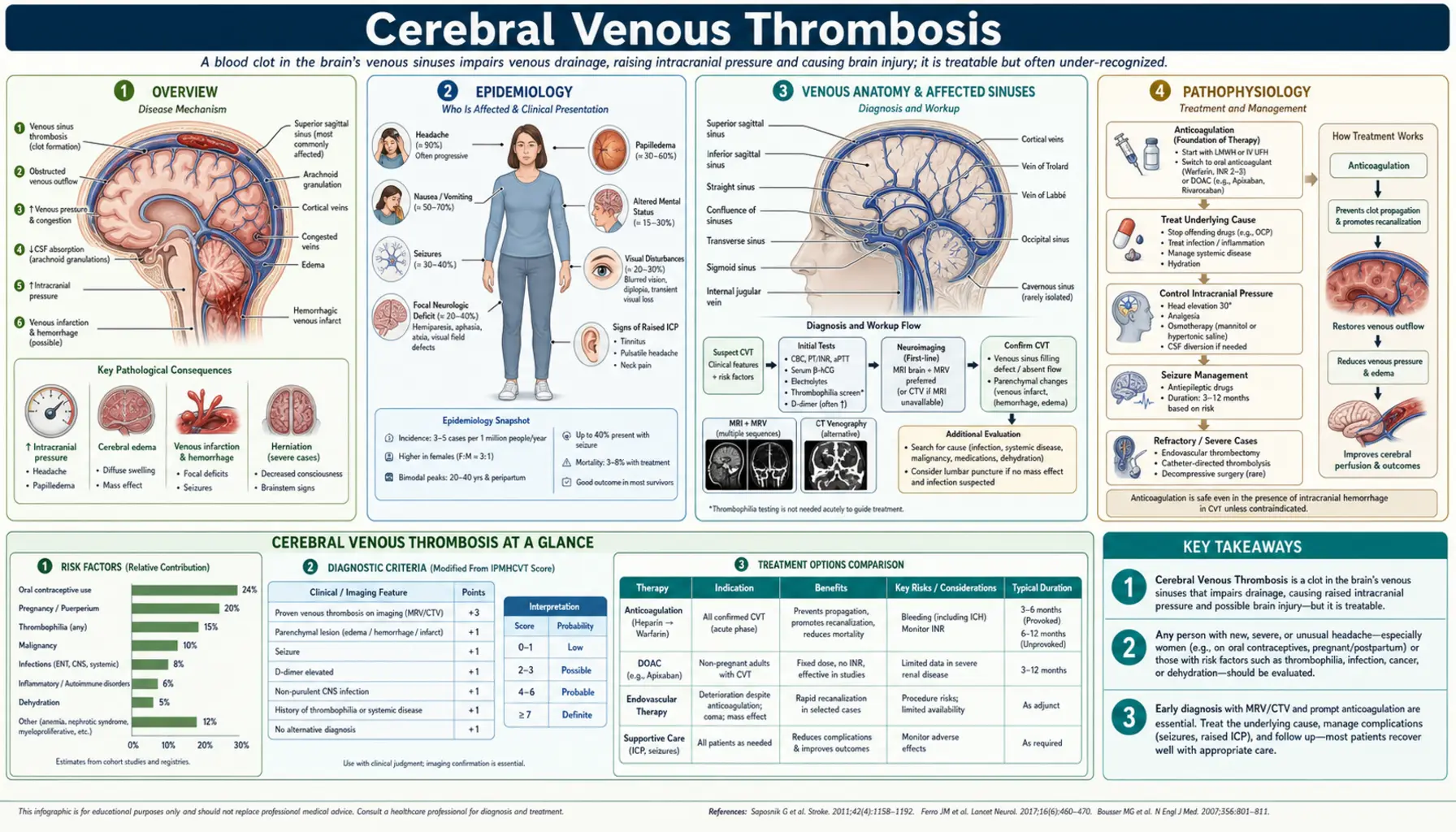
Table of Contents
1. Overview
CVST encompasses thrombosis affecting the large dural venous sinuses (superior sagittal sinus, lateral/transverse sinuses, straight sinus, cavernous sinus) as well as the smaller cortical veins draining into them. The superior sagittal sinus is involved in more than 50% of cases, followed by the lateral sinuses (44%), straight sinus (18%), and cortical veins (17%); multiple sinus involvement occurs in approximately 30% of cases. The condition is distinguished from idiopathic intracranial hypertension (IIH/pseudotumor cerebri), arterial stroke, encephalitis, and brain tumor — all of which can present with headache, papilledema, or focal deficits.
A landmark shift in CVST understanding occurred in 2021 with the recognition of vaccine-induced immune thrombocytopenia and thrombosis (VITT), a rare but severe CVST syndrome triggered by adenovirus-vector COVID-19 vaccines. VITT-associated CVST is characterized by antibodies against platelet factor 4 (anti-PF4). It clinically mimics heparin-induced thrombocytopenia (HIT type 2) and is treated differently — heparin is contraindicated, and intravenous immunoglobulin (IVIG) is first-line. This discovery has reinforced the importance of checking platelet count in every CVST patient before starting anticoagulation.
2. Epidemiology
The annual incidence of CVST is approximately 1.32–1.57 cases per 100,000 population, making it far less common than arterial stroke but with similar or greater diagnostic challenge due to variable presentation. Women are disproportionately affected, with a female-to-male ratio of approximately 3:1 overall, driven by oral contraceptive use and pregnancy/puerperium risk. The peak incidence is in young adults aged 20–35 years, though CVST occurs at all ages including neonates and children (where infection and dehydration are the leading causes) and the elderly (where malignancy and hyperviscosity predominate).
The American Heart Association's 2024 statement says CVST accounts for 0.5% to 3% of all strokes. Its share among strokes in women under 45 years of age is sometimes given as up to 15%; no source for that figure was found. Despite its rarity, CVST is important because it disproportionately affects young productive adults and, with prompt treatment, carries substantially better outcomes than arterial stroke (case fatality 8–10% in modern series, compared to 25–30% for large-vessel arterial stroke). In-hospital mortality in contemporary cohorts is 3–4% with appropriate anticoagulation.
3. Venous Anatomy and Affected Sinuses
The dural venous sinuses are endothelium-lined venous channels within the dura mater, lacking valves (allowing bidirectional flow) and receiving drainage from cortical veins, diploic veins (skull), and emissary veins (scalp). Key sinuses and their clinical correlates:
- Superior sagittal sinus: Runs in the superior dural fold from the crista galli to the torcular Herophili (confluence of sinuses); drains most of the cerebral convexity via cortical veins. superior sagittal sinus thrombosis → bilateral cortical venous infarction (often parasagittal), bilateral leg weakness, seizures, raised intracranial pressure, and in severe cases herniation. Most commonly affected sinus (>50% of cases).
- Lateral (transverse + sigmoid) sinuses: Each transverse sinus runs laterally from the torcular; the sigmoid sinus is its S-shaped continuation leading to the jugular bulb. Thrombosis causes headache, papilledema, and isolated intracranial hypertension mimicking IIH — particularly common with lateral sinus thrombosis. Left lateral sinus thrombosis may cause aphasia via involvement of draining Labbé vein territory.
- Straight sinus: Receives the great cerebral vein of Galen; drains deep structures (basal ganglia, thalami). Straight sinus thrombosis → deep venous infarction with bilateral thalamic edema — associated with altered consciousness, coma, and poor prognosis. The most dangerous single-sinus involvement.
- Cavernous sinus: Paired sinus flanking the sella turcica, receiving drainage from the orbit and superior ophthalmic vein. Cavernous sinus thrombosis (CST) is usually septic (from facial infection, sinusitis, dental abscess) and presents with orbital pain, chemosis, proptosis, ophthalmoplegia (palsies of cranial nerves III, IV and VI), and periorbital edema — often bilateral due to sinus intercommunication.
- Cortical vein thrombosis: Isolated cortical vein thrombosis without sinus involvement is underdiagnosed; presents with focal seizures or focal deficits corresponding to the draining territory; visible on MRI FLAIR/T1 (cord sign = hyperdense vein) + susceptibility-weighted imaging (SWI).
4. Pathophysiology
CVST causes brain injury through two simultaneous mechanisms that often coexist in the same patient, accounting for the heterogeneous magnetic resonance imaging (MRI) appearance:
Venous Hypertension and Edema
Thrombotic obstruction of a venous sinus raises venous pressure in draining cortical veins. When venous pressure exceeds capillary perfusion pressure, cerebral blood flow in the drained territory falls — a mechanism fundamentally different from arterial occlusion. The resulting edema is initially vasogenic (blood-brain barrier disruption from venous hypertension) and, as ischemia deepens, becomes cytotoxic. Critically, venous collateral drainage (leptomeningeal veins, diploic channels) often develops rapidly, limiting infarction — this explains why large superior sagittal sinus thromboses can present with only headache and papilledema with no parenchymal injury, while smaller thrombi in poor-collateral territories cause massive hemorrhagic infarction.
Hemorrhagic Venous Infarction
When venous hypertension exceeds the capacity of collateral drainage, venules rupture into the parenchyma, producing hemorrhagic venous infarction. These lesions do not follow arterial territories, are often bilateral and parasagittal (for superior sagittal sinus thrombosis), and typically show surrounding edema exceeding the hemorrhage volume. The hemorrhagic nature of CVST lesions historically led to withholding of anticoagulation — a practice later studies did not support: the heparin trials reported no new symptomatic brain hemorrhages with anticoagulation, even in patients who already had a hemorrhage, and fewer deaths and less dependency that did not reach statistical significance when pooled.
Raised Intracranial Pressure
Impaired cerebrospinal fluid (CSF) absorption at arachnoid granulations (which drain into the superior sagittal sinus) — combined with reduced venous capacitance — raises intracranial pressure (ICP). This mechanism is responsible for the IIH-like syndrome of isolated intracranial hypertension seen in lateral sinus thrombosis, with headache and papilledema but no focal deficits or parenchymal injury.
5. Etiology and Risk Factors
Hormonal and Pregnancy-Related (Young Women)
- Combined oral contraceptives (OCP): Estrogen-containing OCPs increase coagulation factor levels (II, VII, VIII, X, fibrinogen) and reduce protein S; relative risk of CVST is approximately 13-fold; risk further amplified by concomitant thrombophilia (factor V Leiden + OCP = 30-fold risk). The most common identifiable cause of CVST in developed countries.
- Pregnancy and puerperium: Third trimester and the first 4 weeks post-partum are the highest-risk period; puerperal CVST (formerly the most common cause historically) still accounts for 5–20% of cases in developing countries with high rates of pregnancy-related infection and dehydration. Pathophysiology: physiological hypercoagulability of pregnancy + venous stasis + local injury from delivery.
- Hormone replacement therapy (HRT): Similar mechanism to OCP though lower estrogen doses than first-generation pills; smaller increase in relative risk.
Thrombophilia
- Factor V Leiden (G1691A): Most common inherited thrombophilia in Europeans (~5% carrier frequency); responsible for activated protein C resistance; increases CVST risk 3–4-fold alone, multiplicatively with OCP.
- Prothrombin G20210A mutation: Second most common inherited thrombophilia (~2% frequency); increases prothrombin (factor II) levels; odds ratio (OR) for CVST approximately 2.5–5.
- Antithrombin, protein C, or protein S deficiency: Rarer but high-risk hereditary thrombophilias; testing is described for young patients with unprovoked CVST and a family history of thrombosis.
- Antiphospholipid syndrome (APS): Acquired thrombophilia; anticardiolipin antibodies, anti-β2-glycoprotein-I, and lupus anticoagulant increase arterial and venous thrombosis risk; treated with long-term anticoagulation.
- Hyperhomocysteinemia: Elevated homocysteine impairs endothelial function; associated with methylenetetrahydrofolate reductase (MTHFR) mutations and B vitamin deficiencies (B6/B9/B12); moderate risk factor for CVST.
Infection
- Otogenic lateral sinus thrombosis: Otitis media/mastoiditis spreads to sigmoid/transverse sinus via emissary veins; formerly called "otitic hydrocephalus"; treated with antibiotics ± anticoagulation ± mastoidectomy.
- Sinusitis-related superior sagittal sinus/cavernous sinus thrombosis: Frontal sinusitis → superior sagittal sinus thrombosis (Pott's puffy tumor); ethmoid/sphenoid sinusitis → cavernous sinus thrombosis; common pathogens: Staphylococcus aureus, Streptococcus spp., gram-negative organisms.
- Meningitis: Bacterial meningitis (especially in neonates and children) can cause CVST as a complication of infectious vasculitis and local thrombogenic inflammation.
- COVID-19: SARS-CoV-2 infection associated with increased CVST risk via endothelial injury, hypercoagulability, and platelet activation.
Malignancy and Hematological Disorders
- Solid tumor compression or direct invasion of venous sinuses (meningioma, metastasis)
- Paraneoplastic hypercoagulability; L-asparaginase chemotherapy depletes antithrombin
- Polycythemia vera, essential thrombocythemia — myeloproliferative neoplasms; JAK2 V617F mutation testing is important in young patients with unprovoked CVST
- Paroxysmal nocturnal hemoglobinuria (PNH) — rare but important cause in young adults with unexplained venous thrombosis in unusual sites
Other Causes
- Head trauma (direct venous laceration or delayed thrombosis)
- Intracranial hypotension (cerebrospinal fluid leak — after lumbar puncture, spontaneous, post-neurosurgery); downward brain shift stretches bridging veins and sinuses promoting thrombosis
- Dehydration (common in neonates and elderly)
- Inflammatory bowel disease (Crohn's, ulcerative colitis) — especially during flares
- Systemic lupus erythematosus, Behçet's disease (vasculitis)
- Nephrotic syndrome (loss of antithrombin and protein C/S in urine)
- VITT (post-adenoviral vector COVID-19 vaccine): anti-PF4 antibody-mediated platelet activation — distinct mechanism requiring non-heparin anticoagulation
6. Clinical Presentation
Headache — The Cardinal Symptom
Headache is the presenting symptom in approximately 90% of CVST patients and is the sole symptom in 25% of isolated intracranial hypertension presentations. Unlike the thunderclap headache of subarachnoid hemorrhage, CVST headache typically has a subacute onset over days to weeks, is diffuse, progressive, and worsening — often positional (worse lying flat due to elevated ICP) or worsening with Valsalva. However, thunderclap CVST onset does occur (15% of cases) and is part of the differential diagnosis of sudden severe headache alongside subarachnoid hemorrhage. The headache may be accompanied by nausea, vomiting, and photophobia, mimicking migraine or tension headache — leading to delayed diagnosis of 1–4 weeks on average.
Focal Neurological Deficits
- Hemiparesis or monoparesis — particularly leg-predominant (parasagittal cortex drained by superior sagittal sinus) or bilateral leg weakness (bilateral parasagittal infarction from superior sagittal sinus thrombosis)
- Aphasia — left hemisphere cortical vein or left lateral sinus thrombosis
- Hemianopia or cortical visual loss
- Sensory deficits in a cortical distribution
- Bilateral thalamic syndrome — altered level of consciousness, memory impairment, movement disorder — from deep venous thrombosis (straight sinus/Galen vein)
Seizures
Seizures occur in approximately 40% of CVST patients — a substantially higher rate than arterial stroke (8–10%), reflecting cortical irritation from hemorrhagic venous infarction and perilesional edema. Focal-onset seizures with secondary generalization are most common; status epilepticus occurs in 5–10% and carries significantly worse prognosis. Early seizures (within 2 weeks) predict higher risk of further seizures but do not independently worsen long-term outcome when controlled. The 2017 European Stroke Organization guideline suggests antiepileptic drugs in people who have had an early seizure and have lesions in the upper part of the brain (supratentorial lesions), to prevent further early seizures. It could make no recommendation on preventing later seizures, and it gives no length of treatment.
Isolated Intracranial Hypertension Syndrome
Approximately 20–40% of CVST — predominantly lateral sinus thrombosis — presents as isolated intracranial hypertension without focal deficits or parenchymal injury, clinically indistinguishable from idiopathic intracranial hypertension (IIH/pseudotumor cerebri). These patients have headache, papilledema, and visual obscurations; cerebrospinal fluid opening pressure is elevated; brain magnetic resonance imaging parenchyma is normal. The 2018 UK consensus guidelines on idiopathic intracranial hypertension call CT or MR venography mandatory to exclude cerebral sinus thrombosis, and call for more in-depth investigation in patients who are not female, not of childbearing age, or have a body mass index below 30.
Cavernous Sinus Thrombosis
Septic cavernous sinus thrombosis presents with periorbital edema, proptosis, chemosis, ophthalmoplegia (cranial nerves III, IV and VI), and retinal venous congestion, often progressing from unilateral to bilateral within 24–48 hours via the intercavernous sinus. Fever, leukocytosis, and identifiable primary infection source (facial cellulitis, sinusitis, dental abscess) differentiate septic CST from aseptic orbital apex syndrome. Urgent antibiotics (with antifungal cover if mucormycosis is suspected in immunocompromised or diabetic patients) are primary therapy.
Encephalopathy and Coma
Progressive encephalopathy, bilateral thalamic dysfunction, coma, or decerebrate posturing indicate deep venous involvement (straight sinus ± Galen vein thrombosis) — the most severe presentation with the worst prognosis. Bilateral thalamic hyperintensity on magnetic resonance imaging in a young patient without arterial risk factors is a recognized pointer to deep CVST.
7. Diagnosis
Brain Computed Tomography — Initial Emergency Scan
Non-contrast computed tomography is typically the first imaging obtained. It may show:
- Dense (hyperdense) cord sign: Hyperdense thrombus in a cortical vein on non-contrast computed tomography, visible as a tubular cord-like density — specific but only present in 25–30% of cases.
- Dense triangle sign: Triangular hyperdensity in the posterior superior sagittal sinus — fresh thrombus; present in 10–20% of cases.
- Empty delta sign (contrast computed tomography): Triangular filling defect in the posterior superior sagittal sinus on contrast-enhanced computed tomography, representing thrombus surrounded by collateral flow — present in 35–40% of cases with superior sagittal sinus thrombosis; classic but not pathognomonic (absent in acute phase, before collateral opacification).
- Hemorrhagic venous infarction: bilateral parasagittal hemorrhages not conforming to arterial territories
- Diffuse cerebral edema, sulcal effacement
- Normal computed tomography (in up to 30–40% of cases) — a normal computed tomography does not exclude CVST
Magnetic Resonance Imaging Brain + magnetic resonance venography — Gold Standard
Magnetic resonance imaging with magnetic resonance venography is the gold standard investigation, with sensitivity approaching 90–95% for CVST when T1, T2, fluid-attenuated inversion recovery (FLAIR), diffusion-weighted imaging (DWI), susceptibility-weighted imaging (SWI), and time-of-flight (TOF) magnetic resonance venography sequences are combined:
- T1-weighted: Isointense (acute, days 1–5) → hyperintense (subacute, days 5–14) thrombus signal within the sinus; hyperintense on T1 is highly specific when combined with magnetic resonance venography absence of flow
- T2/FLAIR: Hyperintense thrombus (subacute); venous edema/infarction in drained territory; cortical/subcortical signal change not following arterial boundaries
- SWI/GRE (susceptibility-weighted imaging): Blooming artifact (hypointense) from hemoglobin breakdown products in thrombus and microhemorrhages; excellent for detecting cortical vein thrombosis and early hemorrhagic change
- DWI: Restricted diffusion in cytotoxic edema component of venous infarction; an apparent diffusion coefficient (ADC) map helps distinguish reversible (high value) from irreversible (low value) injury
- TOF magnetic resonance venography/phase-contrast magnetic resonance venography: Demonstrates absent or markedly reduced flow in thrombosed sinuses; TOF has false positives from flow-related saturation in slow flow; gadolinium-enhanced magnetic resonance venography improves specificity
- Computed tomography venography (CTV): Excellent alternative to magnetic resonance venography, particularly in acute setting or when magnetic resonance imaging is contraindicated; fast acquisition; filling defects in sinuses on CTV match magnetic resonance venography sensitivity for major sinuses; requires iodinated contrast and radiation
Laboratory Investigations
- D-dimer: Elevated in most CVST patients but non-specific; a normal D-dimer makes CVST unlikely (negative predictive value ~99% in low-pretest-probability patients) — useful to guide imaging decision in low-probability headache workup. However, D-dimer may be normal in isolated headache/papilledema CVST presentations.
- Complete blood count (CBC) with platelet count: Critical before anticoagulation — thrombocytopenia suggests VITT or HIT; polycythemia or thrombocytosis suggest myeloproliferative neoplasm
- Anti-PF4 antibody (enzyme-linked immunosorbent assay or chemiluminescence immunoassay): Used in patients with a recent adenovirus-vector COVID-19 vaccine (within 4–28 days) + thrombocytopenia
- Coagulation screen: prothrombin time, activated partial thromboplastin time, fibrinogen; lupus anticoagulant (an activated partial thromboplastin time-based assay)
- Thrombophilia workup: The 2017 European Stroke Organization guideline suggests not routinely screening people with this clot for thrombophilia or cancer. When screening is done, the tests are: antithrombin activity, protein C activity, protein S free antigen (defer protein S/C/antithrombin testing until off anticoagulation; levels unreliable during acute thrombosis); factor V Leiden polymerase chain reaction (PCR); prothrombin G20210A polymerase chain reaction; anticardiolipin immunoglobulin G (IgG)/immunoglobulin M (IgM); anti-β2-GP1 immunoglobulin G/IgM; JAK2 V617F mutation (if clinical suspicion for myeloproliferative neoplasm)
- Homocysteine: Elevated in vitamin B12/B9/B6 deficiency, methylenetetrahydrofolate reductase mutations
- Blood cultures + inflammatory markers (C-reactive protein (CRP), erythrocyte sedimentation rate (ESR)): If septic CVST or cavernous sinus thrombosis suspected
- PNH screen (flow cytometry): In young patients with unexplained CVST + cytopenias or hemolysis
Lumbar Puncture
Lumbar puncture can be performed safely after imaging confirms no mass lesion or significant midline shift. Cerebrospinal fluid findings in CVST: elevated opening pressure (often >25 cmH2O), mildly elevated protein (50–100 mg/dL), mild pleocytosis (<50 cells/mm³), or xanthochromia (if subarachnoid blood). Lumbar puncture is also therapeutic for severe papilledema with visual deterioration (reduces ICP acutely). Cerebrospinal fluid analysis also helps exclude meningitis/encephalitis, which may coexist with or mimic CVST.
8. Treatment
Anticoagulation — First-Line Therapy
Anticoagulation is the cornerstone of CVST treatment, despite the frequent presence of hemorrhagic venous infarction. It rests on two small but influential randomized controlled trials (Einhäupl 1991; de Bruijn 1999). A 2011 Cochrane review pooled them (79 patients): the relative risk of death was 0.33 (95% confidence interval 0.08 to 1.21) and of death or dependency 0.46 (0.16 to 1.31), a potentially important reduction that did not reach statistical significance. No new symptomatic brain hemorrhages were seen, and one major gastrointestinal hemorrhage occurred after anticoagulant treatment. The pathophysiological rationale is compelling: anticoagulation arrests thrombosis propagation and allows natural thrombolysis to restore venous drainage, reducing venous hypertension and preventing further infarction. The hemorrhagic component of venous infarction does not worsen significantly with anticoagulation — it is a consequence of venous hypertension, not the anticoagulant.
- Acute phase: Low molecular weight heparin (LMWH, e.g., enoxaparin 1 mg/kg twice daily) or unfractionated heparin (UFH) IV — UFH preferred when rapid reversal may be needed (severe hemorrhage, need for urgent surgery). LMWH is generally preferred for its predictable pharmacokinetics and lower HIT risk.
- VITT exception: Standard heparin (UFH or LMWH) is contraindicated in VITT-associated CVST (anti-PF4 antibody positive + thrombocytopenia). Non-heparin anticoagulation is used: argatroban, fondaparinux, or direct oral anticoagulants (apixaban/rivaroxaban). intravenous immunoglobulin 1 g/kg/day × 2 days is first-line to neutralize anti-PF4 antibodies and halt platelet activation. Platelet transfusion contraindicated (worsens thrombosis by providing fuel for anti-PF4-driven platelet activation).
- Transition and duration: After acute LMWH/UFH, transition to oral anticoagulation:
- Warfarin (target international normalized ratio (INR) 2.0–3.0): customary practice is 3–6 months for provoked (reversible risk factor) CVST; 6–12 months for unprovoked; indefinite if thrombophilia (APS, antithrombin deficiency) or recurrent thrombosis. These schedules are practice, not trial results: the 2017 European Stroke Organization guideline could make no recommendation on how long to continue after the acute phase.
- Dabigatran: the exploratory RE-SPECT CVT trial (120 patients) saw no repeat clots in either group and major bleeding in 1 person (1.7%) on dabigatran and 2 (3.3%) on warfarin. Its authors concluded both may be safe and effective. It was not a non-inferiority trial. The 2017 European Stroke Organization guideline, written before it, suggests not using direct oral anticoagulants in the acute phase.
- Rivaroxaban: SECRET (2023), a small Canadian trial of 55 people testing feasibility, compared it with standard blood thinners. There was 1 brain bleed, 2 lesser bleeds and 1 repeat clot by day 180, all in the rivaroxaban group; overall rates were low, and everyone had at least partial reopening of the vein.
- Direct oral anticoagulants as a group: DOAC-CVT (2025) followed 619 adults in 23 countries. New clots or major bleeding within 6 months occurred in 3% on these drugs and 3% on warfarin-type drugs (weighted odds ratio 0.99), and deaths were 1% in each. It was observational, not randomized, and excluded pregnancy and breastfeeding.
Endovascular Thrombectomy and Local Thrombolysis
Endovascular treatment — mechanical thrombectomy (aspiration, stent-retriever) ± local catheter-directed thrombolysis (urokinase, tissue plasminogen activator) via the venous sinuses — is reserved for patients with severe CVST deteriorating despite therapeutic anticoagulation. The ISCVT registry identifies factors predicting poor outcome with anticoagulation alone: coma (Glasgow Coma Scale below 9), deep venous thrombosis, intracranial hemorrhage at presentation, and bilateral motor deficits. Uncontrolled case series report recanalization in 70–80% of treated patients. The one randomized trial, TO-ACT (67 patients with severe disease, published 2020), was halted early for futility: a good outcome at 12 months was seen in 67% with endovascular treatment and 68% with standard care (relative risk 0.99), and deaths were 12% against 3%, a difference that was not statistically significant (P = .20). Its authors say the treatment did not appear to improve outcome, and that the small size leaves open the possibility that future studies show better recovery. The 2017 European Stroke Organization guideline could make no recommendation on thrombolysis or thrombectomy. Endovascular treatment is reserved for ≤10% of CVST cases.
Antiepileptic Drugs (AEDs)
The 2017 European Stroke Organization guideline suggests antiepileptics in patients with an early seizure and supratentorial lesions, to prevent further early seizures. It does not extend this to people who have had no seizure, although seizures are common in this disease (about 40%). Levetiracetam is often chosen (minimal drug interactions, no hepatic induction). Valproate and phenytoin are alternatives. Duration: the guideline made no recommendation on later seizures or on a length of treatment; in practice some patients with cortical lesions continue for 1 year, with earlier stopping described in those without seizures and with lesion resolution. Routine prophylaxis in patients without cortical lesions is not strongly supported by evidence.
Intracranial Pressure Management
- Head elevation (30°) and keeping blood carbon dioxide from rising (hypercapnia)
- Acetazolamide (250–500 mg twice daily) reduces cerebrospinal fluid production; used for the isolated intracranial hypertension phenotype. The 2017 European Stroke Organization guideline suggests not using acetazolamide or steroids to reduce death or dependency.
- Repeated therapeutic lumbar punctures for severe papilledema with visual deterioration; the 2017 European Stroke Organization guideline could make no recommendation on them
- Osmotic therapy (mannitol, hypertonic saline) for acute ICP crises
- Decompressive craniectomy: lifesaving in malignant cerebral edema (large hemorrhagic venous infarction + uncal herniation); may be required as a bridge to anticoagulation-mediated recanalization
- Cerebrospinal fluid diversion (ventriculostomy, ventriculoperitoneal shunt) for obstructive hydrocephalus
Infection Treatment (Septic CVST)
Intravenous antibiotics covering the causative pathogen are the core of treatment and are started immediately upon suspicion of septic CVST, before culture results return. Empiric coverage: vancomycin + ceftriaxone ± metronidazole (for anaerobes from dental/sinus source). Duration typically 4–6 weeks intravenous (IV) followed by oral completion. Surgical source control (mastoidectomy, sinus drainage, dental extraction) is required for ongoing infectious foci — failure to control the source leads to refractory septic thrombosis. Anticoagulation in septic CVST remains controversial but is used at most centers for deteriorating patients without contraindication.
9. Prognosis and Outcomes
Modern CVST prognosis is substantially better than historical series, reflecting improved recognition and universal anticoagulation. The International Study on Cerebral Vein and Dural Sinus Thrombosis (ISCVT, n=624) established the best available natural history data:
- Complete recovery: approximately 57% of patients at 16 months
- Minor residual symptoms (modified Rankin Scale 1): 22%; mild impairment (scale 2): 7.5%; moderate impairment (scale 3): 2.9%; severe handicap (scale 4 or 5): 2.2%
- In-hospital mortality: 3.4–8%; total mortality at 16 months: 8.3%
- Over a median of 16 months: recurrent sinus thrombosis 2.2%; other thrombotic events 4.3%
- Seizures over a median of 16 months: 10.6% of all patients; driving restriction typically 1 year seizure-free
Recovery can leave problems these numbers do not show. In the SECRET trial, people on average reported reduced quality of life, low mood, fatigue and headache, with impaired thinking on testing, at enrollment; all of these improved markedly by day 180.
The American Heart Association's 2024 statement says death or dependence occurs in 10% to 15% of patients despite intensive medical treatment.
Predictors of Poor Outcome (ISCVT Data)
- Central nervous system infection — hazard ratio 3.3, the highest for death or dependence
- Deep venous thrombosis (straight sinus/Galen vein involvement) — hazard ratio 2.9
- Malignancy as underlying cause — hazard ratio 2.9
- Coma at presentation — hazard ratio 2.7
- Age >37 years — hazard ratio 2.0
- Mental status disorder — hazard ratio 2.0
- Intracranial hemorrhage at presentation — hazard ratio 1.9
- Male sex — hazard ratio 1.6
VITT-Associated CVST (Separate UK Studies)
- Among 70 UK patients with VITT-associated CVST, 47% died or became dependent, against 16% of patients without VITT. The figure was 36% with non-heparin anticoagulants against 75% without them, and 40% with immunoglobulin against 73% without it. These were observational comparisons.
- In a UK cohort of VITT overall (220 definite or probable cases), overall mortality was 22%, and the odds of death were 2.7 times higher with cerebral venous sinus thrombosis.
Recanalization
Partial or complete sinus recanalization is commonly given as approximately 80% of patients with anticoagulation, most within 3–6 months of treatment. The RE-SPECT CVT trial measured 60.0% on dabigatran and 67.3% on warfarin at 25 weeks, and in the SECRET trial every participant had at least partial reopening by day 180, so the number depends on how partial reopening is counted. Recanalization correlates with better functional outcomes but is not universal; partial or absent recanalization does not preclude good functional recovery if collateral drainage is adequate. Magnetic resonance venography at 3–6 months after treatment is used to assess recanalization and guide anticoagulation duration decisions.
10. Special Populations
Pregnancy and Puerperium
LMWH is safe in pregnancy (does not cross the placenta) and is the anticoagulant of choice throughout pregnancy. In customary practice, UFH replaces it at 36 weeks to allow neuraxial anesthesia planning. Post-partum: LMWH restarts within 12–24 hours of delivery, with transition to warfarin after 6–12 weeks. The DOAC-CVT study treated pregnancy and lactation as an absolute contraindication to direct oral anticoagulants as a class, not to dabigatran alone. The 2017 European Stroke Organization guideline suggests that subsequent pregnancies are safe and that prophylactic LMWH be considered throughout pregnancy and the puerperium. It also suggests that women who have had this clot avoid contraceptives containing estrogen; its wording covers estrogen-containing products, not all contraception.
Pediatric CVST
Neonatal CVST: most commonly presents with seizures in the first week of life; superior sagittal sinus and straight sinus predominantly affected; dehydration, perinatal asphyxia, infection are common causes; treatment with LMWH is reasonable but evidence base is weaker than adults; outcome generally good with appropriate treatment. Childhood/adolescent CVST: head/neck infection (otitis media, mastoiditis), leukemia, L-asparaginase chemotherapy, and iron-deficiency anemia are important causes not seen in adults.
Elderly CVST
Malignancy, myeloproliferative neoplasms, dehydration from intercurrent illness, and direct oral anticoagulant interruption or under-dosing are the primary etiologies. Presentation may be atypical (encephalopathy, confusion) without prominent headache. Higher hemorrhagic risk with anticoagulation in elderly; balance individualized.
11. Key Research and Recent Advances
The RE-SPECT CVT trial (2019, n=120) was an exploratory randomized trial of dabigatran (150 mg twice daily) against warfarin after CVST. No repeat clots occurred in either group, major bleeding was 1.7% against 3.3%, and recanalization was 60.0% against 67.3%; its authors concluded both drugs may be safe and effective. It was the first randomized trial of a direct oral anticoagulant in CVST. Rivaroxaban has since been tested in the small SECRET feasibility trial (2023), and the DOAC-CVT cohort (2025) found no difference in clots or major bleeding against warfarin-type drugs.
The identification of VITT as a CVST-specific syndrome in 2021 (Greinacher et al., NEJM; Schultz et al., NEJM) transformed understanding of immune-mediated thrombosis. VITT mimics HIT type 2 mechanistically (anti-PF4 immunoglobulin G antibodies triggering FcγRIIA-mediated platelet activation) but without prior heparin exposure. The clinical lesson drawn from this work is that a platelet count in every CVST patient, and anti-PF4 antibody testing after a recent adenovirus-vector vaccine, can identify this syndrome, in which heparin is contraindicated and immunoglobulin was linked to better outcomes (see Prognosis).
The TO-ACT randomized trial (2020, 67 patients) found no apparent improvement from endovascular treatment in severe CVST (see Treatment). The American Heart Association updated its scientific statement in 2024, replacing its 2011 statement: this clot causes 0.5% to 3% of strokes, and death or dependence occurs in 10% to 15% of patients despite intensive medical treatment.
Emerging biomarker research focuses on serum glial fibrillary acidic protein (astrocyte injury), neurofilament light chain (axonal injury), and D-dimer trajectory as predictors of CVST severity and recanalization. Artificial intelligence analysis of MRI-magnetic resonance venography for automated sinus signal abnormality detection is under active development to reduce diagnostic delay.
Key Research Papers
- Ferro JM, Canhão P, Stam J, Bousser MG, Barinagarrementeria F; ISCVT Investigators. Prognosis of cerebral vein and dural sinus thrombosis: results of the International Study on Cerebral Vein and Dural Sinus Thrombosis (ISCVT). Stroke. 2004;35(3):664–670. — PubMed PMID: 14976332
- Coutinho JM, Ferro JM, Canhão P, et al. Cerebral venous and sinus thrombosis in women. Stroke. 2009;40(7):2356–2361. — PubMed PMID: 19478226
- Einhäupl KM, Villringer A, Meister W, et al. Heparin treatment in sinus venous thrombosis. Lancet. 1991;338(8767):597–600. — PubMed PMID: 1679154
- de Bruijn SF, Stam J. Randomized, placebo-controlled trial of anticoagulant treatment with low-molecular-weight heparin for cerebral sinus thrombosis. Stroke. 1999;30(3):484–488. — PubMed PMID: 10066840
- Ferro JM, Coutinho JM, Dentali F, et al. Safety and efficacy of dabigatran etexilate vs dose-adjusted warfarin in patients with cerebral venous thrombosis: a randomized clinical trial. JAMA Neurol. 2019;76(12):1457–1465. — PubMed PMID: 31479105
- Greinacher A, Thiele T, Warkentin TE, et al. Thrombotic thrombocytopenia after ChAdOx1 nCov-19 vaccination. N Engl J Med. 2021;384(22):2092–2101. — PubMed PMID: 33835769
- Stam J. Thrombosis of the cerebral veins and sinuses. N Engl J Med. 2005;352(17):1791–1798. — PubMed PMID: 15858188
- Saposnik G, Barinagarrementeria F, Brown RD Jr, et al. Diagnosis and management of cerebral venous thrombosis. Stroke. 2011;42(4):1158–1192. — PubMed PMID: 21293023
- Silvis SM, de Sousa DA, Ferro JM, Coutinho JM. Cerebral venous thrombosis. Nat Rev Neurol. 2017;13(9):555–565. — PubMed PMID: 28820187
- Dentali F, Gianni M, Crowther MA, Ageno W. Natural history of cerebral vein thrombosis: a systematic review. Blood. 2006;108(4):1129–1134. — PubMed PMID: 16609071
- Ferro JM, Bousser MG, Canhão P, et al. European Stroke Organization guideline for the diagnosis and treatment of cerebral venous thrombosis - endorsed by the European Academy of Neurology. Eur J Neurol. 2017;24(10):1203–1213. — PubMed PMID: 28833980
- Devasagayam S, Wyatt B, Leyden J, Kleinig T. Cerebral venous sinus thrombosis incidence is higher than previously thought: a retrospective population-based study. Stroke. 2016;47(9):2180–2182. — PubMed PMID: 27435401
- Coutinho JM, Zuurbier SM, Bousser MG, et al. Effect of endovascular treatment with medical management vs standard care on severe cerebral venous thrombosis: the TO-ACT randomized clinical trial. JAMA Neurol. 2020;77(8):966–973. — PubMed PMID: 32421159
- Coutinho J, de Bruijn SF, Deveber G, Stam J. Anticoagulation for cerebral venous sinus thrombosis. Cochrane Database Syst Rev. 2011;2011(8):CD002005. — PubMed PMID: 21833941
- Saposnik G, Bushnell C, Coutinho JM, et al. Diagnosis and management of cerebral venous thrombosis: a scientific statement from the American Heart Association. Stroke. 2024;55(3):e77–e90. — PubMed PMID: 38284265
- van de Munckhof A, van Kammen MS, Tatlisumak T, et al. Direct oral anticoagulants versus vitamin K antagonists for cerebral venous thrombosis (DOAC-CVT): an international, prospective, observational cohort study. Lancet Neurol. 2025;24(3):199–207. — PubMed PMID: 39986309
- Field TS, Dizonno V, Almekhlafi MA, et al. Study of rivaroxaban for cerebral venous thrombosis: a randomized controlled feasibility trial comparing anticoagulation with rivaroxaban to standard-of-care in symptomatic cerebral venous thrombosis. Stroke. 2023;54(11):2724–2736. — PubMed PMID: 37675613
- Perry RJ, Tamborska A, Singh B, et al. Cerebral venous thrombosis after vaccination against COVID-19 in the UK: a multicentre cohort study. Lancet. 2021;398(10306):1147–1156. — PubMed PMID: 34370972
- Pavord S, Scully M, Hunt BJ, et al. Clinical features of vaccine-induced immune thrombocytopenia and thrombosis. N Engl J Med. 2021;385(18):1680–1689. — PubMed PMID: 34379914
- Mollan SP, Davies B, Silver NC, et al. Idiopathic intracranial hypertension: consensus guidelines on management. J Neurol Neurosurg Psychiatry. 2018;89(10):1088–1100. — PubMed PMID: 29903905
PubMed Topic Searches
Connections
- Neurology
- Transient Ischemic Attack (TIA)
- Stroke
- Intracranial Hypertension (IIH)
- Normal Pressure Hydrocephalus
- Migraine
- Epilepsy
- Vascular Dementia
- All Conditions
- Lab Tests
- Vitamin B12 (Homocysteine)
- Folate / Vitamin B9
- Headache — the cardinal presenting symptom of CVST, present in about 90% of cases and often the only one.
- Covered in depth on its own page: Hypertension