Lupus Nephritis
- Overview
- Epidemiology
- Pathophysiology
- ISN/RPS Classification
- Clinical Presentation
- Diagnosis
- Treatment — Induction Therapy
- Treatment — Maintenance and New Agents
- Special Considerations
- Prognosis
- Key Research Papers
Overview
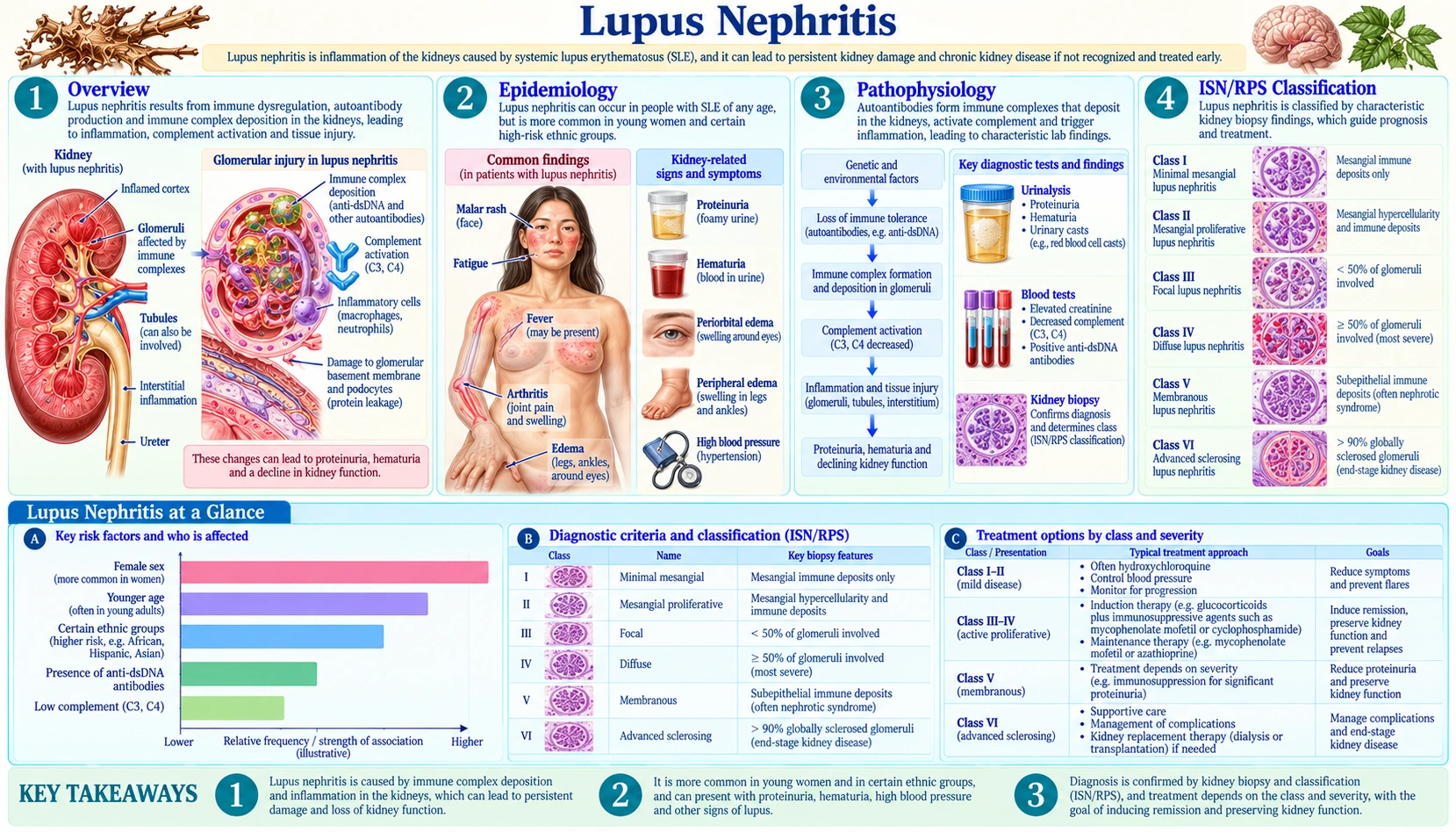
Lupus nephritis (LN) is renal involvement in systemic lupus erythematosus (SLE), the most common and serious organ complication of the disease. Approximately 50% of systemic lupus erythematosus patients develop clinically significant LN during their lifetime, and 10–30% of those progress to end-stage renal disease (ESRD). The central mechanism involves deposition of immune complexes — driven by autoantibodies including anti-dsDNA, anti-C1q, and anti-nucleosome antibodies — within glomerular structures. These deposits activate the classical complement pathway, generating C3a and C5a, which recruit inflammatory cells and drive progressive glomerular injury. Lupus nephritis is a leading cause of morbidity and mortality in systemic lupus erythematosus, and early recognition combined with aggressive immunosuppression has dramatically improved outcomes over the past six decades.
Epidemiology
LN develops in approximately 50% of all systemic lupus erythematosus patients, with marked racial and ethnic disparities. Rates are highest in Black patients (60–70%) and Hispanic patients (50–60%), and lower in White patients (30–40%). Asian patients also experience higher rates and more severe renal disease compared to White patients. The female-to-male ratio in systemic lupus erythematosus is approximately 9:1, reflecting hormonal and genetic contributions. Systemic lupus erythematosus incidence in the United States ranges from 5–10 per 100,000 per year, with considerably higher rates in minority women of childbearing age.
LN onset typically occurs within 3–5 years of the initial systemic lupus erythematosus diagnosis, though it can present at disease onset or emerge decades later. Black and Hispanic patients tend to develop LN earlier and with more severe histological class at presentation. Ten-year survival has improved dramatically — from below 20% in the 1950s to over 90% in contemporary cohorts in developed countries — largely due to the introduction of cyclophosphamide, mycophenolate mofetil, and biological agents. Despite this, 10–30% of LN patients still reach ESRD, with higher rates in Black patients.
Pathophysiology
Systemic lupus erythematosus is characterized by failure of central and peripheral immune tolerance, leading to production of pathogenic autoantibodies against nuclear self-antigens. The most disease-specific are anti-dsDNA antibodies; anti-Sm, anti-C1q, and anti-nucleosome antibodies are also important. These autoantibodies bind self-antigens in circulation (forming circulating immune complexes) and within the kidney itself — nucleosomes and chromatin fragments released from apoptotic cells bind to glomerular matrix components, creating in situ immune complex formation.
Immune complex deposition occurs in distinct compartments depending on LN class: the mesangium (Class I/II), subendothelial space (Class III/IV), and subepithelial space (Class V). Subendothelial deposits are particularly inflammatory because they are accessible to circulating complement and inflammatory cells. Activation of the classical complement pathway generates C3a and C5a, which are potent chemoattractants and activators of neutrophils, monocytes, and macrophages. The resulting cytokine storm (interleukin-6, interleukin-1 beta (IL-1β), TNF-α, IFN-α) drives glomerular endothelial injury, mesangial expansion, podocyte loss, and tubulointerstitial inflammation.
B-cell survival factors BAFF (BLyS) and APRIL are elevated in systemic lupus erythematosus and perpetuate autoreactive B-cell survival and autoantibody production — the basis for belimumab therapy. The type I interferon signature (IFN-α overproduction from plasmacytoid dendritic cells stimulated by nucleic-acid-containing immune complexes via TLR7/TLR9) is a hallmark of systemic lupus erythematosus pathogenesis and correlates with disease activity, justifying anifrolumab (anti-IFN receptor) as a therapeutic target.
ISN/RPS Classification
Kidney biopsy remains the gold standard for LN classification. The International Society of Nephrology / Renal Pathology Society (ISN/RPS) classification, originally published in 2003 (Weening et al.) and revised in 2018 (Bajema et al.), categorizes LN by light microscopy, immunofluorescence, and electron microscopy findings:
- Class I — Minimal Mesangial LN: Normal light microscopy; immunofluorescence shows only mesangial immune deposits. Clinically silent or mild. No immunosuppression needed; treat the underlying systemic lupus erythematosus.
- Class II — Mesangial Proliferative LN: Mesangial hypercellularity and matrix expansion on light microscopy; mesangial deposits on IF. Mild proteinuria and hematuria. Usually managed conservatively; low-dose prednisone for persistent activity.
- Class III — Focal LN: Active or inactive focal, segmental or global endocapillary or extracapillary glomerulonephritis involving fewer than 50% of glomeruli. Subendothelial deposits on EM. Active (A), chronic (C), or mixed (A/C) modifiers. Significant proteinuria and hematuria; requires immunosuppression.
- Class IV — Diffuse LN: Active or inactive diffuse, segmental (IV-S) or global (IV-G) endocapillary or extracapillary glomerulonephritis involving 50% or more of glomeruli. The most common and most severe class. Active lesions include "wire loop" capillary walls (massive subendothelial deposits), hyaline thrombi, fibrinoid necrosis, and cellular crescents. Without treatment, progression to ESRD is nearly universal. Always requires aggressive immunosuppression.
- Class V — Membranous LN: Global or segmental subepithelial immune deposits on EM, with or without mesangial changes. Presents as heavy proteinuria and often nephrotic syndrome. Lower rate of crescents. May co-exist with Class III or IV (mixed LN — classified as III+V or IV+V), which worsens prognosis. Managed with immunosuppression; calcineurin inhibitors (voclosporin, tacrolimus) are particularly effective for the proteinuric component.
- Class VI — Advanced Sclerosing LN: More than 90% of glomeruli are globally sclerosed without residual activity. Irreversible renal damage; ESRD is imminent. Immunosuppression is not beneficial and may be harmful; management focuses on dialysis preparation or transplant evaluation.
The revised 2018 classification introduced standardized Activity Index (AI, 0–24) and Chronicity Index (CI, 0–12) scores. High AI indicates active, potentially reversible inflammation; a high Chronicity Index indicates fibrosis and irreversible damage. These indices inform prognosis and treatment decisions independently of histological class.
Clinical Presentation
Clinical features depend on histological class and disease activity. Class III/IV LN typically presents with:
- Hematuria with dysmorphic red blood cells and RBC casts — pathognomonic for glomerulonephritis
- Proteinuria, often sub-nephrotic in Class III but may be heavy in severe Class IV
- Hypertension — common and contributes to accelerated chronic kidney disease (CKD) progression
- Declining estimated glomerular filtration rate (eGFR) and acute renal failure in severe or crescentic disease
Class V LN presents predominantly as nephrotic syndrome: proteinuria greater than 3.5 g/day, hypoalbuminemia, peripheral edema, and hyperlipidemia. Hematuria may be absent.
Systemic lupus erythematosus features often accompany LN flares: malar rash, photosensitivity, arthritis, serositis (pleuritis, pericarditis), Raynaud phenomenon, cytopenias, and neurological involvement. The classic triad of an LN flare is rising anti-dsDNA titer + falling complement (C3, C4) + worsening urinalysis (RBC casts, increasing proteinuria).
Antiphospholipid antibodies are present in 30–40% of SLE/LN patients and confer risk of arterial and venous thrombosis, thrombocytopenia, and recurrent pregnancy loss (antiphospholipid syndrome, APS). Their presence complicates LN management and worsens renal prognosis. Distinguishing an active LN flare from a superimposed bacterial or viral infection is clinically important: both can raise creatinine; erythrocyte sedimentation rate (ESR) rises in both; C-reactive protein (CRP) rises more prominently in infection than in systemic lupus erythematosus flares.
Diagnosis
Kidney biopsy is required for definitive LN classification and to guide treatment intensity. Guidelines describe early biopsy when: (1) proteinuria exceeds 0.5 g/day combined with hematuria or RBC casts; (2) creatinine is rising unexplained by other causes; or (3) clinical suspicion is high in a known systemic lupus erythematosus patient with active urinary sediment. Biopsy can be deferred only in Class I/II suspected from mild labs or when biopsy risk is prohibitive.
Key diagnostic tests:
- Urinalysis with microscopy: RBC casts + dysmorphic RBCs indicate active glomerulonephritis; granular casts suggest tubular injury; heavy proteinuria on dipstick.
- Quantitative proteinuria: Spot urine protein-to-creatinine ratio or 24-hour urine protein collection; repeat serially to monitor response.
- Anti-dsDNA antibodies: Most specific serological marker for systemic lupus erythematosus; titers correlate with LN flare activity and are used for monitoring (serial measurement every 3–6 months).
- Complement C3 and C4: Both are LOW during active LN due to complement consumption by immune complex activation of the classical pathway. Normal complement makes active Class III/IV LN less likely.
- Anti-C1q antibodies: Particularly associated with LN activity; can supplement anti-dsDNA and complement monitoring.
- Complete blood count (CBC): Cytopenias (leukopenia, lymphopenia, hemolytic anemia, thrombocytopenia) indicate systemic lupus erythematosus activity.
- Antiphospholipid antibody panel: Lupus anticoagulant, anti-cardiolipin immunoglobulin G (IgG)/immunoglobulin M (IgM), anti-β2-glycoprotein I immunoglobulin G/immunoglobulin M — relevant for thrombosis risk stratification and anticoagulation decisions.
- Serum creatinine / estimated glomerular filtration rate: Baseline and serial monitoring for treatment response.
- Infection screening: Before initiating immunosuppression — tuberculosis (an interferon-gamma release assay or PPD skin test), hepatitis B surface antigen and core antibody, hepatitis C antibody, human immunodeficiency virus (HIV) — to prevent reactivation.
Treatment — Induction Therapy
Induction therapy targets rapid suppression of active glomerular inflammation in Class III, IV, and V (or mixed) LN. Current regimens are based on landmark randomized controlled trials and are codified in the 2024 American College of Rheumatology (ACR) lupus nephritis guideline, the KDIGO 2024 lupus nephritis guideline and the EULAR 2023 update of its lupus recommendations.
The 2024 ACR guideline (28 graded recommendations: 7 strong, 21 conditional) treats lupus nephritis therapy as continuous and ongoing rather than as separate "induction" and "maintenance" phases. It describes pulse glucocorticoids followed by an oral glucocorticoid taper and two additional immunosuppressive medicines for 3–5 years in people who reach a complete renal response. The EULAR 2023 update lists glucocorticoids, mycophenolate or low-dose intravenous cyclophosphamide as anchor drugs for active lupus nephritis, with add-on belimumab or a calcineurin inhibitor (voclosporin or tacrolimus) to be considered.
Glucocorticoids
High-dose glucocorticoids remain the backbone of induction. For severe or rapidly deteriorating LN (rising creatinine, crescents, nephrotic syndrome), methylprednisolone 500–1000 mg intravenous pulse dosing for 3 consecutive days is given first, followed by oral prednisone 0.5–1 mg/kg/day (maximum 60–80 mg/day) with gradual taper over 12–24 months. Guidelines aim to keep cumulative glucocorticoid exposure low to reduce metabolic and infectious complications; a common target is prednisone ≤7.5 mg/day by 6 months where possible, and the EULAR 2023 update describes reducing maintenance glucocorticoids to 5 mg/day or less and, when possible, stopping them.
Mycophenolate Mofetil (MMF)
MMF 2–3 g/day is now the preferred induction agent for most Class III/IV LN, particularly in non-Asian patients. The landmark Ginzler 2005 NEJM trial established non-inferiority of MMF to intravenous cyclophosphamide for induction, with a superior safety profile. MMF is strongly preferred over cyclophosphamide in women of reproductive age because cyclophosphamide causes premature ovarian failure in 30–60% of patients depending on cumulative dose and age at treatment.
Cyclophosphamide (CYC)
CYC remains an option for severe, crescentic, or refractory LN. The EuroLupus low-dose protocol (500 mg intravenous every 2 weeks for 6 doses; total 3 g) proved as effective as the original U.S. National Institutes of Health (NIH) high-dose protocol (0.5–1 g/m² monthly for 6 months) in European cohorts (Houssiau 2002, 10-year follow-up 2010), with substantially less gonadotoxicity. The U.S. National Institutes of Health high-dose protocol is reserved for life- or organ-threatening LN, severe crescentic disease, or failure of low-dose approaches. Gonadal protection: egg or sperm banking before therapy is one option; GnRH agonist co-administration may help preserve ovarian reserve.
Voclosporin Triple Therapy
Voclosporin, a novel calcineurin inhibitor with a more favorable nephrotoxicity profile than tacrolimus or cyclosporine, was evaluated in the AURORA 1 trial (Rovin, Lancet 2021). Added to MMF plus low-dose steroids (triple therapy), voclosporin achieved complete renal remission in 40.8% of patients at 52 weeks versus 22.5% in the control arm. The U.S. Food and Drug Administration (FDA) approved voclosporin for active LN (Class III, IV, V, and mixed) in January 2021. It is particularly effective for Class V (membranous) LN with heavy proteinuria and for achieving rapid proteinuria reduction.
In the AURORA 2 extension (216 patients treated for 2 more years after AURORA 1), the lower protein in the urine persisted over 3 years, and complete renal response was more frequent with voclosporin (50.9% vs 39.0%). Kidney filtration (eGFR) stayed stable: the 2-year slope was −0.2 mL/min/1.73 m² with voclosporin versus −5.4 mL/min/1.73 m² in the control group. Investigator-reported falls in filtration (10.3% vs 5.0%) and high blood pressure (8.6% vs 7.0%) were more common with voclosporin.
Belimumab
Belimumab (anti-BAFF/BLyS monoclonal antibody) was evaluated in the BLISS-LN trial (Furie, NEJM 2020) as add-on therapy to standard induction (MMF + steroids or cyclophosphamide + steroids). The primary renal response was achieved in 43% of belimumab patients versus 32% of controls at 104 weeks. Belimumab became the first U.S. Food and Drug Administration-approved treatment for active lupus nephritis on 16 December 2020, for both intravenous and subcutaneous use in adults receiving standard therapy. It is available in both intravenous and subcutaneous formulations and is particularly beneficial for Class III/IV/V LN with persistent systemic lupus erythematosus activity or incomplete response to standard therapy alone.
Treatment — Maintenance and New Agents
Maintenance therapy aims to sustain remission, prevent flares, and minimize cumulative immunosuppression toxicity. It typically continues for a minimum of 3–5 years after achieving complete remission.
Mycophenolate Mofetil (Preferred Maintenance)
MMF 1–2 g/day is the preferred maintenance agent. The ALMS maintenance trial (Dooley 2011, N Engl J Med; 227 patients over 36 months) demonstrated MMF superiority over azathioprine for maintaining remission and preventing treatment failure (hazard ratio 0.44; treatment failure in 16.4% vs 32.4%), establishing MMF as the first-choice maintenance agent in most patients.
Azathioprine
Azathioprine 1.5–2.5 mg/kg/day is a well-established alternative maintenance agent, preferred in patients planning pregnancy because MMF is teratogenic (it causes birth defects) and is typically stopped 6 weeks before conception. AZA is considered the maintenance immunosuppressant of choice during pregnancy in LN when immunosuppression cannot be withdrawn.
Hydroxychloroquine
Hydroxychloroquine (HCQ) 5 mg/kg/day (maximum 400 mg/day) is recommended by the EULAR 2023 update for all patients with lupus (target dose 5 mg/kg real body weight per day, weighing flare risk against retinal toxicity), and is continued throughout LN treatment and maintenance. HCQ reduces the frequency of LN flares, lowers cardiovascular event rates, reduces thrombosis risk (including in APS), and independently improves long-term survival. Annual eye screening is standard because of the risk of retinal toxicity at high cumulative doses.
Belimumab (Maintenance)
Belimumab is usually continued after a successful induction response and can be maintained indefinitely if tolerated. It reduces flare frequency and may allow glucocorticoid tapering.
Obinutuzumab
Obinutuzumab, a glycoengineered anti-CD20 antibody with superior B-cell depletion compared to rituximab, was evaluated in the NOBILITY trial (Furie 2022, Ann Rheum Dis). Added to MMF plus steroids in 125 patients, obinutuzumab achieved complete renal response in 35% versus 23% at week 52 (the primary end point; p=0.115, within the trial's prespecified alpha of 0.2) and in 41% versus 23% at 104 weeks.
The phase 3 REGENCY trial (Furie 2025, N Engl J Med) enrolled 271 adults with active lupus nephritis on mycophenolate and prednisone. A complete renal response at week 76 occurred in 46.4% with obinutuzumab versus 33.1% with placebo. More serious adverse events, mainly infections and COVID-19-related events, occurred with obinutuzumab. The current U.S. label (DailyMed) lists obinutuzumab for adults with active lupus nephritis who are receiving standard therapy, an indication added in October 2025.
Rituximab
Rituximab (anti-CD20) is used off-label for refractory LN. The LUNAR trial did not meet its primary endpoint, but substantial real-world evidence supports rituximab benefit in Class III/IV LN refractory to MMF and cyclophosphamide. It is widely used in clinical practice for refractory disease.
Anifrolumab
Anifrolumab (anti-IFN-α receptor type I) is approved for active non-renal SLE (TULIP trials). Its role in LN is under investigation in the IRIS trial, with promising signals in interferon-high systemic lupus erythematosus patients who have concurrent renal involvement.
Sodium-Glucose Cotransporter-2 Inhibitors
Sodium-glucose cotransporter-2 (SGLT2) inhibitors (dapagliflozin, empagliflozin) are not immunosuppressants but are increasingly used as kidney-protective add-on therapy in LN-associated chronic kidney disease. They reduce proteinuria, slow glomerular filtration rate (GFR) decline, and lower cardiovascular risk. Their use in LN-CKD mirrors their established role in diabetic and non-diabetic chronic kidney disease.
Special Considerations
Antiphospholipid Syndrome
Antiphospholipid antibodies are present in approximately 30–40% of SLE/LN patients. Those with clinical APS (thrombosis or pregnancy morbidity plus persistent antiphospholipid antibodies) are treated with anticoagulation: warfarin targeting international normalized ratio (INR) 2–3 for arterial thrombosis; international normalized ratio 2–3 for venous thrombosis (some guidelines suggest 3–4 for recurrent arterial events). Hydroxychloroquine provides additional antithrombotic protection and is routinely included. APS independently worsens LN prognosis and is associated with a distinct vascular renal lesion (thrombotic microangiopathy superimposed on LN).
Pregnancy and LN
Pregnancy in LN is high-risk and requires multidisciplinary management (rheumatology, maternal-fetal medicine, nephrology). Pre-pregnancy counseling: the usual aim is complete renal remission for at least 6 months before conception. Drugs to stop before conception: MMF (teratogenic; stop 6 weeks before conception) and cyclophosphamide. Hydroxychloroquine is safe throughout pregnancy and is continued. A 2026 review states that low-dose aspirin is recommended universally to reduce preeclampsia risk, and that voclosporin and obinutuzumab have no pregnancy safety data. Azathioprine plus low-dose prednisone can manage LN flares during pregnancy. Risks include preeclampsia, fetal loss, preterm delivery, intrauterine growth restriction, and neonatal lupus.
Hypertension Control
Angiotensin-converting enzyme inhibitors (ACE inhibitors) or angiotensin receptor blockers (ARBs) are the antihypertensive agents of choice in LN: they reduce proteinuria by lowering intraglomerular pressure independently of blood pressure effects. Target blood pressure (BP) is <130/80 mmHg. Angiotensin-converting enzyme inhibitors and angiotensin receptor blockers are stopped during pregnancy. Sodium-glucose cotransporter-2 inhibitors add complementary renoprotection.
LN Recurrence after Renal Transplantation
LN recurs in approximately 10% of renal transplants, substantially less commonly than recurrence of immunoglobulin A (IgA) nephropathy or FSGS. Pre-transplant systemic lupus erythematosus activity is ideally controlled; HCQ continued post-transplant reduces flare risk. Standard transplant immunosuppression (tacrolimus + MMF + prednisone) also treats recurrent LN.
Infection Prophylaxis
Patients receiving cyclophosphamide or high-dose MMF with steroids commonly receive Pneumocystis jirovecii pneumonia (PCP) prophylaxis: trimethoprim-sulfamethoxazole 1 single-strength tablet 3 times per week is standard. Influenza and pneumococcal vaccination before immunosuppression initiation. Live vaccines (live-attenuated influenza, MMR, varicella) are contraindicated during active immunosuppression. Immunosuppression is commonly held or reduced in the setting of serious bacterial or opportunistic infections, with close monitoring for LN flare on de-escalation.
Prognosis
Overall 10-year survival in LN exceeds 90% in developed countries, a transformation from the less than 20% seen in the 1950s before modern immunosuppression. However, ESRD occurs in 10–30% at 10 years, with substantially higher rates in Black and Hispanic patients, reflecting both biological (higher rates of severe Class IV, higher antiphospholipid antibody burden) and socioeconomic (delayed diagnosis, access to specialty care) disparities.
Predictors of ESRD include:
- Black or Hispanic race/ethnicity
- Class IV LN at biopsy, particularly with high Chronicity Index (>4)
- Failure to achieve complete or partial remission by 12 months of induction therapy
- Persistent proteinuria >1 g/day at 6–12 months
- Elevated creatinine at LN diagnosis
- Antiphospholipid antibody positivity
- Hypertension inadequately controlled
- Frequent LN flares or relapsing-remitting course
The leading causes of death in LN are cardiovascular disease (accelerated atherosclerosis from chronic inflammation, glucocorticoid-induced dyslipidemia and diabetes, and renal-driven hypertension) and infections (from chronic immunosuppression). Newer agents — belimumab and voclosporin — have improved rates of complete renal remission in clinical trials; their long-term impact on ESRD prevention is under active study. Complete remission at 12 months remains the strongest modifiable predictor of long-term renal survival.
Key Research Papers
- Furie R, et al. Two-Year, Randomized, Controlled Trial of Belimumab in Lupus Nephritis (BLISS-LN). N Engl J Med. 2020;383(12):1117–1128. — PubMed PMID: 32937045 · doi:10.1056/nejmoa2001180
- Rovin BH, Teng YKO, Ginzler EM, Arriens C, Caster DJ, Romero-Diaz J, et al. Efficacy and safety of voclosporin versus placebo for lupus nephritis (AURORA 1): a double-blind, randomised, multicentre, placebo-controlled, phase 3 trial. Lancet. 2021;397(10289):2070–2080. — PubMed PMID: 33971155 · doi:10.1016/S0140-6736(21)00578-X
- Ginzler EM, et al. Mycophenolate mofetil or intravenous cyclophosphamide for lupus nephritis. N Engl J Med. 2005;353(21):2219–2228. — PubMed PMID: 16306519 · doi:10.1056/NEJMoa043731
- Houssiau FA, et al. Immunosuppressive therapy in lupus nephritis: the Euro-Lupus Nephritis Trial. Arthritis Rheum. 2002;46(8):2121–2131. — PubMed PMID: 12209517 · doi:10.1002/art.10461
- Dooley MA, et al. Mycophenolate versus azathioprine as maintenance therapy for lupus nephritis (ALMS). N Engl J Med. 2011;365(20):1886–1895. — PubMed PMID: 22087680 · doi:10.1056/NEJMoa1014460
- Furie RA, Aroca G, Cascino MD, Garg JP, Rovin BH, Alvarez A, et al. B-cell depletion with obinutuzumab for the treatment of proliferative lupus nephritis: a randomised, double-blind, placebo-controlled trial. Ann Rheum Dis. 2022;81(1):100–107. — PubMed PMID: 34615636
- Furie RA, Rovin BH, Garg JP, Santiago MB, Aroca-Martínez G, Zuta Santillán AE, et al. Efficacy and safety of obinutuzumab in active lupus nephritis. N Engl J Med. 2025;392(15):1471–1483. — PubMed PMID: 39927615
- Bajema IM, et al. Revision of the ISN/RPS classification for lupus nephritis: clarification of definitions, and modified NIH activity and chronicity indices. Kidney Int. 2018;93(4):789–796. — Search PubMed · doi:10.1016/j.kint.2017.11.023
- Weening JJ, et al. The classification of glomerulonephritis in systemic lupus erythematosus revisited. J Am Soc Nephrol. 2004;15(2):241–250. — PubMed PMID: 14747370 · doi:10.1097/01.asn.0000108969.21691.5d
- Hochberg MC. Updating the American College of Rheumatology revised criteria for the classification of systemic lupus erythematosus. Arthritis Rheum. 1997;40(9):1725. — PubMed PMID: 9324032 · doi:10.1002/art.1780400928
- Fanouriakis A, Kostopoulou M, Alunno A, et al. 2019 update of the EULAR recommendations for the management of systemic lupus erythematosus. Ann Rheum Dis. 2019;78(6):736–745. — PubMed PMID: 30926722 · doi:10.1136/annrheumdis-2019-215089
- Saxena A, Ginzler EM, Gibson K, Satirapoj B, Santillán AEZ, Levchenko O, et al. Safety and efficacy of long-term voclosporin treatment for lupus nephritis in the phase 3 AURORA 2 clinical trial. Arthritis Rheumatol. 2024;76(1):59–67. — PubMed PMID: 37466424 · doi:10.1002/art.42657
- Sammaritano LR, Askanase A, Bermas BL, Dall'Era M, Duarte-García A, Hiraki LT, et al. 2024 American College of Rheumatology (ACR) guideline for the screening, treatment, and management of lupus nephritis. Arthritis Rheumatol. 2025;77(9):1115–1135. — PubMed PMID: 40331662
- Kidney Disease: Improving Global Outcomes (KDIGO) Lupus Nephritis Work Group. KDIGO 2024 clinical practice guideline for the management of lupus nephritis. Kidney Int. 2024;105(1S):S1–S69. — PubMed PMID: 38182286
- Fanouriakis A, Kostopoulou M, Andersen J, Aringer M, Arnaud L, Bae SC, et al. EULAR recommendations for the management of systemic lupus erythematosus: 2023 update. Ann Rheum Dis. 2024;83(1):15–29. — PubMed PMID: 37827694
- Liu T, Neuner R, Thompson A, Pottackal G, Petullo D, Liu J, et al. Clinical pharmacology considerations for the approval of belimumab for the treatment of adult patients with active lupus nephritis: a regulatory perspective. Lupus. 2022;31(4):424–432. — PubMed PMID: 35238725
- Golubović S, Živković S, Veselinov V, Ćelić D, Popović M, Ilić T, et al. Navigating the therapeutic landscape: biologics and immunosuppressive therapy in lupus nephritis during pregnancy. Lupus. 2026;35(12):1217–1226. — PubMed PMID: 42420832
- Anders HJ, et al. Lupus nephritis. Nat Rev Dis Primers. 2020;6(1):7. — PubMed PMID: 31974366 · doi:10.1038/s41572-019-0141-9
PubMed Topic Searches
- Lupus nephritis treatment clinical trials — PubMed
- Lupus nephritis mycophenolate mofetil vs cyclophosphamide — PubMed
- Belimumab lupus nephritis BLISS-LN trial — PubMed
- Voclosporin lupus nephritis AURORA trial — PubMed
- ISN/RPS classification lupus nephritis histology — PubMed
- Lupus nephritis ESRD outcomes and prognosis — PubMed
- Anti-dsDNA complement lupus nephritis monitoring — PubMed
Connections
- Nephrology & Hepatology
- Lupus Nephritis and Kidney Involvement
- Glomerulonephritis
- Nephrotic Syndrome
- Immunoglobulin A Nephropathy
- Membranous Nephropathy
- Focal Segmental Glomerulosclerosis
- Rapidly Progressive GN
- Chronic Kidney Disease
- Acute Kidney Injury
- Alport Syndrome
- Kidney Function Tests
- Urinalysis
- Systemic Lupus Erythematosus