Nephrotic Syndrome
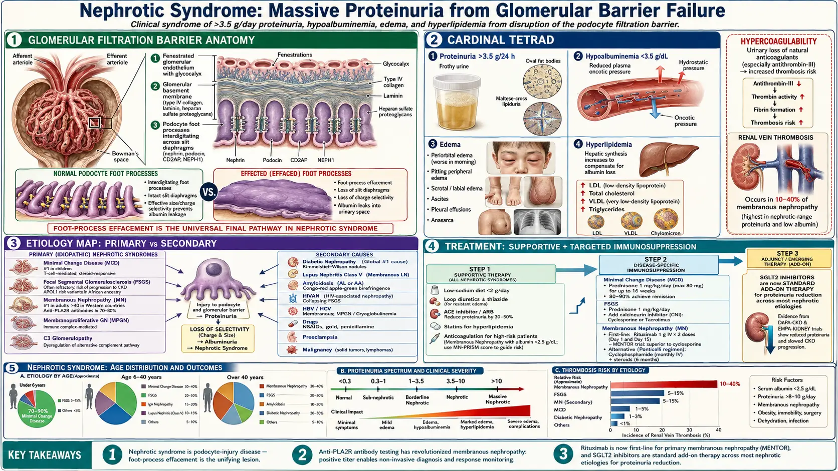
Nephrotic Syndrome is a clinical syndrome defined by heavy proteinuria (>3.5 g/day per 1.73 m²), hypoalbuminemia (<3.5 g/dL), and edema. Hyperlipidemia and lipiduria are commonly considered fourth and fifth components of the full syndrome. It results from podocyte injury disrupting the glomerular filtration barrier, leading to protein loss in urine and a cascade of secondary metabolic consequences including coagulopathy, infection susceptibility, and cardiovascular risk.
Table of Contents
1. Overview
Nephrotic syndrome is not itself a disease but a final common presentation of diverse glomerular diseases that share podocyte injury as their primary insult. The glomerular filtration barrier consists of three layers: fenestrated glomerular endothelium, glomerular basement membrane (GBM), and the podocyte slit diaphragm. Damage to any layer — particularly the podocyte — leads to protein leak. The syndrome can manifest at any age. In children, Minimal Change Disease (MCD) accounts for ~90% of cases; in adults, Focal Segmental Glomerulosclerosis (FSGS), Membranous Nephropathy, and Diabetic Nephropathy predominate. Prompt identification of the underlying cause guides specific treatment, while general measures target proteinuria reduction, edema, and complication prevention.
2. Epidemiology
Annual incidence: 3 cases per 100,000 adults; 2–7 cases per 100,000 children. Prevalence: 15–30 cases per 100,000. FSGS is the most common primary cause in Black and Hispanic Americans (40% of adult nephrotic syndrome in the US); Membranous Nephropathy is the most common cause in White adults (30–40%); MCD predominates in children (80–90% in ages 1–8 years). Diabetic nephropathy is the most common overall cause when secondary conditions are included, accounting for 30–40% of ESRD in developed nations. Male predominance in adults; FSGS has 4-fold higher incidence in Black Americans (APOL1 variants).
3. Pathophysiology
The glomerular filtration barrier maintains a charge-selective and size-selective filter. Anionic glycoproteins on endothelial and podocyte surfaces repel negatively charged albumin. Loss of charge selectivity (early in MCD) or structural disruption (FSGS, membranous nephropathy) leads to protein leakage:
Step 1 — Podocyte injury: Loss of slit diaphragm proteins (nephrin, podocin), cytoskeletal disruption (actin), and foot process effacement. Circulating factors (CLCF1 in primary FSGS), immune complexes (membranous nephropathy anti-PLA2R), or direct metabolic injury (diabetic nephropathy, hyperglycemia → PKC activation → GBM thickening).
Step 2 — Proteinuria: Albumin, immunoglobulin G (IgG), transferrin, coagulation proteins, hormone-binding proteins lost in urine.
Step 3 — Hypoalbuminemia: Hepatic albumin synthesis increases but cannot compensate for urinary loss (>3.5 g/day). Malnutrition, catabolism, protein redistribution all contribute.
Step 4 — Edema (two mechanisms):
- Underfill (classical): Low oncotic pressure → capillary fluid shift to interstitium → reduced effective circulating volume → RAAS/ADH activation → sodium/water retention
- Overfill (primary sodium retention): Primary sodium retention in collecting duct (independent of oncotic pressure); contributes in MCD where renin/aldosterone are often suppressed
Step 5 — Compensatory hyperlipidemia: Low albumin → reduced oncotic signal → hepatic lipoprotein synthesis upregulation (low-density lipoprotein (LDL), very-low-density lipoprotein (VLDL), lipoprotein(a)); reduced lipoprotein lipase activity (lost in urine); elevated low-density lipoprotein, triglycerides, lipoprotein(a).
Step 6 — Lipiduria: Filtered lipoproteins appear as oval fat bodies (free cholesterol in epithelial cells showing Maltese cross birefringence under polarized light) and fatty casts — a pathognomonic finding.
4. Causes: Primary Glomerular Diseases
Minimal Change Disease (MCD / Nil Disease)
- Characteristic: no abnormality on light microscopy; IF negative; EM shows diffuse foot process effacement only (“nil” structural change beyond podocyte)
- Most common cause in children (80–90% of ages 1–8); 10–15% of adult nephrotic syndrome
- Abrupt onset; severe edema; very heavy proteinuria (often >10 g/day); normal or near-normal complement; normal renal function usually preserved
- Pathogenesis: T-cell dysregulation → circulating lymphokine → podocyte slit diaphragm dysfunction; rarely associated with Hodgkin lymphoma, nonsteroidal anti-inflammatory drugs (in adults)
- Steroid-responsive in >90% of children; >80% of adults; excellent prognosis if treated; relapse common
Focal Segmental Glomerulosclerosis (FSGS)
- Most common primary glomerular disease causing nephrotic syndrome in US adults (particularly Black Americans)
- Primary (circulating permeability factor), secondary (obesity/hypertension/human immunodeficiency virus (HIV)/drugs), genetic (NPHS2, ACTN4, TRPC6)
- Columbia classification (tip, perihilar, cellular, collapsing, NOS)
- Steroid response lower (~30–40%) than MCD; often steroid-resistant; calcineurin inhibitors second-line; 50% ESRD at 10 years without remission
Membranous Nephropathy (MN)
- Most common primary cause of nephrotic syndrome in White adults (30–40% of adult nephrotic syndrome in Europe/North America)
- Primary MN: anti-PLA2R (phospholipase A2 receptor) antibodies in 70–80%; detected in serum; correlates with disease activity; anti-THSD7A in ~3%; newer antigens (NELL-1, SEMA3B, EXT1/EXT2 in lupus-MN)
- Pathogenesis: subepithelial immune complex deposition → complement activation (C5b-9 MAC) → podocyte injury → GBM thickening with “spikes” on silver stain; “spike and dome” pattern on EM
- Light microscopy: GBM thickening (no proliferation, no crescents); IF: granular immunoglobulin G/C3 along GBM
- Natural history: “rule of thirds” 1/3 spontaneous complete remission, 1/3 partial remission, 1/3 progressive chronic kidney disease (CKD)
- Treatment: KDIGO 2021 — wait 6 months on supportive therapy for spontaneous remission; if high-risk (anti-PLA2R titer high, glomerular filtration rate (GFR) declining, heavy persistent proteinuria): Ponticelli protocol (an alkylating agent, chlorambucil or cyclophosphamide, alternating with steroids) or rituximab (MENTOR trial: noninferior to cyclosporine for remission at 12 months, superior in maintaining remission up to 24 months); tacrolimus alternative; belimumab under investigation
Membranoproliferative GN (MPGN)
- Immune complex-mediated (infections/autoimmune) or complement-mediated (C3 GN, dense deposit disease)
- Light microscopy: mesangial proliferation + GBM thickening + “tram-track” (double contour) from new GBM laid on subendothelial deposits
- Hypocomplementemia (low C3 ± C4)
- Presents with mixed nephritic-nephrotic syndrome
5. Causes: Secondary Nephrotic Syndrome
Diabetic Nephropathy (most common worldwide secondary cause)
- Hyperglycemia → AGE formation → PKC activation → TGF-β overexpression → mesangial expansion → GBM thickening → Kimmelstiel-Wilson nodules (pathognomonic nodular glomerulosclerosis)
- Microalbuminuria (30–300 mg/day) → frank proteinuria (>300 mg/day) → nephrotic-range proteinuria
- Typically associated with >10 years of diabetes and evidence of other microvascular complications (retinopathy, neuropathy); absence of these features raises question of alternative GN
Lupus Nephritis (LN)
- ISN/RPS Class V MN: presents as nephrotic syndrome; subepithelial deposits
- Class III/IV diffuse proliferative LN: mixed nephritic-nephrotic with hematuria and RBC casts
- Characterized by “full house” IF (IgG, immunoglobulin M (IgM), immunoglobulin A (IgA), C3, C1q) — pathognomonic for lupus
Viral
- Hepatitis B: membranous nephropathy (especially in children in endemic areas)
- Hepatitis C: MPGN (Type I), cryoglobulinemic GN
- HIV: FSGS collapsing pattern (HIVAN); strongly APOL1-associated
Drug-Induced
- Nonsteroidal anti-inflammatory drugs (NSAIDs): MCD (interstitial nephritis + nephrotic syndrome), membranous nephropathy
- Gold salts, penicillamine: membranous nephropathy
- Heroin: FSGS
- Lithium: MCD, FSGS
- Pamidronate: collapsing FSGS
- Captopril: membranous nephropathy (rare with modern angiotensin-converting enzyme inhibitor (ACEi))
Malignancy-Associated
- Solid tumors (colon, lung, stomach): membranous nephropathy (anti-NELL-1, anti-THSD7A); new-onset nephrotic MN in elderly men is a recognised trigger for malignancy screening
- Lymphoma (especially Hodgkin’s): MCD
- Multiple myeloma/plasma cell dyscrasia: AL amyloidosis, light-chain deposition disease, MPGN
Amyloidosis
- AL amyloidosis: Congo red–positive deposits (apple-green birefringence with polarized light); SAP scan; Lambda > Kappa light chains; treat with plasma cell–directed therapy (bortezomib, daratumumab)
- AA amyloidosis: serum amyloid A (chronic inflammation: rheumatoid arthritis, inflammatory bowel disease (IBD), familial Mediterranean fever)
Other: pre-eclampsia, immunoglobulin A vasculitis (HSP), sickle cell disease, congenital/infantile nephrotic syndrome
6. Clinical Presentation
Classic presentation: pitting edema (periorbital edema worst in morning — characteristic), pedal/ankle edema, ascites (in children), pleural effusions; frothy urine (heavy proteinuria creates foam); weight gain (fluid retention); fatigue, dyspnea.
Physical examination: pitting edema ≥2+ (ankle, pretibial, periorbital); xanthelasma/xanthomata in chronic hyperlipidemia; pleural effusion dullness to percussion; ascites (fluid wave, shifting dullness); nail signs: leukonychia (white nails from hypoalbuminemia), Muehrcke’s lines (paired white transverse bands).
Laboratory findings: 24h urine protein >3.5 g (or spot urine PCR >3.5 mg/mg); serum albumin <3.5 g/dL (often <2.5 g/dL); total protein low; lipid panel: elevated low-density lipoprotein, triglycerides, lipoprotein(a), total cholesterol (often >300 mg/dL); urinalysis: lipiduria (oval fat bodies, Maltese crosses), fatty casts, granular casts; coagulation: elevated fibrinogen, low antithrombin III.
7. Diagnosis
Clinical diagnosis confirmed by urinalysis + labs. Kidney biopsy required in adults for etiology and to guide treatment (not required in children with classic MCD presentation who respond to steroids).
Stepwise workup:
- Quantify proteinuria: 24h urine protein (gold standard) or spot urine protein/creatinine ratio (PCR) — values ≥3.5 mg/mg confirm nephrotic-range
- Serum albumin, total protein, creatinine, estimated glomerular filtration rate (eGFR), blood urea nitrogen (BUN)
- Lipid panel, complete blood count (CBC) (normocytic anemia from hypoalbuminemia), coagulation studies (PT, antithrombin III)
- Exclude secondary causes:
- Antinuclear antibody (ANA), anti-dsDNA, complement (C3, C4): lupus nephritis
- Anti-PLA2R antibody (serum): primary membranous nephropathy — positive in 70–80%
- Hepatitis B surface antigen, anti-HBc, anti-HCV, human immunodeficiency virus serology
- Fasting glucose, hemoglobin A1c (HbA1c): diabetic nephropathy
- SPEP, UPEP, serum free light chains: myeloma/amyloidosis
- Antinuclear antibody, antineutrophil cytoplasmic antibody (ANCA) (if nephritic features coexist)
- Age >60: malignancy screen (PSA, CEA, mammography)
- Kidney biopsy (adults): light microscopy + IF + EM; provides definitive etiology; contraindicated if solitary kidney, coagulopathy, uncontrolled hypertension
- Chest X-ray/echocardiogram: pleural effusion, pericardial effusion, cardiac function (low output can mimic/worsen nephrotic edema)
Urinalysis findings:
- Heavy proteinuria (protein 3+ to 4+ on dipstick — note: dipstick detects albumin only)
- Lipiduria: oval fat bodies (epithelial cells engorged with cholesterol showing Maltese crosses on polarized microscopy)
- Fatty casts: lipid in cast matrix
- Absence of RBC casts (distinguishes from nephritic syndrome); if RBC casts present → consider overlap/mixed syndrome
8. Complications
Thromboembolism (most dangerous acute complication)
Pathogenesis: urinary loss of anticoagulant proteins (antithrombin III, protein C, protein S); increased hepatic synthesis of procoagulant factors (fibrinogen, factor VIII, vWF); reduced fibrinolysis; hyperviscosity from hyperlipidemia; immobility from edema.
Manifestations: Renal vein thrombosis (RVT) — classic in membranous nephropathy (30–50% prevalence); presents as flank pain, worsening edema, hematuria; diagnosed by computed tomography (CT) venography/Doppler; Pulmonary embolism (PE) — most dangerous; deep vein thrombosis (DVT) in lower extremities.
Anticoagulation threshold: prophylactic anticoagulation recommended if serum albumin <2.5–3.0 g/dL in membranous nephropathy; therapeutic anticoagulation for confirmed VTE; warfarin preferred over DOACs (risk of rapid protein binding changes).
Infection
Pathogenesis: urinary loss of immunoglobulin G and complement (factor B, properdin) → impaired opsonization → susceptibility to encapsulated organisms (Streptococcus pneumoniae, Gram-negative bacteria).
Risk: Spontaneous bacterial peritonitis in children with ascites; pneumococcal pneumonia/sepsis; cellulitis in edematous skin.
Prevention: pneumococcal vaccination, annual influenza vaccination; prompt treatment of infections.
Acute Kidney Injury
Pathogenesis: Severe hypoalbuminemia → reduced effective circulating volume → prerenal acute kidney injury (AKI); renal vein thrombosis → acute venous congestion; tubular toxicity from lipid-loaded casts; nephrotoxin exposure.
Cardiovascular Disease
Severe dyslipidemia (low-density lipoprotein >200 mg/dL, lipoprotein(a) elevation, high-density lipoprotein (HDL) low) accelerates atherosclerosis; nephrotic syndrome itself is an independent cardiovascular risk factor.
Hormonal and Nutritional Deficiencies
- Hypothyroidism (urinary loss of thyroid-binding globulin → low total T4; usually normal free T4 and thyroid-stimulating hormone (TSH))
- Vitamin D deficiency: urinary loss of vitamin D–binding protein → low 25-OH vitamin D → hypocalcemia
- Iron-deficiency anemia: urinary loss of transferrin
- Zinc/copper deficiency: urinary loss of binding proteins
- Malnutrition from protein wasting (net negative protein balance when proteinuria >3.5 g/day)
9. Treatment: General Measures
Proteinuria Reduction (cornerstone)
- Angiotensin-converting enzyme inhibitor or angiotensin receptor blocker (ARB): standard therapy for nephrotic patients; reduce intraglomerular pressure and proteinuria by 30–40%; slow chronic kidney disease progression; usually titrated to the maximum tolerated dose
- Sodium-glucose cotransporter-2 (SGLT2) inhibitors: dapagliflozin/empagliflozin — reduce proteinuria via tubuloglomerular feedback; recommended in addition to RAAS blockade for estimated glomerular filtration rate ≥20; demonstrated chronic kidney disease benefit in DAPA-CKD and EMPA-KIDNEY trials
- Dietary sodium restriction (<2 g/day): reduces edema and improves response to diuretics
- Dietary protein: moderate restriction (0.8 g/kg/day); high protein intake worsens proteinuria
Edema Management
- Loop diuretics: furosemide 40–120 mg/day orally; intravenous (IV) in severe resistance; thiazide addition in refractory cases
- Albumin + furosemide intravenous: for severe hypoalbuminemia with diuretic resistance (intravenous albumin transiently raises oncotic pressure, allowing furosemide to reach tubular lumen); short-term measure
- Fluid restriction in severe hyponatremia or massive edema
Hyperlipidemia
- HMG-CoA reductase inhibitors (statins): reduce low-density lipoprotein; tolerated; may reduce proteinuria modestly; anti-inflammatory pleiotropic effects; standard of care in persistent nephrotic dyslipidemia
- Fibrates: for severe hypertriglyceridemia (>500 mg/dL); use with caution with statins (myopathy risk; fenofibrate preferred over gemfibrozil)
Anticoagulation
- Prophylactic in membranous nephropathy with albumin <2.5–3.0 g/dL or additional risk factors
- Therapeutic anticoagulation for renal vein thrombosis, PE, DVT; international normalized ratio (INR) target 2–3 (warfarin); unfractionated heparin is generally not favoured in severe hypoalbuminemia (altered protein binding)
Blood Pressure
The KDIGO 2021 blood pressure guideline proposes a systolic target of less than 120 mmHg (standardized office reading) for most people with chronic kidney disease not on dialysis, except children and kidney transplant recipients (older guidance used <125/75 mmHg in proteinuric chronic kidney disease); angiotensin-converting enzyme inhibitor/angiotensin receptor blocker first-line; calcium channel blocker or thiazide added if needed.
Vaccination
Pneumococcal (PCV20/PPSV23), influenza, hepatitis B vaccination before immunosuppression.
10. Treatment: Disease-Specific
Minimal Change Disease
- Prednisone 1 mg/kg/day (max 80 mg): first-line; 90% remission in children within 4–8 weeks; in adults 80% remission within 16 weeks
- Taper after remission over 3–6 months; relapse in 75% of children (frequently relapsing/steroid-dependent)
- Steroid-sparing: cyclophosphamide (8–12 weeks); cyclosporine/tacrolimus; MMF; rituximab (Iijima Lancet 2014)
- Risks of prolonged high-dose steroids: growth impairment in children, osteoporosis, diabetes, infections
Primary Membranous Nephropathy
- First 6 months: conservative (angiotensin-converting enzyme inhibitor/angiotensin receptor blocker + SGLT2i) with anti-PLA2R monitoring — 30–35% spontaneous remission
- High-risk triggers for treatment (any of): estimated glomerular filtration rate decline >30%, persistent heavy proteinuria >4 g/day despite 6 months supportive therapy, severe nephrotic complications
- Rituximab (preferred per KDIGO 2021, MENTOR trial): 375 mg/m² × 4 doses or 1000 mg × 2 doses; anti-PLA2R titer decline predicts remission; fewer side effects than cyclophosphamide
- Ponticelli protocol: methylprednisolone 1 g/day intravenous × 3 days then prednisone 0.5 mg/kg/day alternating monthly with chlorambucil 0.2 mg/kg/day × 6 months; effective (60–70% remission) but significant toxicity
- Cyclosporine/tacrolimus: effective but high relapse rate on cessation
- Rituximab treatment is paired with monitoring of the anti-PLA2R titer response
FSGS
Prednisone 1 mg/kg/day × 16–24 weeks; calcineurin inhibitors for steroid-resistant; rituximab; sparsentan; angiotensin-converting enzyme inhibitor/angiotensin receptor blocker + SGLT2i always. (See FSGS page for full detail.)
Lupus Nephritis
High-dose corticosteroids + cyclophosphamide (EURO-LUPUS) or MMF induction; hydroxychloroquine in all systemic lupus erythematosus (SLE); belimumab (BLISS-LN) + voclosporin (AURORA-1) as adjunct therapies for classes III/IV/V; azathioprine/MMF maintenance.
Diabetic Nephropathy
Tight glycemic control (hemoglobin A1c <7%); angiotensin-converting enzyme inhibitor/angiotensin receptor blocker; sodium-glucose cotransporter-2 inhibitors (finerenone [FIDELIO-DKD] in addition for residual proteinuria); semaglutide/glucagon-like peptide-1 (GLP-1) agonists (FLOW trial: kidney protection); RAAS optimization.
Amyloidosis
AL amyloidosis — plasma cell–directed therapy (CyBorD/bortezomib-based, daratumumab); AA amyloidosis — treat underlying inflammatory disease; interleukin-1 (IL-1) blockers in FMF.
Key Research Papers
- Ronco P, Debiec H. Pathogenesis of membranous nephropathy: recent advances and future challenges. Nat Rev Nephrol. 2012;8(4):203–213. — PubMed PMID: 22371247 · doi:10.1038/nrneph.2012.35
- Sethi S, Fervenza FC. Membranoproliferative glomerulonephritis: a new look at an old entity. N Engl J Med. 2012;366(12):1119–1131. — PubMed PMID: 22435371 · doi:10.1056/NEJMra1108178
- Fervenza FC, et al. Rituximab or Cyclosporine in the Treatment of Membranous Nephropathy (MENTOR). N Engl J Med. 2019;381(1):36–46. — PubMed PMID: 31269364 · doi:10.1056/NEJMoa1814427
- Heerspink HJL, Stefánsson BV, Correa-Rotter R, et al. Dapagliflozin in Patients with Chronic Kidney Disease (DAPA-CKD). N Engl J Med. 2020;383(15):1436–1446. — PubMed PMID: 32970396 · doi:10.1056/NEJMoa2024816
- Iijima K, Sako M, Nozu K, et al. Rituximab for childhood-onset, complicated, frequently relapsing nephrotic syndrome or steroid-dependent nephrotic syndrome: a multicentre, double-blind, randomised, placebo-controlled trial. Lancet. 2014;384(9950):1273–1281. — PubMed PMID: 24965823 · doi:10.1016/S0140-6736(14)60541-9
- EMPA-KIDNEY Collaborative Group. Empagliflozin in Patients with Chronic Kidney Disease. N Engl J Med. 2023;388(2):117–127. — PubMed PMID: 36331190 · doi:10.1056/NEJMoa2204233
- Kidney Disease: Improving Global Outcomes (KDIGO) Glomerular Diseases Work Group. KDIGO 2021 Clinical Practice Guideline for the Management of Glomerular Diseases. Kidney Int. 2021;100(4S):S1–S276. — PubMed PMID: 34556256 · doi:10.1016/j.kint.2021.05.021
- Cheung AK, Chang TI, Cushman WC, et al. Executive summary of the KDIGO 2021 Clinical Practice Guideline for the Management of Blood Pressure in Chronic Kidney Disease. Kidney Int. 2021;99(3):559–569. — PubMed PMID: 33637203 · doi:10.1016/j.kint.2020.10.026
- Branten AJ, Reichert LJ, Koene RA, Wetzels JF. Oral cyclophosphamide versus chlorambucil in the treatment of patients with membranous nephropathy and renal insufficiency. QJM. 1998;91(5):359–366. — PubMed PMID: 9709470 · doi:10.1093/qjmed/91.5.359
- Sethi S, Fervenza FC. Standardized classification and reporting of glomerulonephritis. Nephrol Dial Transplant. 2019;34(2):193–199. — PubMed PMID: 30124958 · doi:10.1093/ndt/gfy220
- Wheeler DC, Toto RD, Stefánsson BV, et al. A pre-specified analysis of the DAPA-CKD trial demonstrates the effects of dapagliflozin on major adverse kidney events in patients with IgA nephropathy. Kidney Int. 2021;100(1):215–224. — PubMed PMID: 33878338 · doi:10.1016/j.kint.2021.03.033
PubMed Topic Searches
- PubMed: Nephrotic syndrome treatment adults
- PubMed: Membranous nephropathy rituximab anti-PLA2R
- PubMed: Minimal change disease steroid resistant
- PubMed: FSGS nephrotic syndrome APOL1
- PubMed: Nephrotic syndrome hypercoagulability renal vein thrombosis
- PubMed: Diabetic nephropathy nephrotic syndrome SGLT2 inhibitors
- PubMed: Nephrotic syndrome children steroid relapsing
Connections
- Nephrology & Hepatology
- Focal Segmental Glomerulosclerosis
- Immunoglobulin A Nephropathy
- Glomerulonephritis
- Rapidly Progressive GN
- Chronic Kidney Disease
- Acute Kidney Injury
- Lupus Nephritis
- Diabetes Mellitus
- Deep Vein Thrombosis
- Kidney Function Tests
- Creatinine
- Urinalysis
- Edema
- Hypertension
- Membranous Nephropathy — the leading cause of nephrotic syndrome in White adults.